2470
Ultrashort Echo Time Magnetization Transfer Imaging of Meniscus After a Marathon1Department of Radiology,Fifth Affiliated Hospital, Sun Yat-Sen University, Zhuhai, China, 2MR Research, GE Healthcare, Beijing, China, 3University of California, San Diego, Department of Radiology, San Diego, CA, United States
Synopsis
Keywords: MSK, Joints
In this study, a 3D UTE-MT preparation will be used to detecting changes in the meniscus of amateur marathon runners before and after a marathon. In this prospective cohort study, 23 amateur marathon runners were enrolled. There were three MRI scans (pre-race, 2 days post-race, and 4 weeks post-race) using the UTE-MT and UTE-T2* sequences. UTE-MTR values decreased 2 days post-race and increased after 4 weeks of rest. Conversely, the UTE-T2* values increased 2 days post-race and increased after 4 weeks of rest. UTE-MTR is a promising biomarker for the detection of changes in the meniscus after a marathon.INTRODUCTION
Long-distance running can cause meniscus damage, resulting in alterations in knee force lines that are linked to the development of osteoarthritis1. Monitoring early alterations of meniscus by using non-invasive MR had an extensive prospect. In this study, a 3D ultrashort echo time MRI sequence with magnetization transfer (UTE-MT) preparation will be used to test the detection of changes in the meniscus of amateur marathon runners before and after a marathon.METHODS
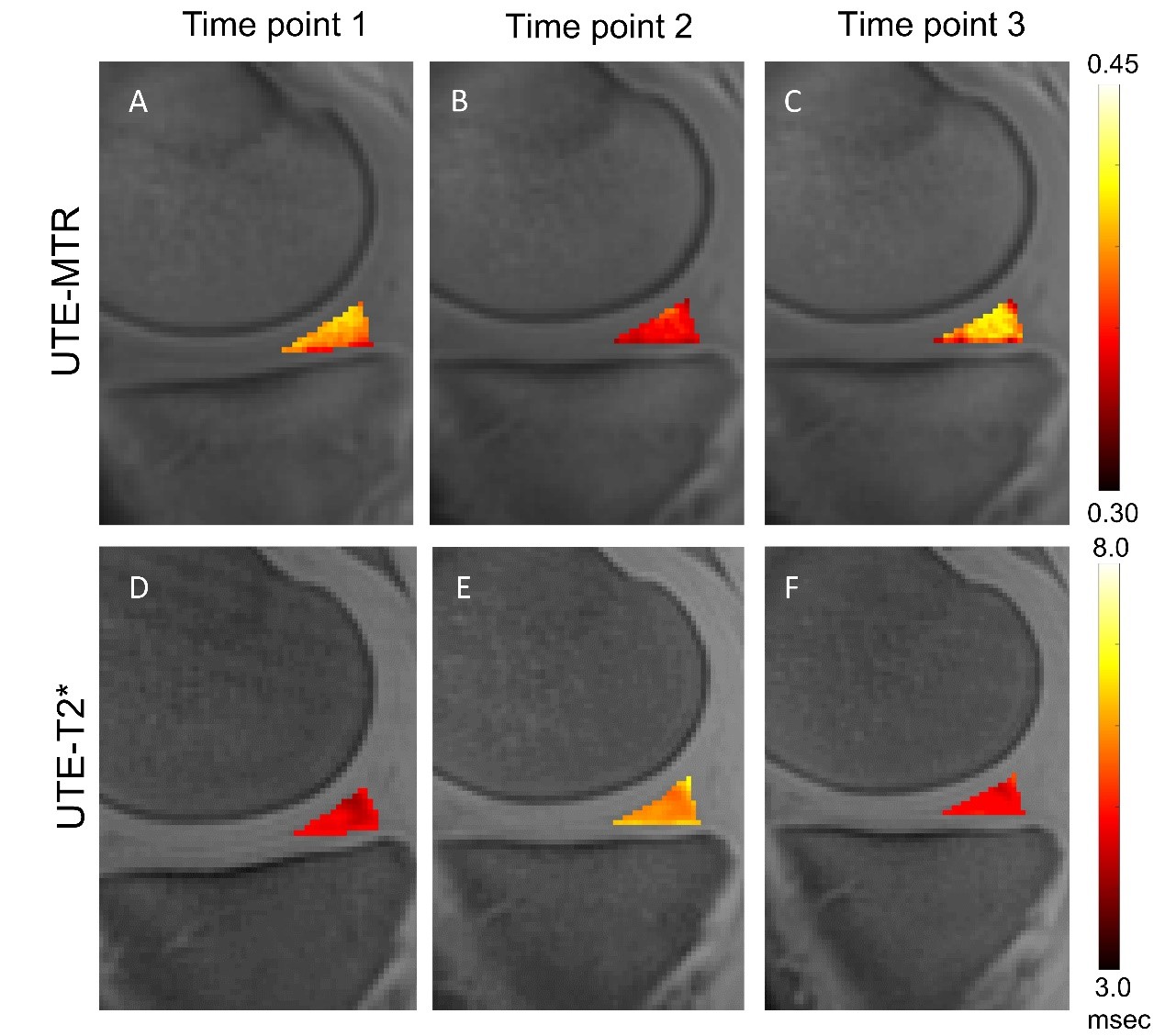
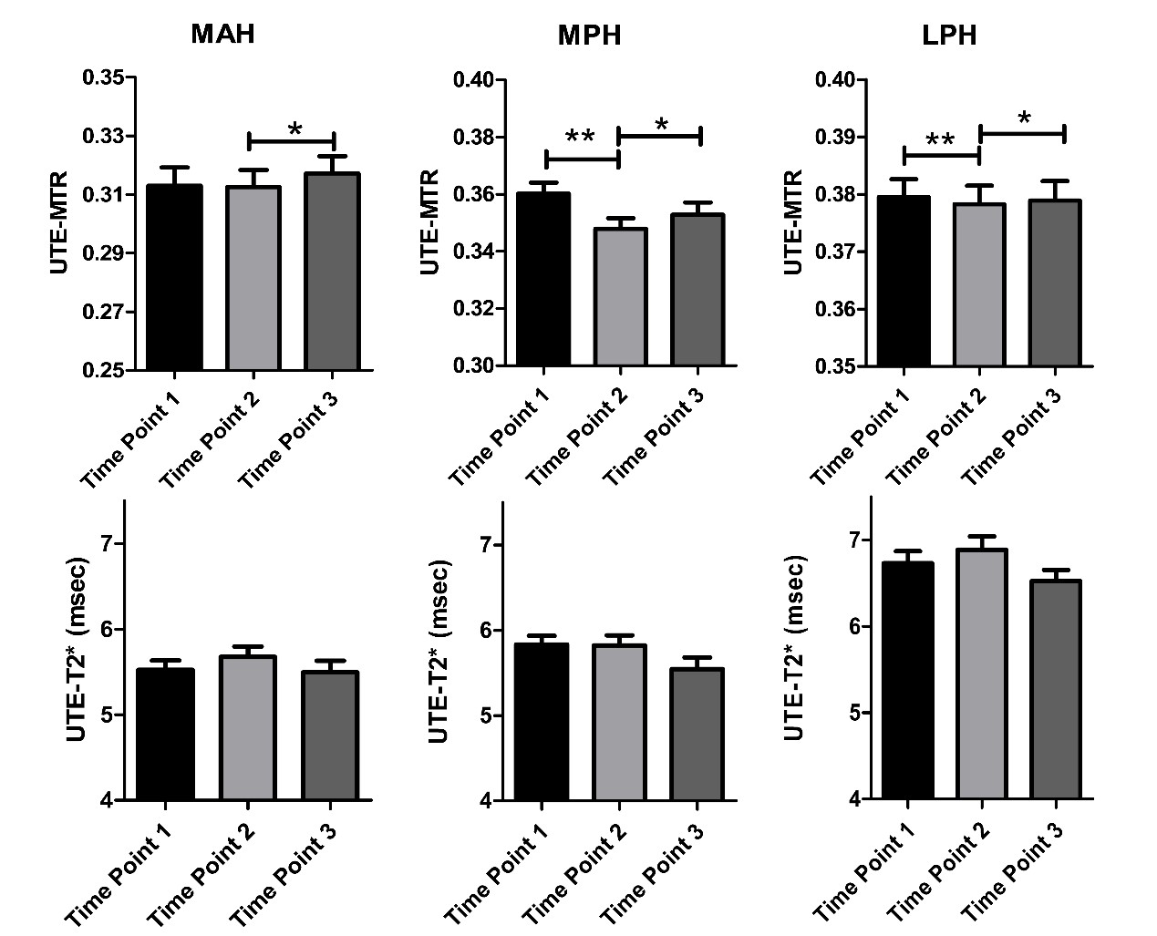
In this prospective cohort study, 23 amateur marathon runners (46 knees) were enrolled. There were three MRI scans (pre-race, 2 days post-race, and 4 weeks post-race) using the UTE-MT and UTE-T2* sequences. Four subregions of the meniscus had their UTE-MT ratio (UTE-MTR) and UTE-T2* assessed before and after running.RESULTS
For most subregions of meniscus, the UTE-MTR values decreased 2 days post-race and increased after 4 weeks of rest. Conversely, the UTE-T2* values increased 2 days post-race and increased after 4 weeks of rest. UTE-MTR values in the medial posterior horn (MPH) and lateral posterior horn (LPH) regions at 2 days post-race were significantly lower than those at pre-race and 4 weeks post-race (P < 0.05). By comparison, only the UTE-T2* values in medial posterior horn showed a significant difference.DISCUSSION
In this study, the UTE-MTR values in meniscus decreased while UTE-T2* values increased after a marathon. These changes may be caused by the mild degenerative damage induced by fatigue loading in meniscus during long-distance running. It has been recognized that in the early stages of meniscal degeneration, there is an increase in water permeability due to breakage of the collagen fibrous network2,3. In the meantime, part of the collagen’s bound water becomes free water as a result of collagen integrity loss, resulting in a decrease of the collagen’s bound water percentage and an increase of the free water percentage. The efficacy of magnetization exchange between the water and collagen pools is thereby reduced, leading to decreased UTE-MTR values4. On the other hand, the increased free water fraction results in a higher UTE-T2* value in the degenerated meniscus2,3.CONCLUSION
UTE-MTR is a promising biomarker for the detection of biochemical changes in the meniscus after a marathon.Acknowledgements
This article is supported by the National Natural Science Found (No. 82101995); the National Natural Science Found(82172053).References
[1] Greene, G.W., X. Banquy, D.W. Lee, et al (2011) Adaptive mechanically controlled lubrication mechanism found in articular joints. Proc Natl Acad Sci U S A 108(13):5255-9.
[2] Eijgenraam, S.M., F.A.T. Bovendeert, J. Verschueren, et al (2019) T(2) mapping of the meniscus is a biomarker for early osteoarthritis. Eur Radiol 29(10):5664-5672.
[3] Zarins, Z.A., R.I. Bolbos, J.B. Pialat, et al (2010) Cartilage and meniscus assessment using T1rho and T2 measurements in healthy subjects and patients with osteoarthritis. Osteoarthritis Cartilage 18(11):1408-16.
[4] Zhang, X., Y.J. Ma, Z. Wei, et al (2021) Macromolecular fraction (MMF) from 3D ultrashort echo time cones magnetization transfer (3D UTE-Cones-MT) imaging predicts meniscal degeneration and knee osteoarthritis. Osteoarthritis Cartilage 29(8):1173-1180.
Figures