2464
Quantitative assessment of the adipose tissue around the knee joint in patients with KOA using IDEAL-IQ and T2 mapping sequences with 3.0 T MRI1Medical Imaging Center, First Affiliated Hospital, Jinan university, Guangzhou, China, 2MR Research, GE Healthcare, Guangzhou, China
Synopsis
Keywords: Cartilage, Osteoarthritis
IDEAL-IQ and T2 mapping sequences are capable of objectively assessing fat and water content in the adipose tissue around the knee joint. The severity of KOA is related to the fat and water content of knee adipose tissue.
purpose
Adipose tissue is now thought of as an endocrine organ that participates in metabolism and inflammation[1]. Adipocytes can synthesize and release physiologically active molecules and adipokines, inflammatory cytokines that contribute to synovial inflammation, matrix metalloproteinase production, cartilage degeneration, and bone remodelling during KOA pathogenesis. Consequently, adipose tissue should be evaluated as a mechanism underlying the development of KOA. The IDEAL-IQ sequence has been used to estimate the proton-density fat fraction (PDFF) and fibrosis of adipose tissue in many organs [2-6]. In addition, the regional quantification of the transverse relaxation time constant (T2 mapping) sequence, as a noninvasive quantitative biochemical MRI technique that provides information concerning the variation of collagen and water in cartilage, is widely used to evaluate articular cartilage injury[7-10]. The main purpose of this study was to evaluate knee adipose tissue in KOA patients and healthy volunteers using T2 mapping and IDEAL-IQ quantified MR sequences and to explore the effect of knee adipose tissue on KOA.Methods
Eighty patients with newly diagnosed KOA and thirty-two healthy volunteers were enrolled in 3.0 T superconducting MR scanner. Sagittal IDEAL-IQ sequence and sagittal T2 mapping sequence were performed. T2 values were obtained from the T2 mapping sequence, and PDFF values were obtained from the FF fraction image of the IDEAL-IQ sequence using the Advantage Workstation version 4.5 image postprocessing workstation (GE Healthcare). Two radiologists manually delineated regions of interest (ROIs) on the median sagittal plane T2 mapping image maps and FF fraction maps. Each radiologist delineated three ROIs on the IPFP, SPFP, subchondral bone marrow (SubcBM) and deep bone marrow (DBM) on consecutive slices for measurements and calculated the average. The cartilage lesions were scored with the modified WORMS system on an eight-point scale[11] . Grade 0 lesions were classified as the normal group; Grade 1 and 2 lesions were classified as the mild cartilage damage group; Grade 2.5 and 3 lesions were classified as the moderate cartilage damage group; and Grade 4 to 6 lesions were classified as the severe cartilage damage group.Statistical analysis was performed using SPSS version 25.0. Univariate analysis was used to compare the PDFF and T2 values among the subgroups. Multivariate analysis was employed to evaluate the effects of different adipose tissues on the severity of KOA. The correlation between the PDFF and T2 values was analysed by the Pearson correlation coefficient.
Results
32 subjects were classified into the normal group, 19 subjects were classified into the mild subgroup, 21 subjects were classified into the moderate subgroup, and 40 subjects were classified into the severe subgroup.Univariate analysis revealed that the PDFF values of the IPFP (F=22.797; p< 0.001), SPFP (F=35.034; p<0.001), and SubcBM (c2=59.489; p<0.001) and the T2 values of the IPFP (c2=30.268; p<0.001) and SPFP (c2=28.777; p<0.001) were significantly associated with KOA. Ordered logistic regression analyses showed that the PDFF values of the IPFP, SPFP and SubcBM were independent protective factors for KOADiscussion
Our study evaluated the efficiency of the IDEAL-IQ sequence in detecting the severity of KOA, which reflected the fat content of different adipose tissues in the knee joint. T2 mapping was also conducted to investigate whether water content was correlated with fat content in KOA.KOA could lead to fibrosis of the IPFP and SPFP, and the fibrosis of adipose tissue could contribute to the decrease in the PDFF values of the IPFP and SPFP.[12, 13]. Moreover ,inflammatory stimulation leads to vascularization increases in the IPFP and SPFP in KOA [14].
In this study, we compared the water content of IPFP, SPFP, SubcBM, and BM tissues in patients with different stages of KOA and a healthy control group using the T2 mapping sequence. The results confirmed that the T2 values of the IPFP and SPFP in the healthy control group were higher than those in the KOA subgroups at different stages. Chronic inflammation is associated with pathological fibrosis[15] [16]. This suggests that the abnormality represented by this signal has a potentially important role in osteoarthritis progression.
In our study, it was found that the fat pad tissue around the knee joint of KOA patients showed a decrease in fat content and water content, and there was a correlation between them. The correlation between the changes in fat content and water content in the SPFP and SubcBM was more obvious. Pathologically, OA is characterized by the progressive degeneration of articular cartilage, the abnormal reconstruction of subchondral bone, and subchondral bone sclerosis[17]. Therefore, SubcBM fat and water contents may be reduced with KOA progression.
Conclusion
IDEAL-IQ and T2 mapping sequences are capable of objectively assessing fat and water content in the adipose tissue around the knee joint. The severity of KOA is related to the fat and water content of knee adipose tissue.Acknowledgements
no.References
1. Whitehead A, Krause FN, Moran A, MacCannell ADV, Scragg JL, McNally BD, et al. Brown and beige adipose tissue regulate systemic metabolism through a metabolite interorgan signaling axis. Nat Commun 2021; 12: 1905.
2. Guo RM, Li QL, Luo ZX, Tang W, Jiao J, Wang J, et al. In Vivo Assessment of Neurodegeneration in Type C Niemann-Pick Disease by IDEAL-IQ. Korean J Radiol 2018; 19: 93-100.
3. Chen Y, Long L, Jiang Z, Zhang L, Zhong D, Huang X. Quantification of pancreatic proton density fat fraction in diabetic pigs using MR imaging and IDEAL-IQ sequence. BMC Med Imaging 2019; 19: 38.
4. Liau J, Shiehmorteza M, Girard OM, Sirlin CB, Bydder M. Evaluation of MRI fat fraction in the liver and spine pre and post SPIO infusion. Magn Reson Imaging 2013; 31: 1012-1016.
5. Chen R, Bai Y, Liu T, Zhang G, Han Y, Chen L, et al. Evaluation of Glypican-3 Expression in Hepatocellular Carcinoma by Using IDEAL IQ Magnetic Resonance Imaging. Acad Radiol 2020.
6. Ren C, Zhu Q, Yuan H. Mono-exponential and bi-exponential model-based diffusion-weighted MR imaging and IDEAL-IQ sequence for quantitative evaluation of sacroiliitis in patients with ankylosing spondylitis. Clin Rheumatol 2018; 37: 3069-3076.
7. Wang D, Yin H, Liu W, Li Z, Ren J, Wang K, et al. Comparative analysis of the diagnostic values of T2 mapping and diffusion-weighted imaging for sacroiliitis in ankylosing spondylitis. Skeletal Radiol 2020; 49: 1597-1606.
8. Endo K, Takahata M, Sugimori H, Yamada S, Tadano S, Wang J, et al. Magnetic resonance imaging T1 and T2 mapping provide complementary information on the bone mineral density regarding cancellous bone strength in the femoral head of postmenopausal women with osteoarthritis. 2019; 65: 13-18.
9. Bristela M, Skolka A, Eder J, Szomolanyi P, Weber M, Piehslinger E, et al. T2 mapping with 3.0T MRI of the temporomandibular joint disc of patients with disc dislocation. Magn Reson Imaging 2019; 58: 125-134.
10. Chen M, Qiu L, Shen S, Wang F, Zhang J, Zhang C, et al. The influences of walking, running and stair activity on knee articular cartilage: Quantitative MRI using T1 rho and T2 mapping. PLoS One 2017; 12: e0187008.
11. Peterfy CG, Guermazi A, Zaim S, Tirman PFJ, Miaux Y, White D, et al. Whole-Organ Magnetic Resonance Imaging Score (WORMS) of the knee in osteoarthritis. OSTEOARTHR CARTILAGE OSTEOARTHRITIS AND CARTILAGE 2004; 12: 177-190.
12. Ioan-Facsinay A, Kloppenburg MJNRR. Osteoarthritis: Inflammation and fibrosis in adipose tissue of osteoarthritic joints. 2017.
13. Barboza E, Hudson J, Chang WP, Kovats S, Towner RA, Silasi‐Mansat R, et al. Profibrotic Infrapatellar Fat Pad Remodeling Without M1 Macrophage Polarization Precedes Knee Osteoarthritis in Mice With Diet‐Induced Obesity. Arthritis & Rheumatology 2017; 69: 1221-1232.
14. Favero M, El-Hadi H, Belluzzi E, Granzotto M, Porzionato A, Sarasin G, et al. Infrapatellar fat pad features in osteoarthritis: a histopathological and molecular study. Rheumatology 2017; 56: 1784-1793.
15. Peng CY, Liao YW, Lu MY, Yang CM, Hsieh PL, Yu CC. Positive Feedback Loop of SNAIL-IL-6 Mediates Myofibroblastic Differentiation Activity in Precancerous Oral Submucous Fibrosis. Cancers (Basel) 2020; 12.
16. Han W, Aitken D, Zhu Z, Halliday A, Wang X, Antony B, et al. Hypointense signals in the infrapatellar fat pad assessed by magnetic resonance imaging are associated with knee symptoms and structure in older adults: a cohort study. Arthritis Research & Therapy 2016; 18.
Figures
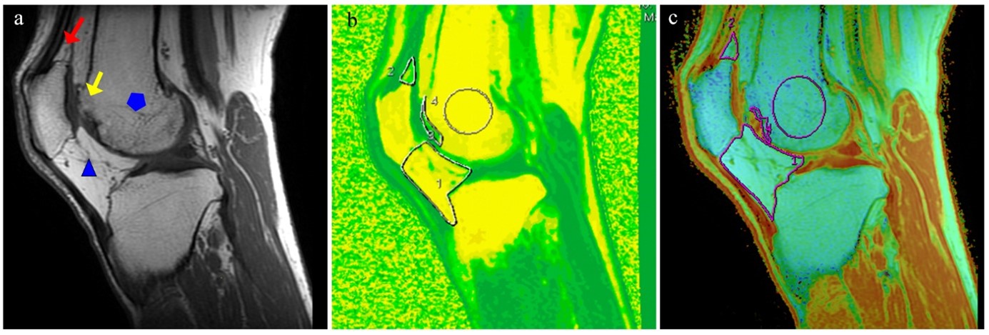
Outline of ROIs of adipose tissue in different parts around the knee joint. a, T1-weighted image showing the fat around the knee joint. red arrow, blue triangle, yellow arrow, and blue star represents SPFP, IPFP, SubcBM and cartilage injury area and DBM of the femur, respectively. b, the measurement of PDFF value ROIs in different parts; c, ROIs measurement of T2 value in different parts.