2462
Evaluation of whole compartment and regional meniscal T2* values within elite athletes
Erin C Argentieri1, Sara E Sacher1, Ek Tsoon Tan1, Garry Gold2, Hollis G Potter1, Sharmila Majumdar3, and Matthew F Koff1
1Radiology and Imaging, Hospital for Special Surgery, New York, NY, United States, 2Stanford University, Stanford, CA, United States, 3University of California San Francisco, San Francisco, CA, United States
1Radiology and Imaging, Hospital for Special Surgery, New York, NY, United States, 2Stanford University, Stanford, CA, United States, 3University of California San Francisco, San Francisco, CA, United States
Synopsis
Keywords: Cartilage, Quantitative Imaging
Results of the current study demonstrate compartment- and region-specific differences in medial and lateral meniscal T2* values. Meniscal T2* values may provide a non-invasive means to assess response of the tissue specific to the loading environment and identify the early onset meniscal degeneration. A better understanding of the early changes in meniscal T2* values may help define the progression of meniscal degeneration, prior to the onset of gross morphologic defects.Introduction
Basketball players, and other cutting and jumping athletes are at increased risk for sustaining acute meniscal injuries, which often result from high compressive forces with concomitant flexion or rotation of the knee joint.1,2,3 As meniscal pathology is strongly correlated with the early onset and progression of degenerative joint disease,4 and elite basketball players have an inherently high risk of sustaining meniscal injuries, evaluation of quantitative magnetic resonance imaging (qMRI) metrics of the meniscus within these athletes may improve clinical insight. Ultra-short echo (UTE) MRI sequences capture the rapid transverse relaxation times associated with the menisci5,6 and allows for quantitative evaluation of tissue microstructure with derived T2* maps of imaged structures. While prior studies have demonstrated that T2* metrics are correlated to meniscal degeneration7,8 no studies have evaluated meniscal T2* metrics within elite athletes over time nor have attempted to evaluate the differences in T2* metrics between weight-bearing (WB) and non-weight bearing (NWB) athletes. Therefore, the objective of this study was to evaluate meniscal T2* metrics within and between collegiate basketball players (WB) and swimmers (NWB) to determine if global (whole meniscus) and regional meniscal T2* metrics change over time.Methods
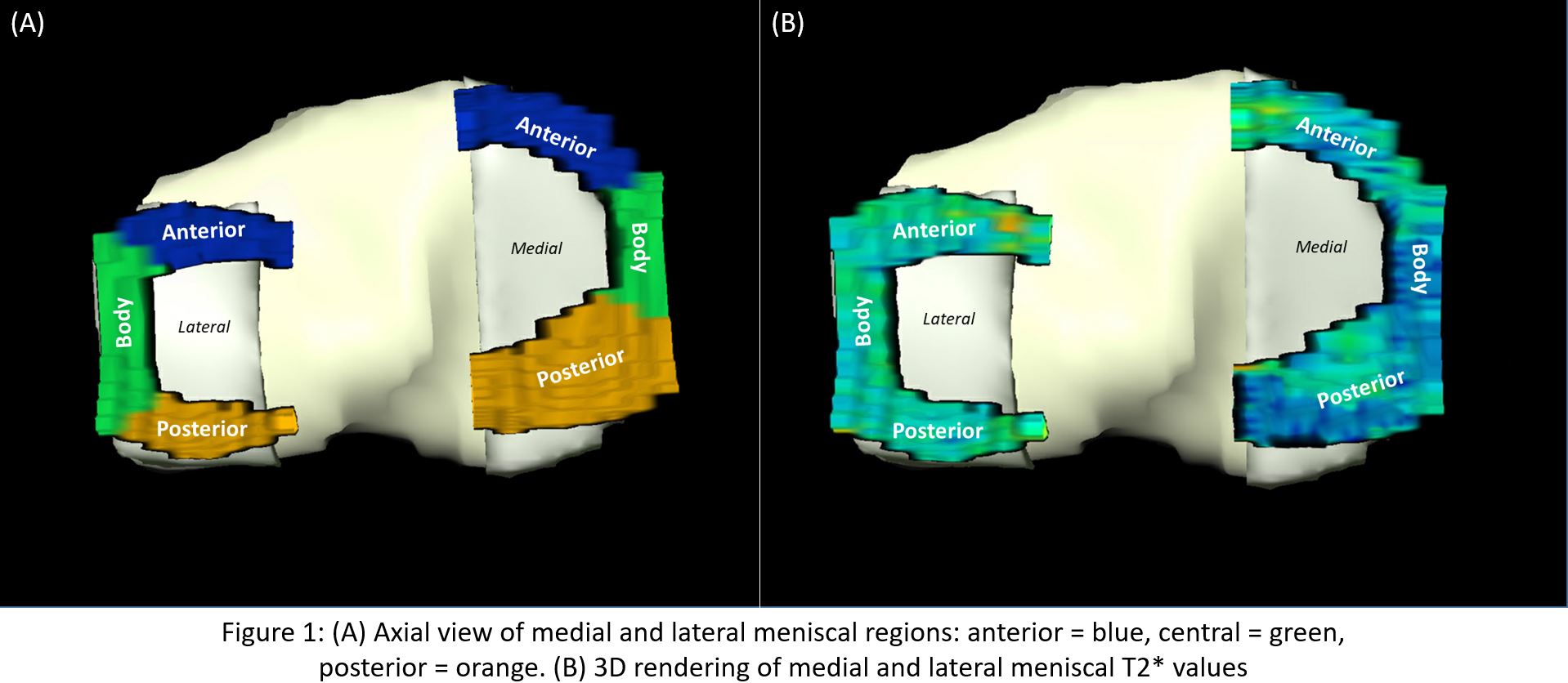
This was an IRB approved longitudinal multi-site study that included 32 collegiate athletes (16=swimmers/16=basketball players). Longitudinal data were collected at 2 pre-season study visits (TP#1 and TP#2) with a 1-year interval. A total of 11/32 subjects (34%, 7=swimmers, 4=basketball players) had data collected at the two time points while the remaining 21/31 subjects (66%) had a single pre-season study visit (TP#1). MRI Acquisition: Bilateral MRI examinations were performed on clinical 3T scanners (GE Healthcare, Waukesha, WI) using an 8-channel phased array knee coil (Invivo). Medial and lateral menisci were manually segmented (MeVisLab) from three-dimensional, Cones UTE sequences (TEs: 5 echoes between 0.03-24ms, TR: 188ms, voxel size: 0.63x0.63x3mm3, RBW: ±83.3kHz, Flip-Angle: 16°). Medial and lateral menisci were further separated into subregions (Anterior/Body/Posterior) by dividing the whole meniscus into thirds. Meniscal T2* metrics were calculated via a mono-exponential fit of signal intensity to corresponding echo time (Matlab, Natick, MA). Statistical Analysis: One- and two-way repeated measures ANOVA and regression analyses were performed to evaluate the effects of WB vs NWB sport (swimming/basketball), knee compartment (medial/lateral meniscus), region (anterior/body/posterior) and timepoint (TP#1 and TP#2) on corresponding T2* values (SAS, V9.3, Cary, NC). All data were evaluated in a side-specific (right/left) manner, with significance at p<0.05.Results
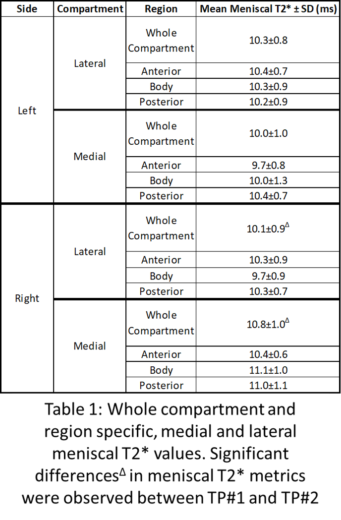
No significant differences in medial or lateral meniscal T2* values were exhibited between WB and NWB athletes at either timepoint (p>0.05). However, significant differences in meniscal T2* values were found by compartment (medial/lateral), and region (anterior, body, posterior) both within and between timepoints. Differences by Compartment & Region: Medial and lateral menisci of both right and left knees exhibited significant differences in whole compartment meniscal T2* values (mean difference: 0.2ms, p≤0.01). Further analysis revealed significant inter-regional differences in meniscal T2 metrics between respective medial and lateral meniscal subregions (Anterior/Body/Posterior). Specifically, within left-sided knees, the posterior region of the medial meniscus (10.4±0.7ms) was significantly prolonged relative to the body (10.0±1.3ms) and anterior (9.7±0.8ms) regions (p≤0.0003). In addition, in right-sided knees, the body of the lateral meniscus had significantly shorter T2* values (9.7±0.9ms, p<0.0001) than the anterior (10.3±0.9ms) and posterior regions (10.3±0.7ms, Table 1).Discussion
Significant differences in compartmental and regional T2* metrics detected between the medial and lateral menisci reflect the differences in structure and function between the medial and lateral menisci10,11 and are likely the result of the disparate loading environments between the medial and lateral compartments.10 Though basketball players are exposed to higher compression and shear forces in their knee during play, no significant differences in meniscal T2* metrics were evident between WB and NWB athletes. The lack of differences of T2* between the basketball players and swimmers may be attributable to limited subjects available for participation at TP#2. Additionally, as TP#1 and TP#2 data were collected 12 months apart, WB and NWB athletes were in the same phase of (pre-season) training for both visits – training phase similarities may have contributed to the lack of differences seen between WB and NWB athletes over time. However, when data from case and control subjects were combined for regional evaluations, significant changes in left sided medial meniscal T2* were revealed, but not for all regions. This furthers the notion of the importance of regional analyses as whole compartment analyses could have averaged out significant findings.Conclusion
These initial findings demonstrate compartment and regional differences in medial and lateral meniscal T2* values and could benefit from concomitant assessment of the adjacent femoral and tibial articular surfaces. Meniscal T2* values may provide a non-invasive means to assess response of the tissue specific to the loading environment and identify the early onset meniscal degeneration. A better understanding of the longitudinal changes in meniscal T2* values can help define the progression of meniscal degeneration in elite athletes.Acknowledgements
The authors acknowledge funding from the GE/NBA Research Consortium, and wish to thank all of the HSS techs and support staff for their assistance with this study.References
1. Zedde 2014; 2. Yeh 2012; 3. Ripani 2012; 4. Lohmander 2007; 5. Gold 1995; 6. Gatehouse 2003; 7. Koff 2014; 8. Williams 2012; 10. Bloecker 2012; 11. Stoller 2007Figures
DOI: https://doi.org/10.58530/2023/2462