2460
Preoperative Femoral Cartilage T2 Relaxation Time Predicts Change in PROs in Patients with Degenerative Meniscal Tears1Radiology, Stanford University, Stanford, CA, United States, 2Mechanical Engineering and Division of Biomedical Engineering, University of Saskatchewan, Saskatoon, SK, Canada, 3Mechanical Engineering, Stanford University, Stanford, CA, United States, 4VA Palo Alto Health Care System, Palo Alto, CA, United States, 5Orthopedic Surgery, Stanford University, Stanford, CA, United States
Synopsis
Keywords: Cartilage, Joints, Meniscus, Knee, Patient Reported Outcomes
Meniscectomies are common but not always successful in treating degenerative meniscus tears. Preoperative patient reported outcomes (PROs) and quantitative MRI could help surgeons decide candidacy for meniscectomy. This study demonstrated that pre-operative T2 relaxation times in the medial central femoral cartilage are correlated to pre-operative PROs in patients with degenerative meniscal tears and are predictive of 1 and 2-year changes in PROs post-operation. This study suggests patients who have longer T2 relaxation times pre-operation might not benefit as much from a meniscectomy.Introduction
The meniscus is a structure that provides rotational and translational stability to the knee1, but it is prone to degeneration and tears with age. Arthroscopic meniscectomy is a surgical treatment for degenerative meniscus tears. However, a meniscectomy is related to an increased risk for developing knee osteoarthritis (OA)2 and is not always successful, as often measured by patient reported outcomes (PROs) surveys, such as the Western Ontario Meniscal Evaluation Tool (WOMET). The WOMET provides comprehensive information on patients’ physical symptoms, daily lifestyles, and emotions regarding their knee3. T2 relaxation times, a quantitative MRI measure that assesses collagen microstructure and hydration in cartilage, have been used to identify early degenerative cartilage changes in the knee as increased T2 relaxation times have been linked to OA disease progression4. Regions of interests in OA have been focused on the medial weight bearing cartilage since relatively high mechanical loads on the medial knee contribute to medial weight bearing cartilage degeneration. Thus, the purpose of this study was to test the hypotheses that longer pre-operative T2 relaxation times in the central medial femoral cartilage 1) are associated with worse PROs at baseline and 2) are predictive of PROs changes from baseline to 1 and 2-years post-meniscectomy.Methods
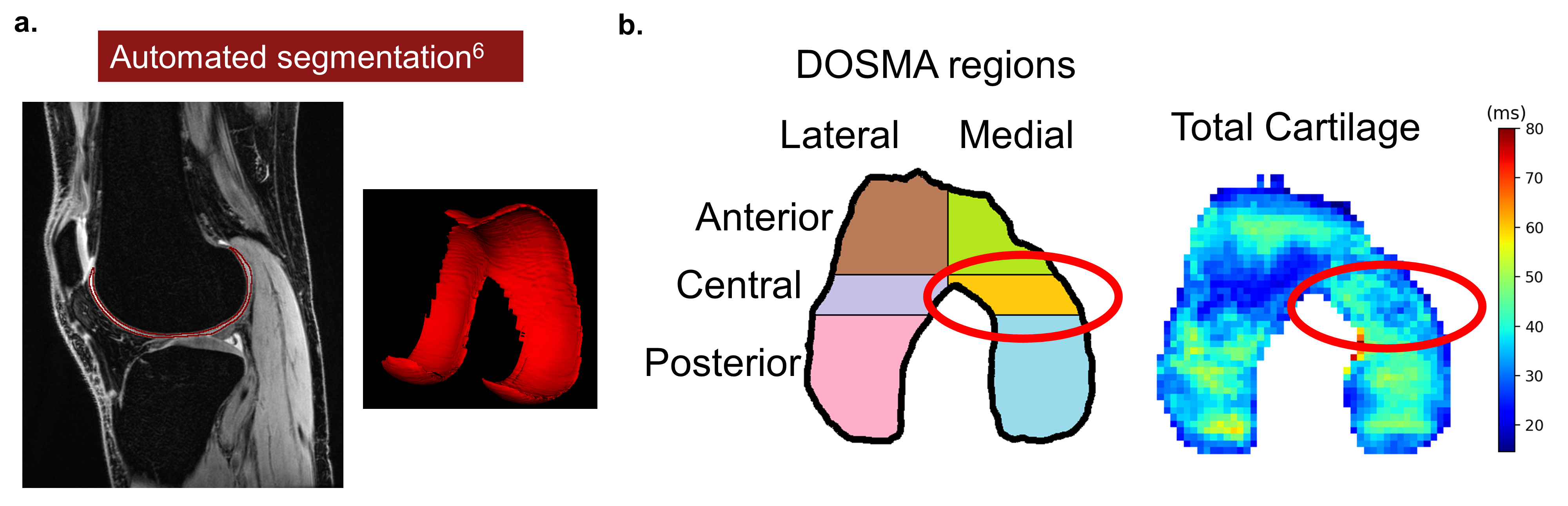
Nine male patients (average±stdev: 58.1±10.6 yrs, 30.3±4.0 kg/m2, 1.4±1.0 KL grade) with degenerative meniscal tears were enrolled in this Institutional Review Board approved study. Pre-operative unilateral MRI scans of their knees (6 left knees) were performed using a whole-body 3 Tesla (3T) MRI scanner (GE Healthcare, Waukesha, WI, USA) with a 16-channel flexible phased-array-receive-only coil (NeoCoil, Pewaukee, WI, USA). The scan sequence used to acquire T2 relaxation times of the knee was qDESS5 (FOV=160mm; TR=29.5ms; TEs=10.6/48.4ms; slice thickness=1.4mm; number of slices=84; matrix size=384x320). Cartilage was automatically segmented using a publicly available deep-learning model provided by DOSMA, an open-source Python framework for musculoskeletal analysis6. All cartilage segmentations were manually reviewed, and no modification to the segmentations were made (Figure 1a). Cartilage T2 relaxation times were determined analytically using previously described methods5. Mean T2 relaxation times were calculated for the full thickness of the cartilage of the central region of the medial femoral condyle using DOSMA6 (Figure 1b).WOMET surveys were conducted pre-meniscectomy (baseline) and at 1- and 2-years post-meniscectomy. WOMET scores were converted to a percentage scale (0-100%) where a lower WOMET score indicates more knee problems3. For prediction of WOMET change, the percent differences between raw scores (out of 1600) at baseline and 1-year and baseline and 2-years WOMET were also calculated.
Data was checked for normality using Shapiro-Wilks tests. A repeated measures ANOVA, with posthoc t-tests, was done to determine differences between baseline and follow-up WOMET measures. The relationship between baseline T2 and baseline Total WOMET, as well as baseline T2 and longitudinal changes in Total WOMET were determined using linear regressions. The significance threshold was P < 0.05.
Results
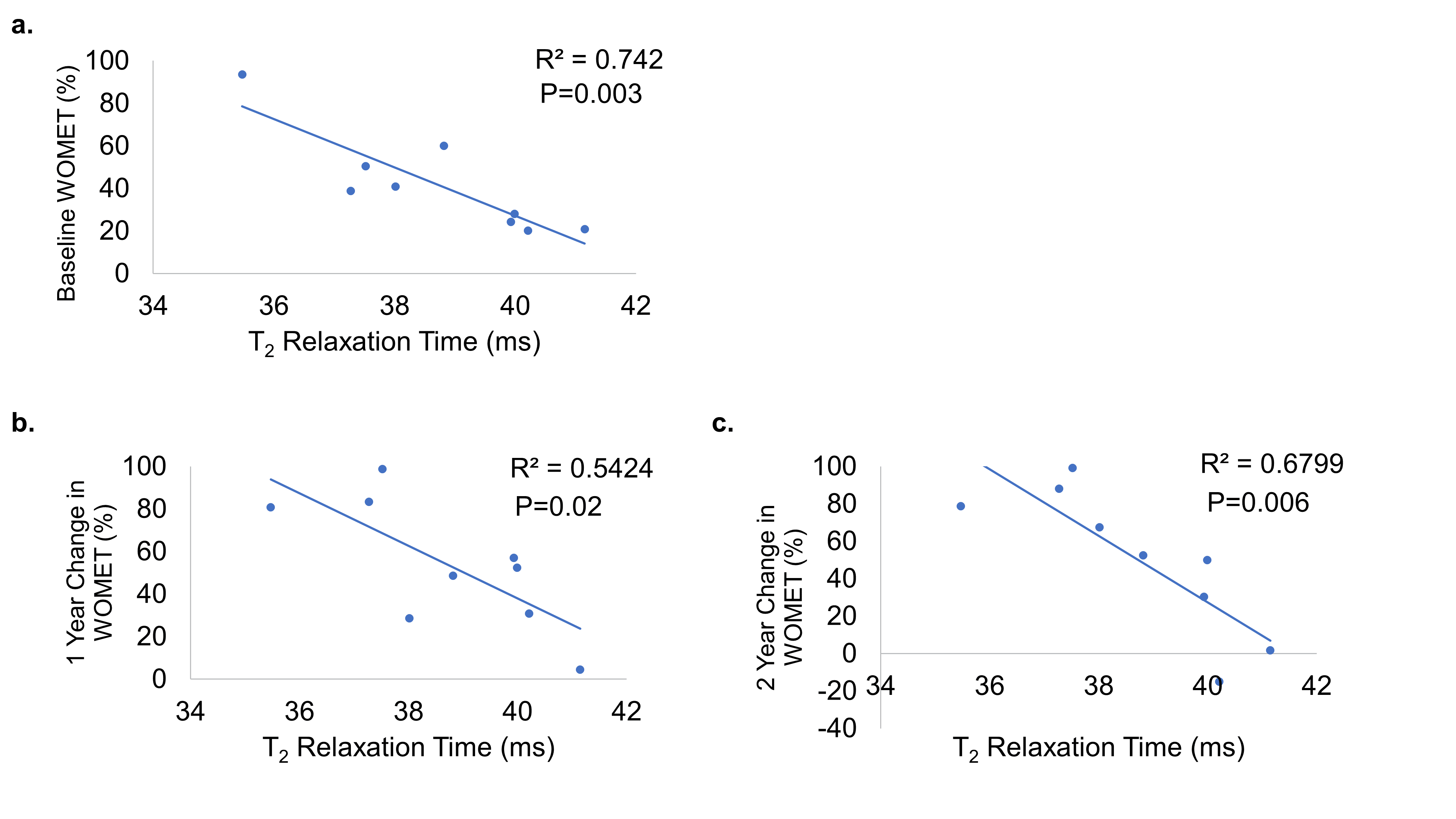
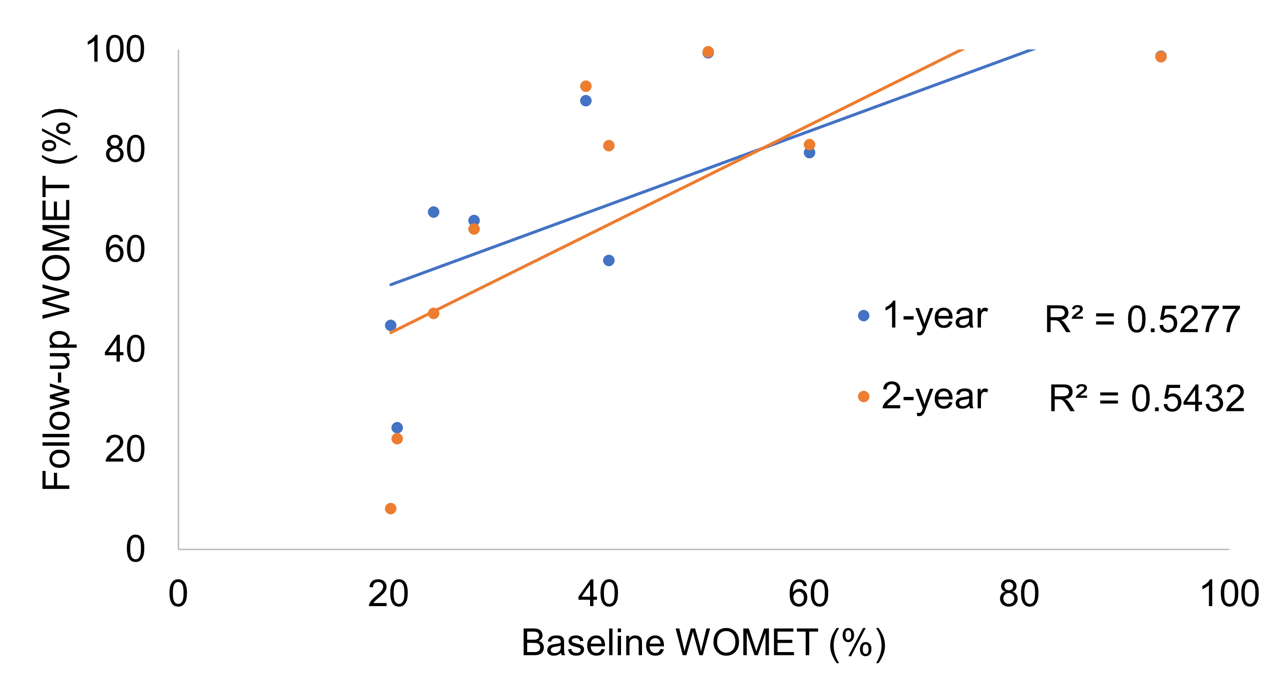
The average (± standard deviation) baseline T2 relaxation time was 41.5±1.5 ms in the medial central femoral cartilage. Average Total WOMET score at baseline was 41.8±23.7%, and there was a significant improvement in Total WOMET from baseline to 1 and 2-years post-meniscectomy: 69.8±25.2% (P=0.002), and 66.0±33.5% (P=0.013), respectively. Mean T2 relaxation time at baseline correlated with baseline Total WOMET (R2=0.74, P=0.003) and the change of Total WOMET from baseline to 1-year (R2=0.54, P=0.02) and 2-years (R2=0.68, P=0.006) post-meniscectomy (Figure 2). Total WOMET at baseline was positively correlated with Total WOMET score at 1-year (R2=0.53, P=0.03) and 2-years (R2=0.54, P=0.02) post-meniscectomy (Figure 3).Discussion
We found that mean baseline T2 relaxation times predicted 1 and 2-year changes in Total WOMET score post-meniscectomy. The T2 relaxation times were also associated with PROs at baseline. Patients with lower T2 relaxation times, had less pre-operative knee symptoms (higher Total WOMET score). Patients with higher T2 relaxation times, an early indicator of cartilage degeneration and OA disease progression4, had more pre-operative knee symptoms (lower Total WOMET score). Patients with lower T2 relaxation times at baseline experienced the greatest relative increase in PROs post meniscectomy. Though as a group, the PROs significantly improved; patients with high WOMET scores pre-operation maintained high WOMET scores at the follow-ups, and patients with low WOMET scores remained low. Recent studies propose preserving the meniscus and not performing meniscectomy as a first resort7. However, our findings suggest that early surgical intervention, before cartilage degeneration, may maximize patient outcomes. Future research with a larger sample size is needed to confirm these results.Conclusion
This work demonstrates that medial central femoral T2 relaxation times are cross-sectionally correlated to PROs in degenerative meniscus patients pre-operation and are predictive of the change in PROs at 1 and 2-years post-meniscectomy. This information may be helpful in deciding when and in what patients to perform a meniscectomy.Acknowledgements
This work was supported by the Veterans Administration (1I01RX000924-01A2), National Institutes of Health (R01EB002524, R01AR077604), GE Healthcare, Wu Tsai Human Performance Alliance, and Canadian Institutes of Health Research Postdoctoral Fellowship.References
1. Levy IM, Torzilli PA, Warren RF. The effect of medial meniscectomy on anterior-posterior motion of the knee. J Bone Joint Surg. 1982. 64: 883-888.
2. Englund M, Roemer FW, Hayashi D, et al. Meniscus pathology, osteoarthritis and the treatment controversy. Nat Rev Rheumatol. 8: 412-419.
3. Kirkley A, Griffin S, Whelan D. The development and validation of a quality of life-measurement tool for patients with meniscal pathology: the Western Ontario Meniscal Evaluation Tool (WOMET). Clin J Sport Med. 2007. 17: 349-356.
4. Prasad AP, Nardo L, Schooler J, et al. T1r and T2 relaxation times predict progression of knee osteoarthritis. Osteoarthr Cartil. 2013; 21(1):69–76.
5. Sveinsson B, Chaudhari AS, Gold GE, Hargreaves BA. A simple analytic method for estimating T2 in the knee from DESS. Magn Reson Imaging. 2017;38:63–70.
6. Desai AD. Barbieri M, Mazzoli V, Rubin E, et al. DOSMA: A deep-learning, open-source framework for musculoskeletal MRI analysis. Proc Intl Soc Mag Reson Med. 2019; 1106(27).
7. Beaufils Ph, Becker R, Kopf S, et al. Surgical management of degenerative meniscus lesions: the 2016 ESSKA meniscus consensus. Knee Surg Sports Traumatol Arthrosc. 2017. 25:335–346
Figures