2454
Preserved patellar cartilage at the expense of anterior femoral cartilage damage in Hip Osteoarthritis: An Inter-Limb Inter-Joint qMRI analysis1Department of Radiology and Biomedical Imaging, University of California, San Francisco (UCSF), San Francisco, CA, United States, 2Department of Physical Therapy and Rehabilitation Science, University of California, San Francisco (UCSF), San Francisco, CA, United States
Synopsis
Keywords: Cartilage, Osteoarthritis, Bilateral, Hip, Knee
It is well documented that osteoarthritis often affects multiple joints in patients with the disease. However, direct associations between limbs and across the joints of the lower extremity remains understudied. With bilateral hip and knee MRI of 18 patients, this study utilizes automated segmentation and VBR-atlas-based methods to explore the associations between twelve femoral and acetabular sub-compartments with trochlear and patellar cartilage T1rho, T2 values. Significant negative correlations between the patellar T2 and anterior hip femoral T1rho and T2 values, suggest a probable ongoing gait dysfunction. The data-distribution indicates possibility of a subgroup of patients having different progression and compensatory trajectories, to be explored further with gait pattern analysis.Introduction
Total-joint-replacement, being the eventual course of action in hip osteoarthritis (HOA), is a temporary solution-in-nature and cost-ineffective overall1. This leads the research focus into direction of preventing joint-health in a holistic way, i.e., identifying early signs of degeneration of the other lower limb joints in terms of proteoglycan loss2 and collagen-network disruption, understanding the effectiveness of altering gait patterns and balancing mechanical loading of the joint etc3. One of the most important steps in this direction, would be to understand the complex relationship in multi-joint disease, including between limb and across the joints of the lower extremity. Direct associations between cartilage health in multiple joints of the lower extremity remains an understudied topic can offer potential for biomechanical intervention depending on the nature of the relationship. Specifically, associations of hip degenerations and the contralateral patellofemoral joint (PFJ) have received limited attention. Literature provides ample examples4 of a significant association between hip strength, abductor weakness, internal rotation, and patellofemoral pain in the same limb. Evidence also suggests5 patellofemoral joint symptoms often are seen as precursors of developing knee OA. We hypothesize, in case of HOA patients, there might exist a direct relationship between the compositional biomarkers, i.e., T1rho and T2 values, of affected hip cartilage and contralateral patellofemoral cartilage, that might explain the effect of biomechanical changes associated with HOA on the knee.Methodology
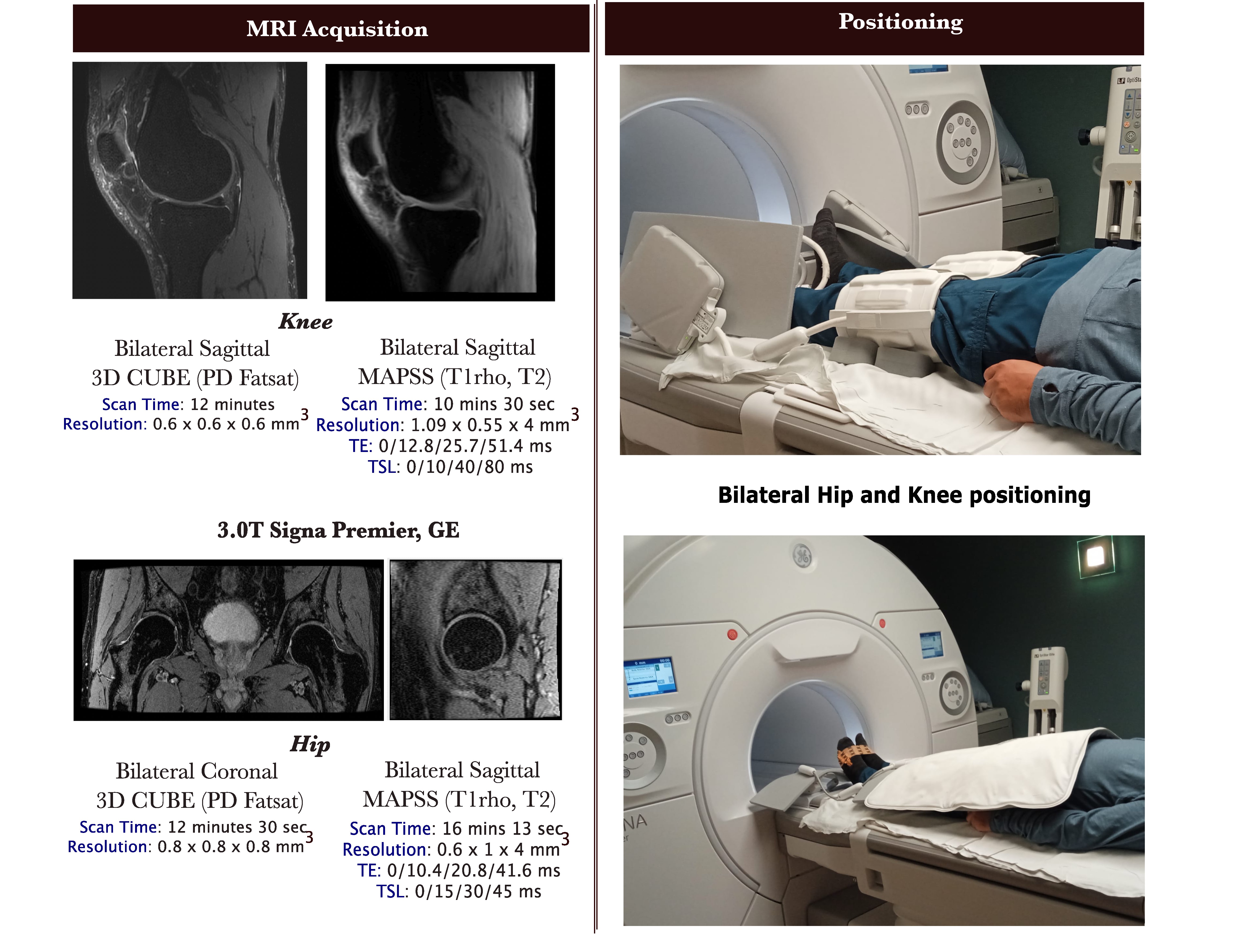
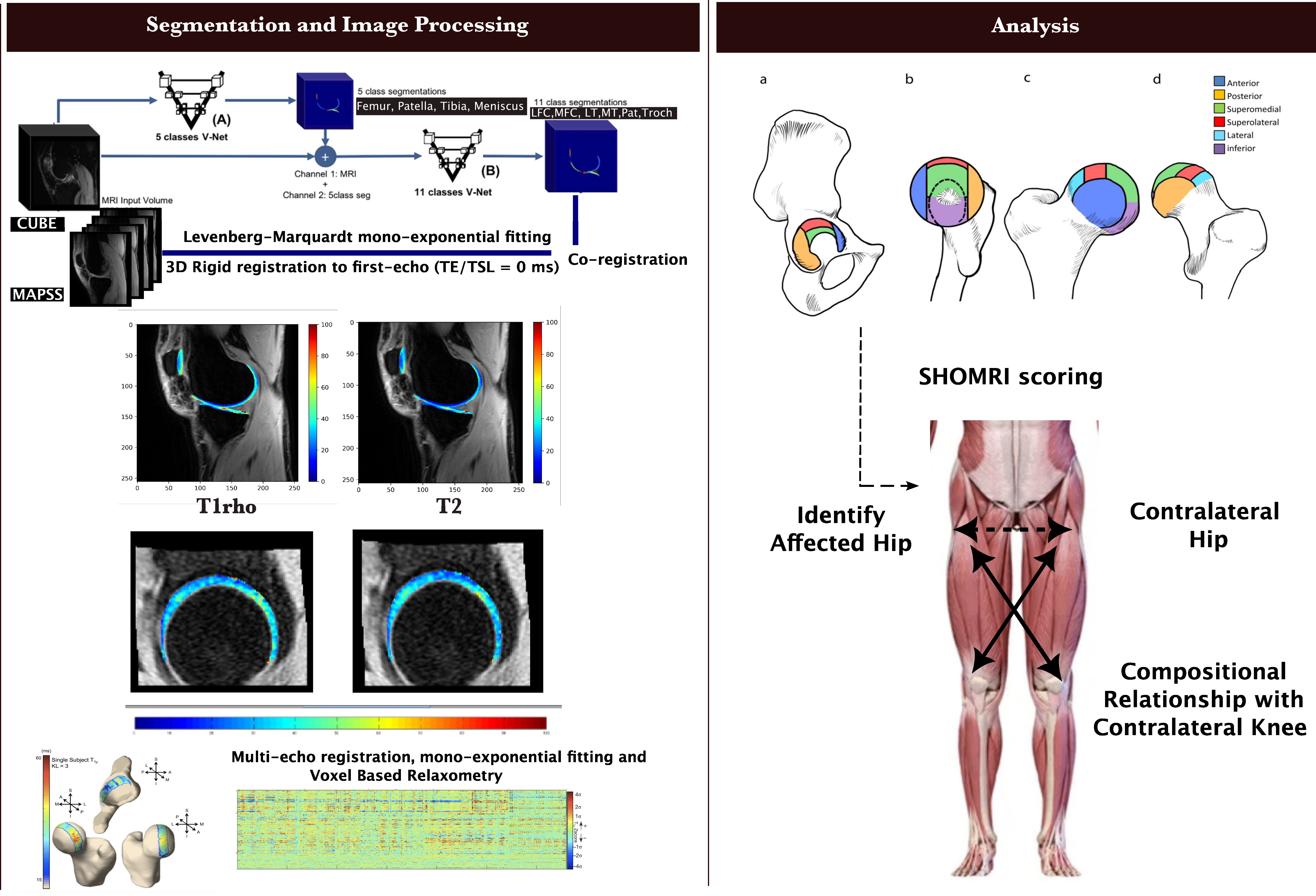
Eighteen radiographic HOA patients (age:57.33±12.34 years, seven females) provided written consents for the IRB-approved ongoing study and underwent MRI scanning in a 3.0T Signa-Premier scanner (GE Healthcare) for bilateral morphological and compositional hip and knee imaging. The scanning protocol with patient positioning is explained in figure-1. As explained in figure-2, the T1rho and T2 values were obtained using a 3D-rigid-registration and Levenberg-Marquardt mono-exponential fitting algorithm6. For region-of-interest (ROI) based analysis of bilateral hip compositional values, a voxel-based-relaxometry approach7 was taken, thereby resulting in six anterior and posterior sub-compartmental T1rho and T2 values for each of the femoral and acetabular cartilages. In the knees, bilateral trochlear and patellar cartilages were segmented using a 3D-V-Net-architecture8, from the morphological images co-registered with the compositional images, to extract the trochlear and patellar T1rho and T2 values for knee. A radiologist, specialized in MSK-imaging, semi-quantitatively scored the hip following the SHOMRI-guidelines9 for the whole-joint HOA evaluation. The SHOMRI-scores of both hips were compared to identify the affected hip joint. HOOS scores were collected for all the patients. Hip-extension maximum isometric torque (Nm) were measured using a HUMAC-NORM-dynamometer (Computer Sports Medicine, inc). Pearson partial correlation matrices were computed between the affected sub-compartmental femoral and acetabular cartilage T1rho and T2 values with those of the contralateral limb trochlear and patellar cartilages, adjusted for age and gender. Statistical significances were accordingly adjusted for partial correlation.Results and Discussion
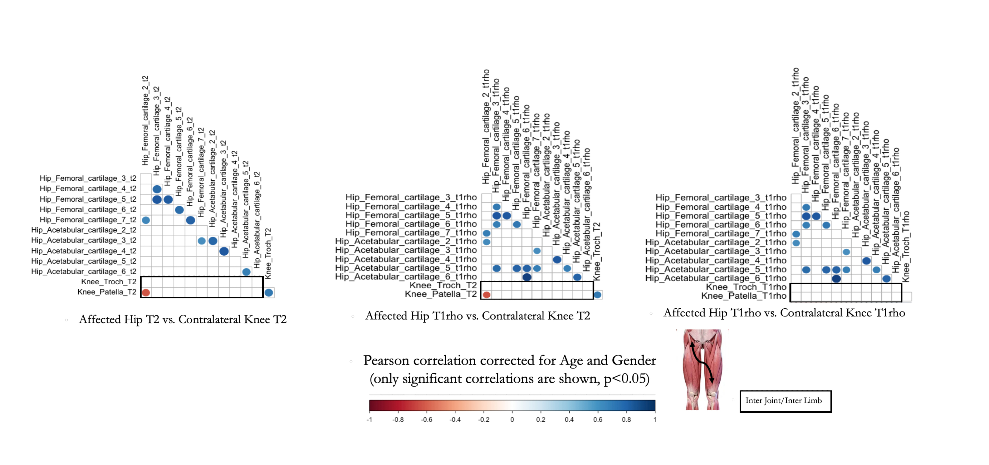
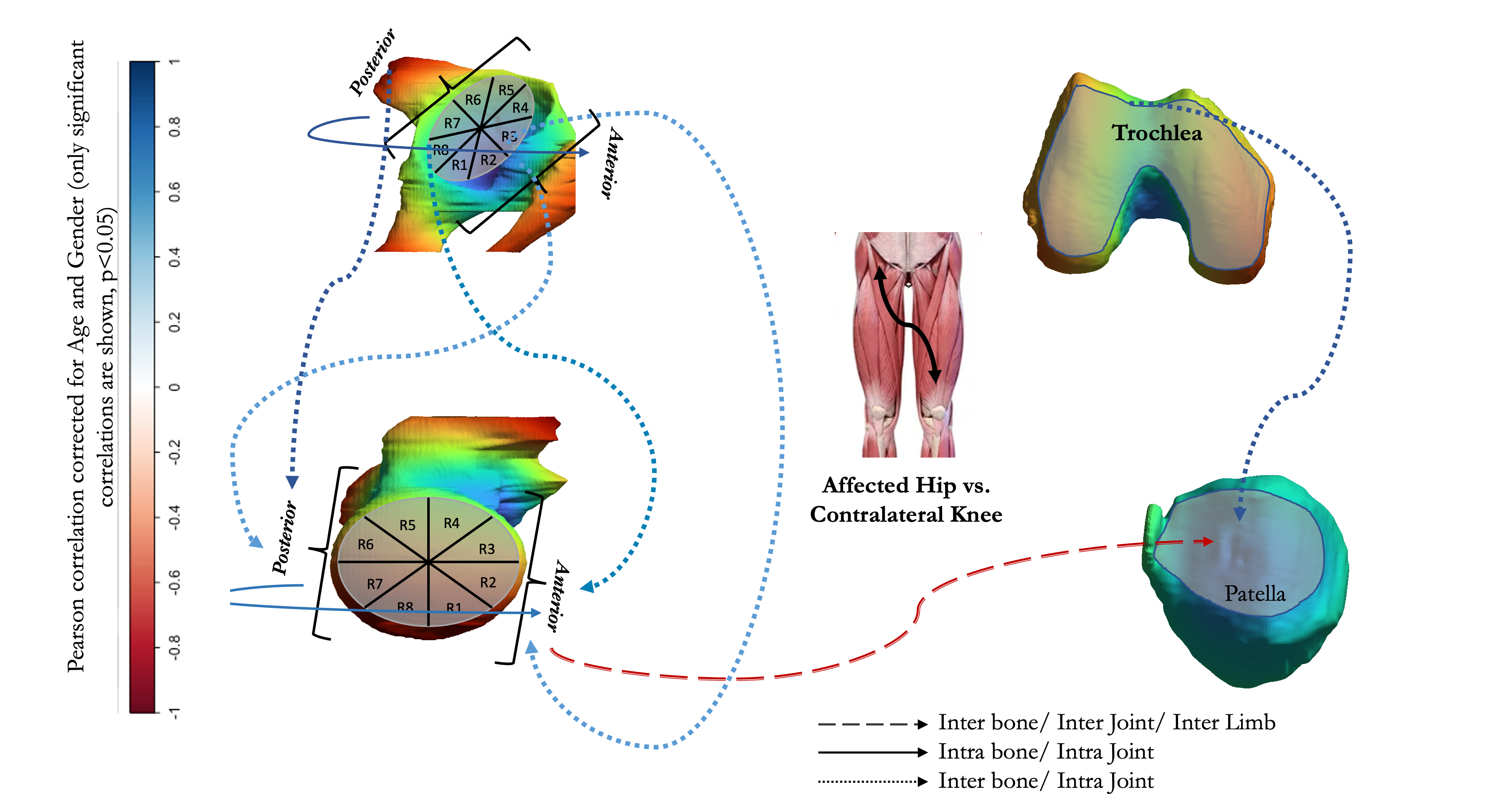
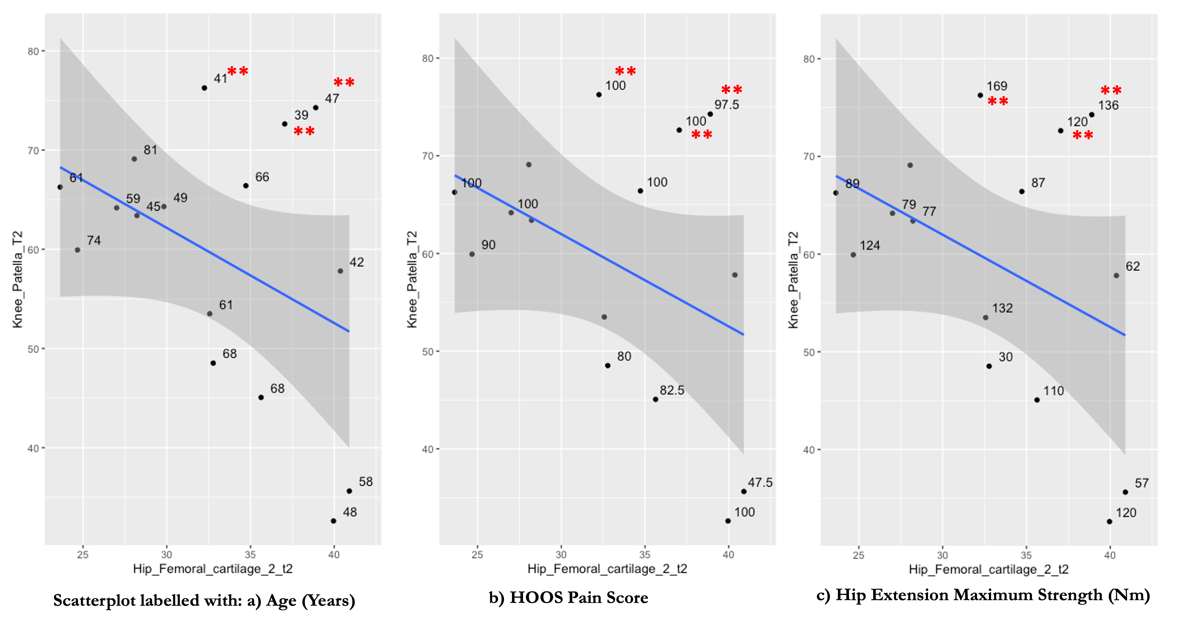
The Pearson’s partial correlation matrix between affected hip and contralateral patellofemoral cartilages, adjusted for age and gender, are shown in figure-3. Significant positive correlations are observed between hip femoral and acetabular sub-compartments (inter-bone/intra-joint and intra-bone/intra-joint) as well as knee trochlear and patellar sub-compartments (inter-bone/intra-joint). These findings are well expected and conforms to previously reported literature10, befitting the understanding of the cartilage compartments progressing or regressing jointly in the degenerative setting of OA. The most noteworthy inter-bone/inter-joint/inter-limb observation, however, is the significant negative correlation (R = -0.57) between the patellar T2 and anterior hip femoral T1rho and T2 values. These findings suggest the potential worsening of the anterior hip femoral cartilage and subsequent prolongation of T1rho and T2 values are associated with lowered patellar T2 values, thereby indicating preserved cartilage health. The associations are depicted in a pictorial representation in figure-4. Although the causality of the relationship cannot be directly inferred from given data, previous associations reported4 between hip biomechanical range of motions and strengths of hip and ipsilateral knee can be referred to speculate an underlying compensatory gait mechanism between the affected hip anterior femoral and the contralateral patellofemoral joint. We also investigated the data distributions of these parameters in the scatterplots, figure-5. Interestingly, young HOA patients with reported no or minimal HOOS pain scores and higher hip extension strengths were found to be drifting from the fitted relationship and were observed to have worse knee cartilage health as compared to the rest of the HOA patients. This may suggest that there are two ongoing trajectories existing in this population of patients, which can be better explored with including higher number of subjects as well as detailed biomechanical analysis to explain the mediated effect.Conclusion
We present one of the first exploratory results between inter-and-intra-bone/inter-and-intra-joint/inter-limb compositional correlation analysis of HOA affected femoral and acetabular T1rho, T2 values with those of the knee patellofemoral cartilage. Significant negative correlations between the patellar T2 and anterior hip femoral T1rho and T2 values, suggest a probable ongoing gait-dysfunction. The data distribution also indicates possibility of HOA patients having different progression and compensatory trajectories, which can be explored further in conjunction with muscle-strength evaluation and gait analysis. HOA being a degenerative disease potentially affecting other joints as well, these findings eventually lead to a better understanding of cross-talks between the two joints and might be useful for predicting disease progression and/or prevention of the overall joint health.Acknowledgements
This project was supported by R01AR069006 (NIH)References
1. Wyatt C, Kumar D, Subburaj K, et al. Cartilage T1ρ and T2 relaxation times in patients with mild-to-moderate radiographic hip osteoarthritis. Arthritis and Rheumatology. 2015;67(6):1548-1556. doi:10.1002/art.39074
2. Stahl R, Luke A, Li X, et al. T1rho, T2 and focal knee cartilage abnormalities in physically active and sedentary healthy subjects versus early OA patients - A 3.0-Tesla MRI study. Eur Radiol. 2009;19(1):132-143. doi:10.1007/s00330-008-1107-6
3. van Drongelen S, Braun S, Stief F, Meurer A. Comparison of Gait Symmetry and Joint Moments in Unilateral and Bilateral Hip Osteoarthritis Patients and Healthy Controls. Front Bioeng Biotechnol. 2021;9. doi:10.3389/fbioe.2021.756460
4. Meira EP, Brumitt J. Influence of the hip on patients with patellofemoral pain syndrome: A systematic review. Sports Health. 2011;3(5):455-465. doi:10.1177/1941738111415006
5. Eijkenboom JFA, Waarsing JH, Oei EHG, Bierma-Zeinstra SMA, van Middelkoop M. Is patellofemoral pain a precursor to osteoarthritis?: Patellofemoral osteoarthritis and patellofemoral pain patients share aberrant patellar shape compared with healthy controls. Bone Joint Res. 2018 Oct 3;7(9):541-547. doi: 10.1302/2046-3758.79.BJR-2018-0112.R1. PMID: 30294426; PMCID: PMC6168714.
6. Han M, Tibrewala R, Bahroos E, Pedoia V, Majumdar S. Magnetization-prepared spoiled gradient-echo snapshot imaging for efficient measurement of R2-R1ρ in knee cartilage. Magn Reson Med. 2022;87(2):733-745. doi:10.1002/mrm.29024
7. Gallo MC, Wyatt C, Pedoia V, et al. T1ρ and T2 relaxation times are associated with progression of hip osteoarthritis. Osteoarthritis Cartilage. 2016;24(8):1399-1407. doi:10.1016/j.joca.2016.03.005
8. Astuto B, Flament I, Namiri NK, et al. Automatic deep learning–assisted detection and grading of abnormalities in knee MRI studies. Radiol Artif Intell. 2021;3(3). doi:10.1148/ryai.2021200165
9. Lee S, Nardo L, Kumar D, et al. Scoring Hip Osteoarthritis with MRI (SHOMRI): A Whole Joint Osteoarthritis Evaluation System. doi:10.1002/jmri.24722
10. Hyun Ro D, Lee J, Lee J, et al. Effects of Knee Osteoarthritis on Hip and Ankle Gait Mechanics. Published online 2019. doi:10.1155/2019/9757369
Figures