2448
Multi-shot Abdomen Diffusion Imaging using external navigators with volume coil1Engineering, Wipro GE HealthCare, Bangalore, India, 2GE HealthCare, Waukesha, WI, United States, 3University of Iowa, Iowa, IA, United States, 4IIT Madras, Chennai, India
Synopsis
Keywords: Liver, Body
Diffusion MRI in liver is key to detecting liver pathology. However, diffusion in abdomen is limited by its inability to use multi-channel surface coil for obese liver patients. Here we present a novel use of external gradient echo and spin echo navigators for diffusion imaging with a single channel volume coil for providing distortion less diffusion images. We also extend and demonstrate this technique on multichannel coil for Diffusion in Brain.Purpose
PURPOSE: Acquire Liver multi-shot diffusion using external navigators in large patients where multichannel coil cannot be used due to narrow space between bore and patient. Here, we present a novel and efficient reconstruction technique to correct the phase mismatch between shots and extend to a multichannel coil case.Introduction
Diffusion weighted MR imaging in Liver plays a key role in qualitative and quantitative assessment of tissue diffusivity. Liver has small T2 values and presence of magnetic field inhomogeneity over liver leads to rapid decay of the diffusion encoded signal in liver EPI-DWI [5]. Liver Diffusion MRI necessitates usage of multi-channel coil acquisition with parallel imaging [7]and/or multi-shot techniques [8].Obesity, a key marker of liver disease [6] limits the adoption of liver diffusion MRI with multi-channel coils due to the narrow space between the magnet bore and abdomen [3]. Here, we present an alternate scheme of acquisition compared to vARC (variable ARC) and enhance the reconstruction previously attempted [9][10]. Here, we extend multi-shot diffusion imaging for the volume coil case by adding external navigators and using a low rank approach to correct for the phase across shots
Methods
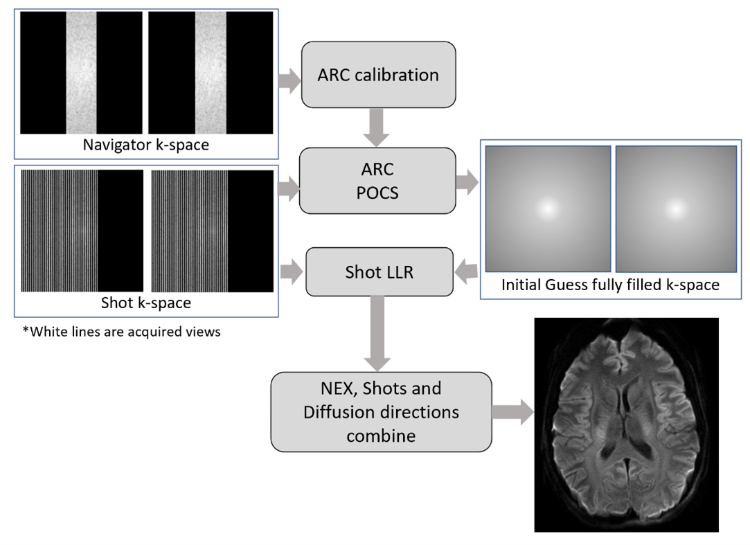
The proposed scheme uses a multi-shot diffusion imaging followed by an external spin echo and gradient echo navigators and uses a low rank approach to correct for the phase mismatch between shots.K-space sampling: Multi-shot Diffusion weighted imaging scheme is extended by adding a gradient echo navigator at the end of each traditional multi-shot (Figure 1). Another scheme called spin echo navigator was used where the multi-shot was extended by adding a second refocusing pulse at the end of every shot [10]. The advantage of external navigators as against inline navigator is the distortion removal is not perturbed due to variable sampling along phase encoding as in [9] and here we use the navigator to correct for phase mismatches across shots.
Image reconstruction: A new shot-LLR based reconstruction approach is proposed for multi-shot diffusion acquisitions with external navigators to reduce aliasing due to motion-induced phase errors. The shot-LLR approach does not require explicit estimations of the phase maps, Instead, it relies on converting smooth phase-modulations between shots as null space-vectors of structured matrix. The acquired K-space data for each shot is stacked together where unacquired data points are filled with zeros. Navigator data is also arranged in similar fashion for each shot. This navigator data is used to fill the unacquired data points in each shot using ARC [7]. An additional POCS recon is done to fill the K-space for partial Fourier acquisitions. This filled K-space is used as initial guess in Shot LLR approach to get an early and accurate convergence. The unacquired data points in original K-space are filled-in using locally low rank algorithm applied on structured shot matrix iteratively with support of initial guess k-space subject to data consistency.
Volunteer scanning: A healthy subject was scanned using the IRB approved study with informed consent on a commercial 1.5T MRI GE Signa HDxt scanner for liver using 12 channel anterior array coil and single channel volume coil. DWI images with b50 and b500 s/mm2 was acquired using the 12-channel surface coil with parallel imaging acceleration of 2. This was compared with volume coil using multi-shot factor of 2 and adding a gradient echo navigator (EXTGRENAV) and a spin echo navigator (EXTSENAV). As an extension of the algorithm to multichannel case, we scanned a patient on 7T GE Signa scanner for brain diffusion with gradient echo navigator. MUSE with factor 2 and ASSET 2 is scanned using 32 channel coil and compared with multi-shot 3 shot followed by a gradient echo navigator (EXTGRENAV). Echo train of 32 was used for both the navigators.
RESULTS and DISCUSSION
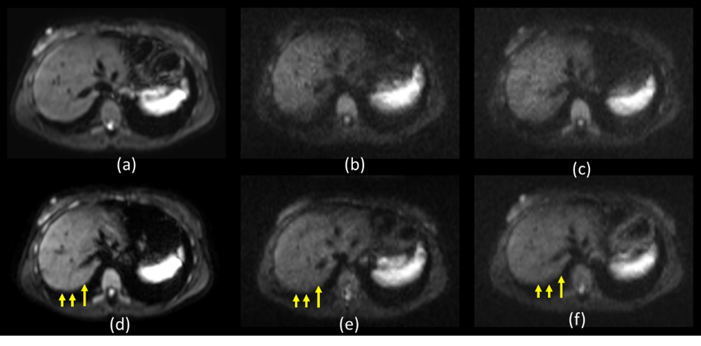
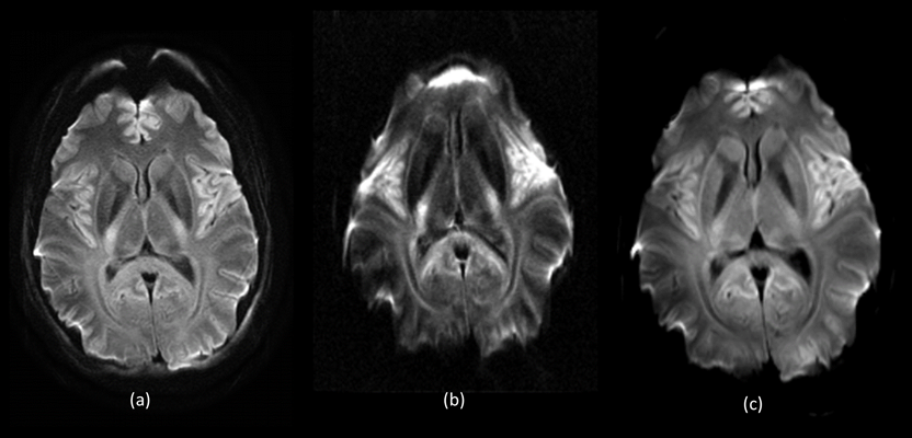
Figure 3 shows results from 1.5T scanner (a) b500 for 12 channel with ASSET 2 ,(b) b500 for single shot DW EPI with volume coil (EXTGRENAV), (c) b500 for multi-shot factor 2 for volume coil with spin echo navigator (EXTSENAV). The Lung liver interface in the diffusion images can be observed to show comparable results as with the multichannel case. The Spin echo navigator shows improved image quality than the GRE navigator as there are more susceptible regions in the abdomen.Figure 4 shows the results from 7T scanner on a 32 multichannel head coil. a) b1000 with MUSE factor 2 and ASSET 2. B) b1000 with single shot DW-EPI and c) b1000 with 2 shot GRE navigator (EXTGRENAV). The single shot DW EPI image shows huge distortion as expected. This is recovered with the GRE navigator and the results are comparable to MUSE [8] proving the extension of the proposed technique.
CONCLUSION:
Multishot diffusion imaging with volume coil can provide distortion less images using the external navigators and the new reconstruction technique. This paves the way to use the volume coil for diffusion images in obese patients and pediatric patients with a smaller volume coil where acceleration and channel sensitivity information is not possible. As an extension, the technique was demonstrated even on multichannel coil on 7T system with gradient echo navigator on brain. The spin echo navigator is effective in abdomen where there are large distortions due to susceptibility.Acknowledgements
We acknowledge scanning help provided by Jeremy Heinlein. We would like to acknowledge financial and technical support from GE Healthcare. The 7T portion of the study is supported from NIH Grant 1S10RR028821-01.References
[1] Kele, P.G. and van derJagt, E.J., 2010. Diffusion weighted imaging in the liver. World journal of gastroenterology: WJG, 16(13), p.1567.
[2] Shenoy-Bhangle, A.,Baliyan, V., Kordbacheh, H., Guimaraes, A.R. and Kambadakone, A., 2017.Diffusion weighted magnetic resonance imaging ofliver: Principles, clinical applications and recent updates. World journal of hepatology, 9(26), p.1081.
[3] Donato, H., França, M.,Candelária, I. and Caseiro-Alves, F., 2017. Liver MRI: from basic protocol to advanced techniques. European journal of radiology, 93, pp.30-39.
[4] Taouli, B. and Koh, D.M.,2010. Diffusion-weighted MR imaging of the liver. Radiology, 254(1), pp.47-66.
[5] Le Bihan, D., Poupon, C.,Amadon, A. and Lethimonnier, F., 2006. Artifacts and pitfalls in diffusion MRI.Journal of Magnetic Resonance Imaging: An Official Journal of the International Society for Magnetic Resonance in Medicine, 24(3), pp.478-488.
[6] Marchesini, G.,Moscatiello, S., Di Domizio, S. and Forlani, G., 2008. Obesity-associated liverdisease. The Journal of Clinical Endocrinology &Metabolism, 93 (11_supplement_1),pp.s74-s80.
[7] Brau, A.C., Beatty, P.J.,Skare, S. and Bammer, R., 2008. Comparison of reconstruction accuracy and efficiency among autocalibrating data‐driven parallel imaging methods. Magnetic Resonance in Medicine: An Official Journal of the International Society for Magnetic Resonance in Medicine,59(2), pp.382-395.
[8] Chen, N.K., Guidon, A.,Chang, H.C. and Song, A.W., 2013. A robust multi-shot scan strategy for high-resolution diffusion weighted MRI enabledby multiplexedsensitivity-encoding (MUSE). Neuroimage, 72, pp.41-47.
[9] Reddy, A.K.P., Agarwal, H.K., Das, R., Sundaresan, R., Ahmed, S., Rajamani, S., Mehta, B., Wu, G., Reddy, M.R. and Venkatesan, R., Multi-Shot Liver Diffusion MRI Using Variable Auto-Calibrating (vARC) Sampling Across Averages, . In Proceedings of the 29th Annual Meeting of ISMRM, London, UK, 2021, p.1146
[10] Taviani, Valentina, Ann Shimakawa, Lloyd Estkowski, Arnaud Guidon, Ersin Bayram, and Robert Peters. "Navigated multi-shot diffusion-weighted imaging with multiplexed sensitivity encoding." In Proc. Intl. Soc. Mag. Reson. Med, vol. 27. 2018.
Figures