2446
High-resolution hepatocyte-specific phase MRI of the liver using golden-angle radial sparse parallel imaging1Department of Radiology, Chongqing University Cancer Hospital, School of Medicine, Chongqing University, Chongqing, China, 2MR Collaborations, Siemens Healthineers Ltd, Shanghai, China
Synopsis
Keywords: Liver, Liver, hepatobiliary phase
Conventional abdominal MRI requires breath-holds in clinical routine which restricts acquisition times. Thus, radial MRI with additional motion compensation is desirable to further improve the performance of free-breathing abdominal exams for routine clinical use. This study aims to evaluate the feasibility of free-breathing high-resolution hepatocyte-specific phase MRI of the liver using golden-angle radial sparse parallel imaging. The results suggest that high-resolution hepatocyte-specific phase MRI with GRASP may offers a flexible alternative to assess liver lesions in patients.Introduction
Dynamic contrast-enhanced (DCE) MRI with liver-specific contrast agents, such as gadoxetic acid disodium, have shown promise in improving the detection and characterization of HCC (1,2), due to its ability to provide an improved contrast on the hepatobiliary phase (HBP). Moreover, both a non-smooth tumor margin and peritumoral hypo-intensity seen on HBP are useful for predicting microvascular invasion of HCC (3,4). However, it should be recognized that the main challenges of conventional live MRI include low speed, sensitivity to motion (ie, respiration) and the requirement of breath-holds in clinical routine. To date, several approaches have been introduced for rapid free-breathing MR acquisitions, such as Golden-angle RAdial Sparse Parallel (GRASP) MRI (5). Stack-of-stars golden-angle acquisition scheme (6,7), it combines the motion robustness and incoherent undersampling behavior of radial sampling, and is well-suited for free-breathing MRI. Therefore, our hypothesis was that free-breathing radial acquisition with GRASP reconstruction can significantly improve image spatial resolution in the HBP. The purpose of this study was to evaluate feasibility of free-breathing hepatocyte-specific phase MRI of the liver using high-resolution golden-angle radial sparse parallel imaging (HR GRASP). The results were compared with conventional T1-weighted imaging breath-hold live MRI images.Methods
Fifty-eight patients (41 males, mean age=52.9±12.9) suspected of having liver lesions were continuously recruited in this study. During hepatocyte-specific phase, each subject underwent a free-breathing DCE-MR scan using a fat-statured T1-weighted stack-of-stars golden-angle radial sequence and a post-contrast breath-held MR scan using a Cartesian volumetric-interpolated imaging sequence (BH-VIBE). GRASP data were obtained at 12 minutes after Gd-EOB-DTPA injection with the following imaging parameters: TR/TE=3.40ms/1.64ms, flip angle=10o, FOV=330×330mm2, Base resolution=320×320mm2, Slice resolution=61%, acquired slice thickness=2.5mm, in-plane spatial resolution=1×1mm2; GRAPPA accelerator factor=2, and bandwidth =610 Hz/voxel. A total of 1000 radial spokes were acquired with a prototype stack-of-stars scheme over 138 seconds. Each data was reconstructed using GRASP and was compared with routine breath-hold VIBE. Two experienced investigators blinded to acquisition schemes independently scored the overall image quality, liver edge sharpness, hepatic vessel clarity, conspicuity of lesion, and overall artifact level of each image-set. The reported scores were averaged over the two readers to yield mean ± standard deviation, and the results were compared using a non-parametric paired two-tailed Wilcoxon signed-rank test. The weighted Kappa was used to evaluate the inter-observer variation for each image quality assessment category. A P-value less than 0.05 indicated statistical significance.Results
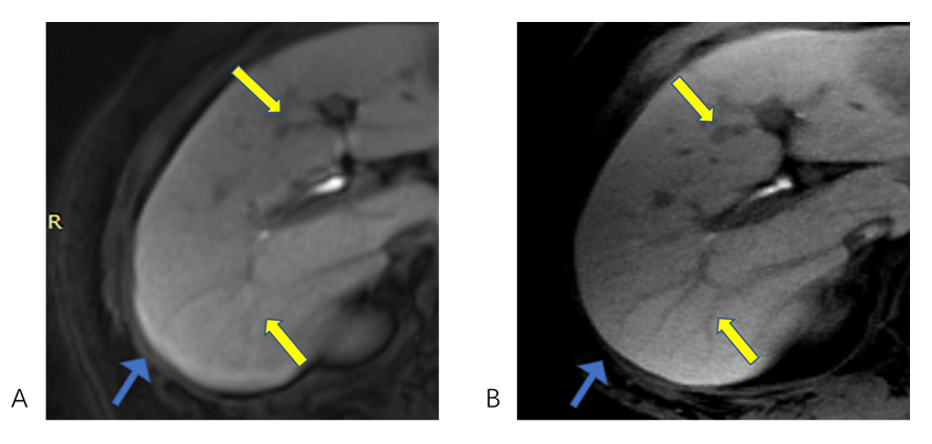
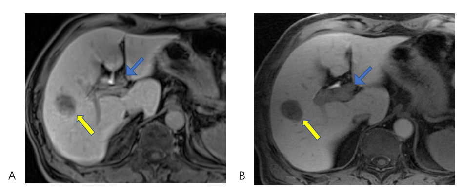
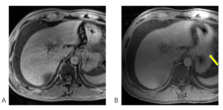
In Figure.1-3, compared to BH-VIBE image-set, HR GRASP image-set achieved higher scores (P<0.05) in liver edge sharpness (4.83±0.45 vs 4.29±0.46), hepatic vessel clarity (4.64±0.67 vs 4.15±0.56), conspicuity of lesion (4.75±0.53 vs 4.31±0.50), but lower scores in overall image quality (4.61±0.50 vs 4.74±0.47). There was no significant difference (P>0.05) between the BH-VIBE and HR GRASP image-set in overall artifact level (4.05±0.61 vs 4.13±0.44).Discussion
Patients with limited breath-holding capacities or in the elderly or children who cannot follow instructions properly during the examination are quite frequently encountered and always difficult to acquire satisfied images in clinical practice. We demonstrated that free-breathing GRASP sequence significantly achieves a higher spatial resolution and improves image quality in HBP as compared to conventional breath-hold Cartesian VIBE sequence. Therefore, we consider free-breathing high-resolution HBP as a valuable approach for clinical routine to achieve improved diagnostic performance.Acknowledgements
No acknowledgement found.References
1. Di Martino M, Marin D, Guerrisi A, et al. Intraindividual comparison of gadoxetate disodium-enhanced MR imaging and 64-section multidetector CT in the Detection of hepatocellular carcinoma in patients with cirrhosis. Radiology 2010;256(3):806-816.
2. Ahn SS, Kim MJ, Lim JS, Hong HS, Chung YE, Choi JY. Added value of gadoxetic acid-enhanced hepatobiliary phase MR imaging in the diagnosis of hepatocellular carcinoma. Radiology 2010;255(2):459-466.
3. Sano K, Ichikawa T, Motosugi U, et al. Imaging study of early hepatocellular carcinoma: Usefulness of gadoxetic acid-enhanced MR imaging. Radiology 2011;261(3):834-844.
4. Costafreda SG, Fu CHY, Picchioni M, et al. Pattern of neural responses to verbal fluency shows diagnostic specificity for schizophrenia and bipolar disorder. Bmc Psychiatry 2011;11.
5. Feng L, Grimm R, Block KT, et al. Golden-angle radial sparse parallel MRI: combination of compressed sensing, parallel imaging, and golden-angle radial sampling for fast and flexible dynamic volumetric MRI. Magnetic resonance in medicine 2014;72(3):707-717.
6. Winkelmann S, Schaeffter T, Koehler T, Eggers H, Doessel O. An optimal radial profile order based on the Golden Ratio for time-resolved MRI. IEEE transactions on medical imaging 2007;26(1):68-76.
7. Chen L, Liu D, Zhang J, et al. Free‐breathing dynamic contrast‐enhanced MRI for assessment of pulmonary lesions using golden‐angle radial sparse parallel imaging. Journal of Magnetic Resonance Imaging Jmri 2018;48(2):459-468.
Figures