2441
Tumor-liver biomechanical interaction investigated by multifrequency MR elastography in patients with HCC1Ruijin Hospital, Shanghai Jiaotong University School of Medicine, Shanhai, China, 2Ruijin Hospital, Shanghai Jiaotong University School of Medicine, Shanghai, China, 3Department of Radiology, Charité–Universitätsmedizin Berlin, Berlin, Germany;Charitéplatz 1, 10117 Berlin, Germany, Berlin, Germany
Synopsis
Keywords: Liver, Cancer
Tumor-liver biomechanical interaction investigated by multifrequency MR elastography in patients with HCCSynopsis
Hepatocellular carcinomas (HCCs) in noncirrhotic livers have a more favorable prognosis than HCCs that grow in cirrhotic livers. However, little is known why HCC aggressiveness is influenced by the properties of the surrounding tissue. Previous work indicated that HCCs favorably grow in stiff livers prompting the hypothesis that liver tumor aggressiveness is influenced by the mechanical properties of the surrounding tissue. Therefore, we here study the biomechanical properties of HCC and their hosting liver using tomoelastography, a multifrequency MR-elastography (MRE) technique. Our results show that HCCs in cirrhotic livers with poorer prognosis presented higher stiffness and increased fluidity, suggesting that the biomechanical properties of the hosting environment influences the aggressiveness the tumor.Introduction
Approximately 20% of HCCs precede the occurrence of cirrhosis through distinct cytogenetic pathways1. Due to the lack of surveillance strategies, HCC in noncirrhotic liver usually manifest with a larger tumor burden and an advanced stage at the time of diagnosis. Nevertheless, these patients present with a more favorable prognosis. To date, little is known about the causes of the differential aggressiveness of HCCs growing in cirrhotic and noncirrhotic livers. The interplay between the tumor entity and the Tumor Surrounding Environment (TSE) plays an important role during tumor progression2. This tumor-TSE interaction potentially changes the composition and the structure of the tissue and alters the biomechanical properties of the affected tissue. Tomoelastography, a multifrequency MR elastography (MRE) technique, can quantitatively map soft tissue viscoelasticity based on shear wave speed (c in m/s) and loss angle (φ in rad). Both parameters are used as surrogate markers of stiffness and viscosity or tissue fluidity, respectively. This study aimed to investigate the tumor-liver biomechanical interaction in patients with HCC.Methods
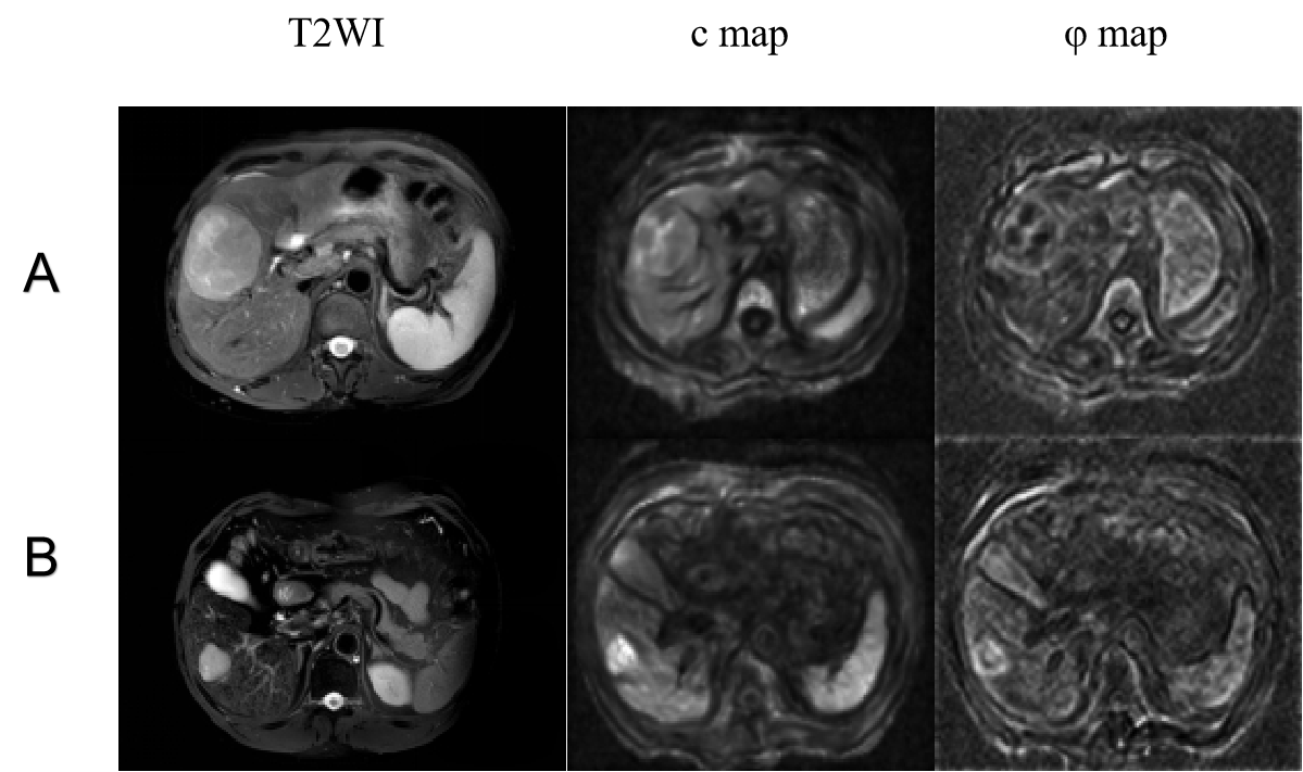
This prospective study included 107 HCC patients (mean age, 59 years ± 12;90 men) who underwent preoperative MRI and tomoelastography. All participants were divided into two groups of patients with non-cirrhotic and cirrhotic livers based on histopathological analysis. The non-cirrhotic group included 51 patients with 52 lesions (mean age, 61 years ± 13;47 men) while the group of patients with cirrhosis included 56 patients with 61 lesions (mean age, 58 years ± 11;43 men).All tomoelastography experiments were performed on a clinical 1.5-Tesla MRI scanner (Magnetom Aera, Siemens, Erlangen, Germany) using four vibration frequencies (30Hz, 40Hz, 50Hz, 60Hz) and the multifrequency processing pipeline available at https://bioqic-apps.com to generate full field-of-view maps of c and φ. Two radiologists independently evaluated the tomoelastography data. Interobserver agreement was assessed by the intraclass correlation coefficient (ICC).Results
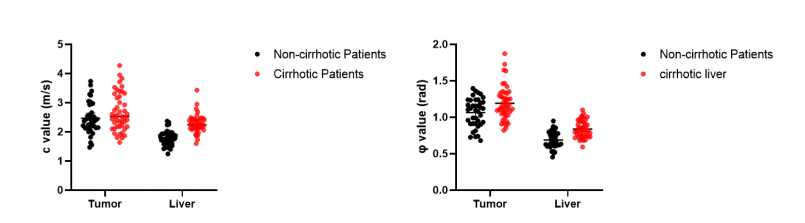
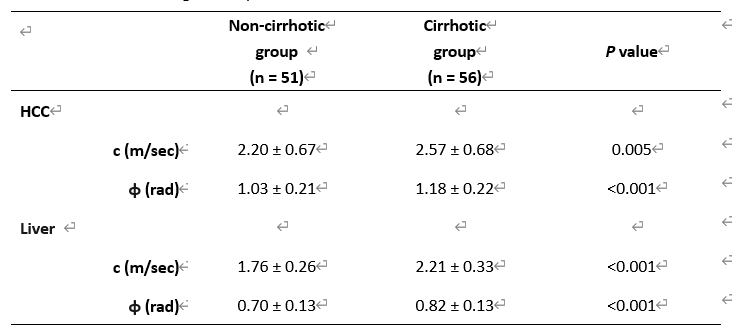
Interobserver reliability as indicated by ICC was 0.95 (95% CI: 0.94, 0.97) for c_tumor, 0.94 (95% CI: 0.91, 0.96) for φ_tumor, 0.96 (95% CI: 0.94, 0.98) for c_liver and 0.96 (95% CI: 0.94, 0.98) for φ_liver, suggesting good concordance and data consistency. The inflammation activities in the livers of the cirrhotic group were higher than those in the non-cirrhotic group (p = 0.042). Cirrhotic livers were distinguishable from non-cirrhotic livers based on higher c- and φ-values (c: 2.21 ± 0.33 m/sec vs. 1.76 ± 0.26 m/sec, p<0.001; φ: 0.82 ± 0.13 rad vs 0.70 ± 0.13 rad, p<0.001). HCCs in both groups were not different in their imaging characteristics such as arterial phase hyperenhancement, non-peripheral washout, enhancing capsule and restricted diffusion (all p > 0.05). However, comparing the biomechanical properties of HCCs between the two groups, we found higher φ and c values in the cirrhotic than non-cirrhotic group (φ: 1.20 ± 0.24 rad vs 1.08 ± 0.19 rad, p<0.001; c: 2.57 ± 0.68 m/s vs. 2.20 ± 0.67 m/s, P=0.005). The biomechanoid properties of the HCC and the livers are plotted in Figure 1 and collected in Table 1.Discussion
Our study showed that biomechanical properties of the HCCs significantly depend on the viscoelasticity of the hosting liver. Cirrhotic livers fostered HCCs with stiffer, yet more fluid-like properties. This seemingly contradictory material’s behavior can be explained with the accumulation of matrix proteins and higher inflammation activity yielding, in cirrhotic livers, an increase in both stiffness and viscoelastic dispersion slope or loss angle φ , respectively3. In agreement to3, we observed that the φ value of non-cirrhotic HCC-hosting liver was lower than π/4 (indicated predominantly solid properties) while the φ of cirrhotic liver was higher than π/4 (indicated predominantly fluid properties). This transition from solid to fluid along with soft to stiff indicates a significant increase in intrinsic mechanical friction, which likely forms a cancerous environment fostering HCCs with more aggressive properties. Furthermore, it is known that cancer cells act collectively. Unjammed cellular streams in HCC could influence the fluid properties of a tumor at the macroscopic level, indicating an increased metastatic potential4. Probably for this reason, higher fluidity has been associated with higher aggressiveness of tumors5,6. The higher fluidity that we observed in the HCCs of the cirrhotic liver is consistent with the clinical observation that HCC patients with liver cirrhosis show lower overall survival rates than those with non-cirrhotic livers. The more aggressive HCCs in the cirrhotic livers also displayed higher stiffness than the HCCs in the non-cirrhotic livers. This observation is in line with previous studies showing that higher tumor stiffness is associated with higher malignancy5-7.Acknowledgements
The authors acknowledge the Department of General Surgery at Ruijin Hospital (Shanghai Jiao Tong University School of Medicine, Shanghai, China) for assistance with patient information, surgical tumor tissue, and the Department of Pathology at Ruijin Hospital (Shanghai Jiao Tong University School of Medicine, Shanghai, China) for assistance with histological analysis. We acknowledge the Department of Radiology at Charité–Universitätsmedizin Berlin (Berlin, Germany) for assistance with technical support of MRE.References
1 Desai, A., Sandhu, S., Lai, J. & Sandhu, D. Hepatocellular carcinoma in non-cirrhotic liver: A comprehensive review. World journal of hepatology 11, 1-18, doi:10.4254/wjh.v11.i1.1 (2019).
2 Mierke, C. et al. The two faces of enhanced stroma: Stroma acts as a tumor promoter and a steric obstacle. NMR in biomedicine 31, e3831, doi:10.1002/nbm.3831 (2018).
3 Reiter, R. et al. Influence of fibrosis progression on the viscous properties of in vivo liver tissue elucidated by shear wave dispersion in multifrequency MR elastography. J Mech Behav Biomed Mater 121, 104645, doi:10.1016/j.jmbbm.2021.104645 (2021).
4 Friedl, P. & Gilmour, D. Collective cell migration in morphogenesis, regeneration and cancer. Nat Rev Mol Cell Biol 10, 445-457, doi:10.1038/nrm2720 (2009).
5 Li, M. et al. Tomoelastography Based on Multifrequency MR Elastography for Prostate Cancer Detection: Comparison with Multiparametric MRI. Radiology, 201852, doi:10.1148/radiol.2021201852 (2021).
6 Shahryari, M. et al. Tomoelastography Distinguishes Noninvasively between Benign and Malignant Liver Lesions. Cancer Res, doi:10.1158/0008-5472.CAN-19-2150 (2019).
7 Zhu, L. et al. Distinguishing pancreatic cancer and autoimmune pancreatitis with in vivo tomoelastography. Eur Radiol, doi:10.1007/s00330-020-07420-5 (2020).
Figures