2431
Identifying the Microvascular Invasion of Hepatocellular Carcinoma with a Continuous Time Random-Walk Diffusion Model
Jiejun Cheng1, Dingxia Liu1, Yunfei Zhang2, Yongming Dai2, and Xiuzhong Yao1
1Shanghai Institute of Medical Imaging, Shanghai, China, 2MR Collaboration, Central Research Institute, United Imaging Healthcare, Shanghai, China
1Shanghai Institute of Medical Imaging, Shanghai, China, 2MR Collaboration, Central Research Institute, United Imaging Healthcare, Shanghai, China
Synopsis
Keywords: Liver, Cancer
DWI is the most widely-used functional magnetic resonance imaging (MRI) technique in clinical practice. It has been widely acknowledged that DWI has the potential to probe tissue cellularity, microstructures, and microvasculature at a subvoxel-level. In this study, we aim to access the clinical application of the continuous-time random-walk (CTRW) model which recognizes intravoxel diffusion heterogeneity in both time and space in the preoperative evaluation of microvascular invasion (MVI) in hepatocellular carcinoma (HCC) patients. Our results showed that diffusion parameters derived from CTRW model can be used as noninvasive quantitative imaging markers for preoperatively predicting the MVI of HCCs.Introduction
HCC is one of the most common malignant tumors worldwide, which account for 75%-85% of primary liver cancer (1). Microvascular invasion (MVI) has been widely acknowledged as one of the most important factors which profoundly affects the prognosis of HCC (2). Preoperative evaluation of the MVI of HCC is crucial to select the optimal strategy and accessing prognosis. With the capability of providing information of tumor cellularity, vascularity and microstructures, DWI can be a promising tool to detect the MVI of HCCs preoperatively without the use of contrast agents (3). However, the clinical acceptance of apparent diffusion coefficient (ADC) calculated from the traditional mono-exponential model was limited because of substantial overlap of ADC values. The diffusion heterogeneities recognized by the CTRW model can directly reflect intravoxel structural heterogeneity, thus reveal the tissue complexity and microenvironment. To the best of our knowledge, no study has included the association of CTRW-derived parameters and the presence of MVI of HCC. Hence, this study aims to access the clinical application of the CTRW diffusion model in the preoperative evaluation of MVI in HCC patients.Methods
A total of 84 patients with HCC were enrolled in this study. Preoperatively MR examination was carried out with a 3.0 T MRI scanner with 10 b-values: 0, 20, 40, 50, 100, 200, 500, 800, 1500 and 2000 s/mm2. Diffusion parameters, including anomalous diffusion coefficient (D), spatial heterogeneity parameter (β), temporal heterogeneity parameter (ɑ) and apparent diffusion coefficient (ADC) were calculated. The diagnostic performance was evaluated by receiver operating characteristic (ROC) analysis.Results
Among all the studied diffusion parameters, significant differences were found in D, β and ADC between the MVI-positive and MVI-negative groups. MVI-positive HCCs showed significantly higher β values (P < 0.001) and lower D and ADC values (both P < 0.001) than those of MVI-negative HCCs. However, the statistical significance of the values of ɑ between MVI-positive and MVI-negative groups was not observed (Figs 1, 2 and 3). According to the ROC analysis, the β demonstrated the largest area under the ROC curve (0.749) compared with other parameters (ADC: 0.732; D: 0.707; α: 0.577) for differentiating MVI-positive from MVI-negative HCCs (Fig 4).Discussion
Aforementioned results demonstrated the feasibility of using the novel non-Gaussian diffusion model which named the CTRW diffusion model to differentiate MVI-positive from MVI-negative HCCs. The results suggested that compared with MVI-negative HCC, MVI-positive HCC demonstrated significantly higher values of β and lower values of D and ADC. Moreover, among all the diffusion parameters, β showed the largest AUC when discriminate MVI-positive from MVI-negative HCCs. The CTRW model is a more comprehensive diffusion model which recognize the diffusion heterogeneity in both time (represented by α) and space (represented by β) and persevere the traditional diffusion coefficient. With different parameters to reveal different biological insights in tumor lesions, the CTRW model can reflect an accurate overview of the process of water molecules diffusion in highly heterogeneous tumor tissues, thus reaching better diagnostic performance.Conclusion
The CTRW-derived parameters can be used as noninvasive quantitative imaging markers for preoperatively predicting the MVI status of HCCs.Acknowledgements
NoneReferences
1. Sung H, Ferlay J, Siegel RL, et al. Global Cancer Statistics 2020: GLOBOCAN Estimates of Incidence and Mortality Worldwide for 36 Cancers in 185 Countries. CA Cancer J Clin 2021;71:209-249.2. Lim KC, Chow PK, Allen JC, et al. Microvascular invasion is a better predictor of tumor recurrence and overall survival following surgical resection for hepatocellular carcinoma compared to the Milan criteria. Ann Surg 2011;254:108-113.
3. Surov A, Pech M, Omari J, et al. Diffusion-Weighted Imaging Reflects Tumor Grading and Microvascular Invasion in Hepatocellular Carcinoma. Liver Cancer 2021;10:10-24.
Figures
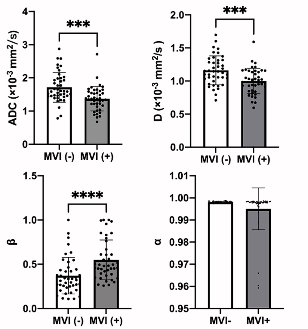
Figure 1 D, β, α and ADC values of MVI-positive and MVI-negative groups.
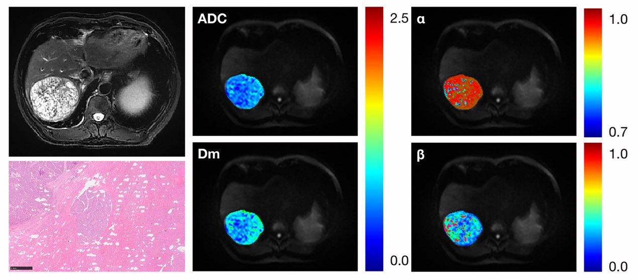
Figure 2 Representative MR Images of a 57-year-old man pathologically verified as HCC with MVI.
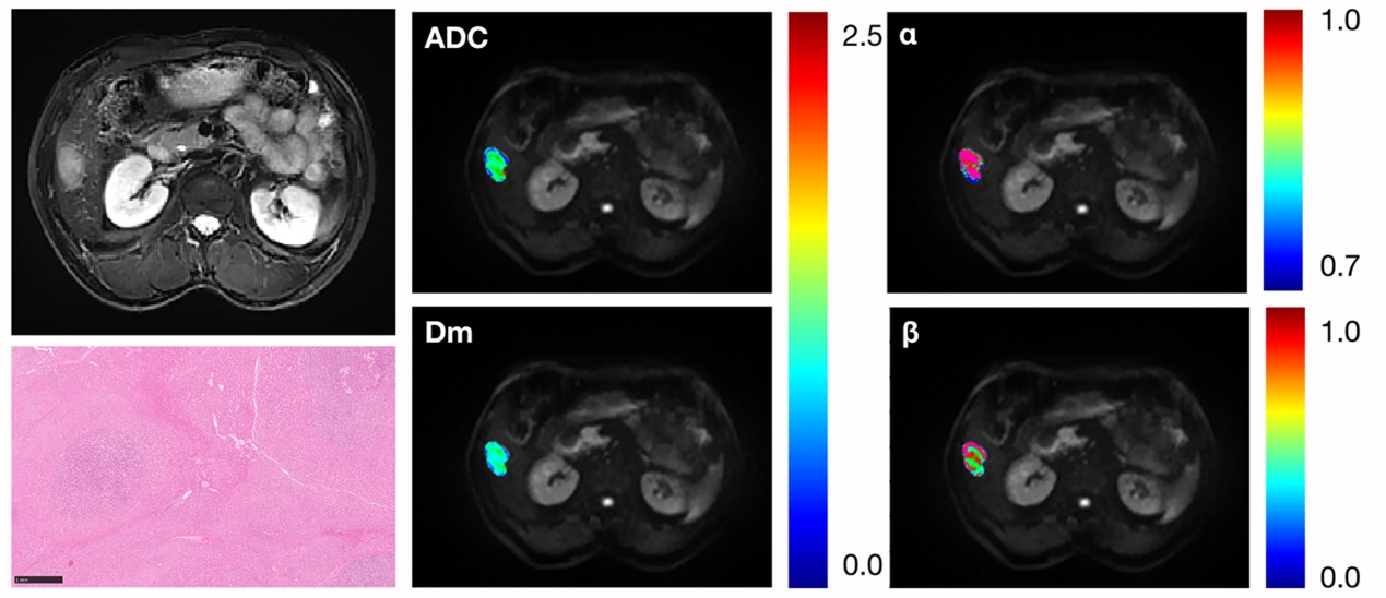
Figure 3. Representative MR Images of a 53-year-old man pathologically verified as HCC without MVI.
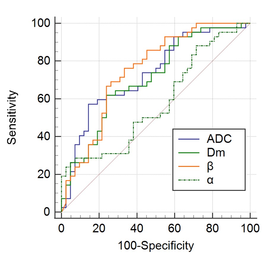
Figure 4. ROC curves of ADC, D, β and α for differentiating MVI-positive and MVI-negative HCCs.
DOI: https://doi.org/10.58530/2023/2431