2430
Adverse effect of non-linearity in gradient on MRI and their impact on localization and morphology of testing sample1Medical Systems Division, S.A.M.E.E.R. Mumbai, Mumbai, India
Synopsis
Keywords: Data Analysis, Artifacts
Study demonstrate the analysis of surface morphology and sample localization with variation from iso-center in the presence of non-linearity in gradient. In these experiments, we have analyzed that gradient non-linearity plays a crucial role in surface morphology and localization of samples. For the end position 200 mm and -200 mm, size of the object appears small. Additionally, the results suggest that when a large sample size is taken into account, gradient non-linearity exhibits greater negative impacts.INTRODUCTION
A non-invasive imaging technique called magnetic resonance imaging (MRI) provides three-dimensional, intricate anatomical pictures [1]. Although the MRI offers precise disease detection, diagnosis, and treatment time monitoring, its cost is higher than that of other imaging modalities due to the high cost of hardware installation, maintenance, and operation [2]. As a result, people in low-income nations like India cannot afford to get an MRI frequently. The development of a low-cost MRI system is required for the provision of essential healthcare so that people can easily and quickly access MRI. In this context, we present the findings of the low-cost Indian Magnetic Resonance Imaging System (IMRI), developed by SAMEER, Mumbai, for MEITY, the Government of India. To optimize the picture performance of samples in MRI, it is necessary to further investigate several issues such as gradient non-linearity, MRI hardware issues, synchronization problems in spectrometers, and imaging algorithm errors [3]. A sample image's geometric distortions can be caused by gradient non-linearity. Additionally, the gradient non-linearity causes an RF shift with an error of up to several millimeters, making it difficult to accurately localize testing samples [4]. In this article, we have discussed how this affects the surface morphology and sample localization of fruit samples like pineapple, orange, and pears.METHODS
Experiments were performed on a 1.5 T IMRI with a body birdcage coil (16 runs, radius: 306.02 mm, length 560 mm, and strip width 53 mm) as transmitter and a 5-inch square loop surface coil (bandwidth ± 400 KHz) as a receiver. Image is acquired using IMRI software developed by C-DAC, Thiruvananthapuram, India. To analyze the size of the sample in the image, MICRO-DICOM VIEWER VERSION 2.9 is used and the graph is plotted using the ORIGIN 2021 Software. GRE sequence has been played for the parameters TR: 40 ms and TE 8 ms. Slice thickness width for the excitation was selected to be 5mm for the scanning. Water is a crucial element of MRI scanning, and fresh fruit samples contain more water than stale fruit samples [5]; as a consequence, fresh fruit samples (orange, pineapple, and pear) of various sizes and surface morphologies have been considered for the MRI image scanning. The samples were scanned for all three orientations axial, sagittal, and coronal. The sample is only moved axially at seven distinct points, including -200 mm, -100 mm, -50 mm, 0 mm, -50 mm, -100, and -200 mm, where 0 represents the iso-center position and - and + directions, respectively, depict the couch's movement toward the inside and outside of the control room.RESULTS
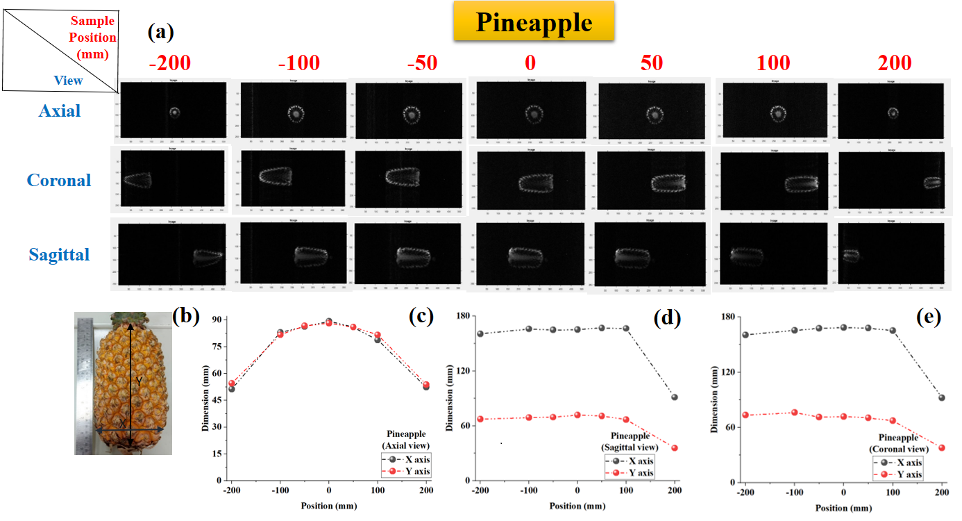
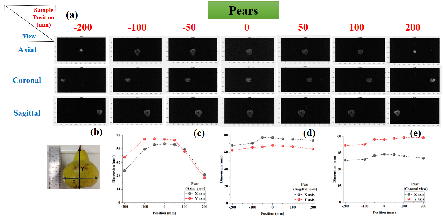
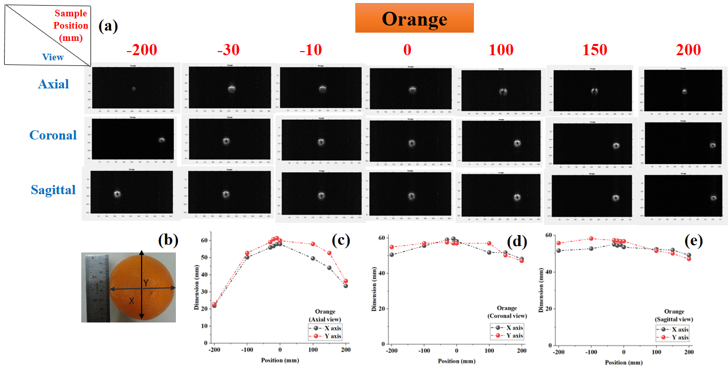
Two distinct sizes of sample pineapple and pear have been considered to distinguish the gradient non-linearity effect on small and big-size samples. In the example of the pineapple, dimensions (length 169.1 mm and width 72.4 mm) have been measured using a scale, and its MRI dimensions are found to be 168.41 mm and 71.88 mm at iso-center as shown in the Figure1. When it comes to iso-center, the size variation is minimal. However, when the sample is displaced to the location + 200 mm, the sample seems small and the measurements provide inconsequential findings (length: 92.14 and width 37.83 mm for Coronal and length: 91.13 and width 35.73 mm for sagittal). These numbers were quite similar to the measurement made using a physical scale (length: 78.42 mm and width: 68.13 mm). The pineapple exhibits geometric distortion in all directions, but the pear only exhibits considerable geometric distortion in the axial view (See Figure 2). These findings suggest that when taking into account lengthy size samples, gradient non-linearity exhibits higher negative consequences. To distinguish between the gradient non-linearity effect on symmetrical and asymmetrical samples, a nearly spherical orange with a diameter of 59.2 mm and 58.3 mm in the x and y axes, respectively, has been measured using the scale, and its dimension using the MRI is found to be 58.17 mm and 57.02 mm in the x and y axes. Size deviations of less than 1 mm indicate iso-center findings that are statistically significant. Analogously, an axial view of an orange or pear reveals a substantial geometric distortion. In addition to affecting size distortion, gradient non-linearity also affects sample intensity.DISCUSSION
In the experiment, the important findings below have been seen.(1) In axial view, for positions 200 mm and -200 mm, the size of the object appears small.
(2) In sagittal and coronal view, the sample slice is not observed at the center however, the RF offset is not given.
(3) As we move to the end position, hosting is observed in the image.
(4) The morphology of the sample disturbs the location at the end. In comparison to sagittal and coronal views, the size distortion of the sample is more pronounced in the axial view.
(5) As we increase the size of the object, the distortion at the end position is high.
CONCLUSION
As demonstrated in our findings, gradient linearity issues commonly result in spatial distortion, blurring, and intensity effects. As we increase the sample dimension, results show that sample distortion also rises. The impact of sample localization disturbs other orientations even when merely shifting the sample in axial view.Acknowledgements
The authors acknowledges the support from MeitY, Government of India for providing the financial support to carry out the Indian MRI research project.References
[1] Moratalla MB, Braun P, Fornas GM. Importance of MRI in the diagnosis and treatment of rhabdomyolysis. European journal of radiology. 2008 Feb 1;65(2):311-5.
[2] Liu, Y., Leong, A.T.L., Zhao, Y. et al. A low-cost and shielding-free ultra-low-field brain MRI scanner. Nat Commun 12, 7238 (2021). https://doi.org/10.1038/s41467-021-27317-1
[3] Yarach U, Luengviriya C, Danishad A, Stucht D, Godenschweger F, Schulze P, Speck O. Correction of gradient nonlinearity artifacts in prospective motion correction for 7T MRI. Magn Reson Med. 2015 Apr;73(4):1562-9. doi: 10.1002/mrm.25283. Epub 2014 May 5. PMID: 24798889; PMCID: PMC4221571
[4] Tao S, Trzasko JD, Shu Y, Huston J 3rd, Bernstein MA. Integrated image reconstruction and gradient nonlinearity correction. Magn Reson Med. 2015 Oct;74(4):1019-31. doi: 10.1002/mrm.25487. Epub 2014 Oct 8. PMID: 25298258; PMCID: PMC4390402.
[5] https://www.fao.org/3/v5030E/V5030E06.HTM (Weblink Date 07/11/2022)
Figures