2428
Post mortem magnetic resonance imaging simulation system
Hiroyuki Kabasawa1, Masatoshi Kojima2, Daisuke Yajima3, and Yohsuke Makino4
1Department of Radiological Sciences, Internationa University of Health and Walfare, Narita, Japan, 2Department of Legal Medicine, Chiba University, Chiba, Japan, 3Department of Forensic Medicine, International University of Health and Welfare, Narita, Japan, 4Department of Forensic Medicine, The University of Tokyo, Tokyo, Japan
1Department of Radiological Sciences, Internationa University of Health and Walfare, Narita, Japan, 2Department of Legal Medicine, Chiba University, Chiba, Japan, 3Department of Forensic Medicine, International University of Health and Welfare, Narita, Japan, 4Department of Forensic Medicine, The University of Tokyo, Tokyo, Japan
Synopsis
Keywords: Software Tools, Software Tools, post mortem imaging
A MR Image simulation system for post mortem subject scanning is developed and validated. The system has MR console like user interface. It has body temperature input as well as tissue fixation condition input, so that post mortem MR imaging can be simulated for various scanning condition. The system was validated for T1 weighted image and FLAIR for low body temperature condition. The result showed the system could simulate the actual acquired image contrast change associated with body temperature change. The proposed system can be used to predict MR contrast change associated with body temperature as well as fixation condition.Introduction
Post mortem forensic magnetic resonance imaging (PM-MRI) has been an alternative, non-invasive method for investigation of deaths. The MRI has been shown useful for the forensic investigation, however, the same scan parameter with normal subject cannot be used because physical condition including deceased body temperature is far different from normal living human subject. The physical condition differences induce MR related parameter changes, such as T1, T2 relaxation time, proton density, and diffusion coefficient. Those changes require MR scan parameter optimization for PM-MRI. Typical challenges were FLAIR inversion time setting and T1 weighted scan parameter setting. PM-MRI scan optimization for specific protocol has been reported [1][2], however, comprehensive PM-MRI scan optimization is still challenging. Here we developed a MR simulator that can help users understand the MRI contrast change associated with deceased body condition. The purpose of the study was to develop MRI simulator for PM-MRI running on the laptop personal computer.Materials and Methods
Python3.8.5 was used for system development. Simple ITK 2.02 was used for 3D image processing and TK 8.6.10 was used for use interface development. Gray matter, whiter matter and CSF tissue probability maps from MNI ICBM152 were used. Fat map was generated by manually segmenting the T1 weighted brain template images. Specific proton density, T1 and T2 relaxation time were allocated for each tissue component. MRI signal was simulated using Bloch equation. Simulation MR image pixel value S(r) was calculated the following equation.$$S(r)=P_{GM}(r)M_{GM}+P_{WM}(r)M_{WM}+P_{CSF}(r)M_{CSF}+P_{Fat}(r+\delta x_{Fat} e_{read})M_{Fat}$$
where $$$P_{GM}$$$, $$$P_{WM}$$$, $$$P_{CSF}$$$ and $$$P_{Fat}$$$ are the tissue probability and $$$M_{GM}$$$, $$$M_{WM}$$$, $$$M_{CSF}$$$ and $$$M_{Fat}$$$ are the magnetization magnitude values of gray matter, white matter, cerebrospinal fluid and fat, respectively. For unfixed brain, previously reported tissue relaxation time temperature dependence data [3] from 23 cadavers (16 male, 7 female; aged 23 to 90 years, mean 58.4 years) were used for simulation. The literature value of proton density and magnetic transfer ratio [4] were also used for PM-MRI simulation. For fixed brain, the literature values of tissue relaxation time temperature dependence for post mortem brain [4] was used. Simulation results were compared with previously reported postmortem MR images [3] from the 1.5T clinical scanner with TR=550ms, TE12.5ms, 256x256 matrix, FOV23cm.
Results
Linear regression result between tissue relaxation time and body temperature was T1=900+32.6T for gray matter and T1=691+3.5T for white matter, respectively. As shown in Figure 1, simulated PM-MR Images at arbitrary temperature could be generated using the proposed system. Arbitrary slice location could be simulated using interactive slice prescription interface. User could simulate both unfixed and fixed PM-MRI scan protocol. As shown in Figure 2, Contrast reverse effect of gray matter and white matter associated with body temperature was able to reproduced with simulation. FLAIR inversion time change with temperature could also be simulated as shown in Figure 3. Fixed and Unfixed brain MR contrast simulation result for T1 weighted imaging is shown in Figure 4.Discussion
PM-MRI contrast change with scan parameters, body temperature and fixation status can be simulated interactively without any waiting time on the standard laptop computer. One can compare the post mortem simulation results with normal living human brain. T1 weighted image contrast reversal effect between gray matter and white matter at low body temperature was able to simulate with the proposed system. It has been shown that the T1 contrast reversal effect was caused by temperature of the body temperature, however, this study visually showed that the reversal effect was associated with the body temperature. The proposed simulation tool can be an effect mean for PM-MRI scan parameters for post mortem subjects.Conclusion
This study showed the proposed MRI simulation system for PM-MRI scanning effective for forensic MRI study. The proposed system can be used to predict MR contrast change associated with body temperature as well as fixation condition for PM-MRI.Acknowledgements
No acknowledgement found.References
[1] Magn Reson Med. 2008; 59(1):190-5.
[2] Magn Reson Med Sci. 2015;14(4):251-5.
[3] Proceedings of the 47th Japanese SMRM, O1-087
[4] Magn Reson Med. 2014 Apr;71(4):1575-80.
Figures
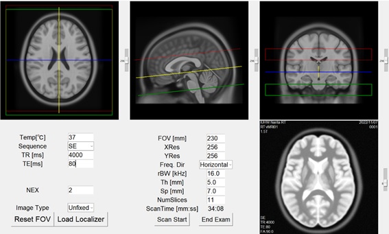
Figure 1 Post mortem (PM) MRI simulator. Body temperature and fixation condition can be
changed on the user interface.
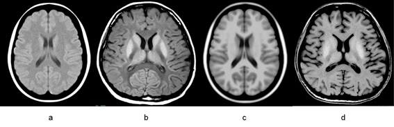
Figure 2 Simulated PM-T1 weighted image. a: simulated spin echo (SE) image with TR 550ms, TE 12.5ms, body temperature 4 degree. b: acquired SE image with the same scan parameter [3]. c: simulated spoiled gradient echo (SPGR) image with TR 25ms, TE 5ms, FA 30 degree, body temperature 4 degree. d: acquired SPGR image with the same scan parameter [3].
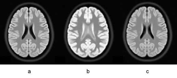
Figure 3 Simulated PM-FLAIR image. a: TR10000ms TE120ms TI 2400ms at body
temperature 37 degree. b: TR10000ms TE120ms TI 2400ms at body
temperature 6 degree. c: TR10000ms TE120ms TI 1500ms at body
temperature 6 degree.
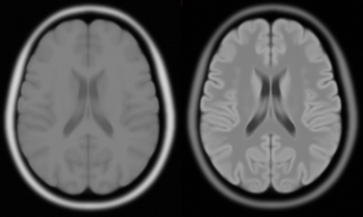
Figure 4 Simulated fixed and unfixed T1 weighted
brain images. Left: unfixed brain T1 weighted image with TR 500ms, TE 10ms, and
body temperature 15 degree. Right: fixed brain T1 weighted image with the same
scan parameter.
DOI: https://doi.org/10.58530/2023/2428