2385
A new pulse sequence to selectively probe the signals of γ-aminobutyric acid
Xue Yang1, Ying Liu2, Caixia Fu3, He Wang2, Ying-Hua Chu4, Da-Xiu Wei1, and Ye-Feng Yao1
1East China Normal University, Shanghai, China, 2Fudan University, Shanghai, China, 3Siemens Shenzhen Magnetic Resonance Ltd., Shanghai, China, 4MR Collaboration, Siemens Healthineers Ltd., Shanghai, China
1East China Normal University, Shanghai, China, 2Fudan University, Shanghai, China, 3Siemens Shenzhen Magnetic Resonance Ltd., Shanghai, China, 4MR Collaboration, Siemens Healthineers Ltd., Shanghai, China
Synopsis
Keywords: Pulse Sequence Design, Spectroscopy
A new pulse sequence was developed to selectively probe the signals of γ-aminobutyric acid (GABA). Different with the previous pulse sequences, the signal selectivity of this pulse sequence is achieved by preparation and reconversion of the 1H spin singlet order (SSO) of GABA. The optimal control method was used in the design of the pulse sequence. By using the developed pulse sequence, the 1H signals of GABA in human brains were selectively probed.Introduction
γ-aminobutyric acid (GABA) is an important inhibitory neurotransmitter in the human brain. It is believed that this molecule has a strong correlation with a variety of neurological and psychiatric disorders. Currently, the technique most used to in vivo probe the GABA molecules in human brains is proton magnetic resonance spectroscopy (1H MRS). MEscher-GArwood Point RESolved Spectroscopy (MEGA-PRESS) [1] and MEscher-Garwood semi-localized by adiabatic selective refocusing (MEGA-sLASER) [2] are the two pulse sequences which are widely used in 3T and 7T scanners to probe the signals of GABA in vivo. However, the subtraction procedure used in the data process in these pulse sequences to remove the overlapped signals from the other metabolites often causes the signal distortion due to the improper phase correction, baseline correction and the mismatching of the two spectra used in the subtraction. Developing new pulse sequences for an easy and accurate probing of GABA in human brains in vivo has attracted a lot of research interests in the field. Thus, the purpose of this study is to develop a new pulse sequence to selectively probe the signals of GABA and to evaluate the performance of pulse sequence on both phantoms and human brains.Materials and Methods
All MRS experiments were performed on a 3T scanner (MAGNETOM Prisma, Siemens Healthcare, Erlangen, Germany) with a 64-channel head receiver coil and a 7T scanner (MAGNETOM Terra, Siemens Healthcare, Erlangen, Germany) with a 32-channel head receiver coil. Figure 1 shows the diagram of the molecularly targeted MRS sequence developed in this work. The sequence consists of three blocks. The first block is to suppress the water signal using WET scheme [3]. The second block is used for preparation and reconversion of the SSO of GABA at 2.28 ppm and 1.89 ppm using the optimal control method [4]. Figure 2 exemplarily shows the amplitude and phase modulation of an optimal control pulse (i.e., Optimal Control I). The third part is the PRESS pulse sequence [5] which is used to localize the volume of interest (VOI) and receive the signal. Two healthy volunteers and a home-made phantom with 400 mg of GABA dissolved in 50 mL of ultrapure water (concentration: 0.08 mol/L, pH =7.3 ± 0.1) were scanned successively on both 3T and 7T scanners using the parameters in Table 1.Results
Figure 3a shows the phantom used in the experiments. Figure 3b shows the T1-weighted MRI images of the phantom. In these images, the regions marked by the pink squares indicate the VOI used in the magnetic resonance spectroscopy experiments. It is observed that in the GABA-targeted MR spectrum (bottom of Figure 3c) acquired from a 7T scanner, only the signals from 2.28 ppm and 1.89 ppm appear, whereas the signals from 3.01 ppm are significantly suppressed. Figure 3d shows the routine and the GABA-targeted MR spectra obtained from a 3T scanner. Similar with the result from 7T, only the targeted signals at 2.28 ppm and 1.89 ppm appear in the GABA-targeted MR spectrum, whereas the signals from 3.01 ppm are significantly suppressed.In the volunteer experiment on 7T, a peak (2.28 ppm) was clearly observed in the GABA-targeted MR spectrum, which matches well with the signals of GABA in the simulated spectrum (indicated by the grey line in Figure 4b). The signal at 1.89 ppm cannot be clearly observed. This can be attributed to the low intensity of the signal and the possible B0 inhomogeneity. Meanwhile, in the GABA-targeted spectrum shown in Figure 4b, the signals of the other metabolites, such as N-acetyl-aspartate, glutamate, glutamine, creatine signals etc., are significantly suppressed. The similar result was achieved in the experiment performed on a 3T scanner (Figure 4c). Comparing the results from the 7T and 3T scanners, the high field seems to facilitate the signal selection in the experiments.
Discussion and Conclusion
In this work, we reported a new pulse sequence which can be used to selectively probe the signals of GABA. The signal selectivity of this pulse sequence was clearly demonstrated by the experimental results on both the phantom and the in vivo study of human brains. Further studies will be conducted to assess the performance of this new sequence on various scanners in clinical practice.Acknowledgements
This work was supported by Xing-Fu-Zhi-Hua Foundation of ECNU and Microscale Magnetic Resonance Platform of ECNU. X.Y. and Y.-F.Y.References
- Mescher M, Merkle H, Kirsch J, Garwood M, Gruetter R. Simultaneousin vivo spectral editing and water suppression. NMR in Biomedicine 1998;11(6):266-272.
- Andreychenko A, Boer VO, Arteaga de Castro CS, Luijten PR, Klomp DW. Efficient spectral editing at 7 T: GABA detection with MEGA-sLASER. Magn Reson Med 2012;68(4):1018-1025.
- Ogg RJ, Kingsley PB, Taylor JS. WET, a T1- and B1-insensitive water-suppression method for in vivo localized 1H NMR spectroscopy. J Magn Reson B 1994;104(1):1-10.
- Tosner Z, Vosegaard T, Kehlet C, Khaneja N, Glaser SJ, Nielsen NC.
Optimal control in NMR spectroscopy: numerical implementation in SIMPSON. J
Magn Reson 2009;197(2):120-134.
Figures
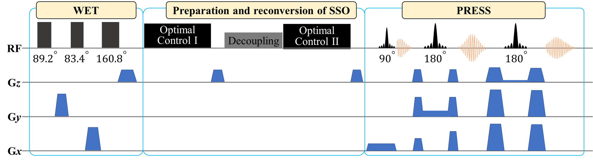
Figure 1. The
diagram of the
molecularly targeted MRS sequence developed to selectively probe the
signals of GABA. This pulse sequence consists of three blocks of different
functions, including water suppression using the WET scheme, preparation
and reconversion the SSO of GABA using the optimal control method, and the PRESS
pulse sequence to
localize and detect MRS signal from the Volume Of Interest.
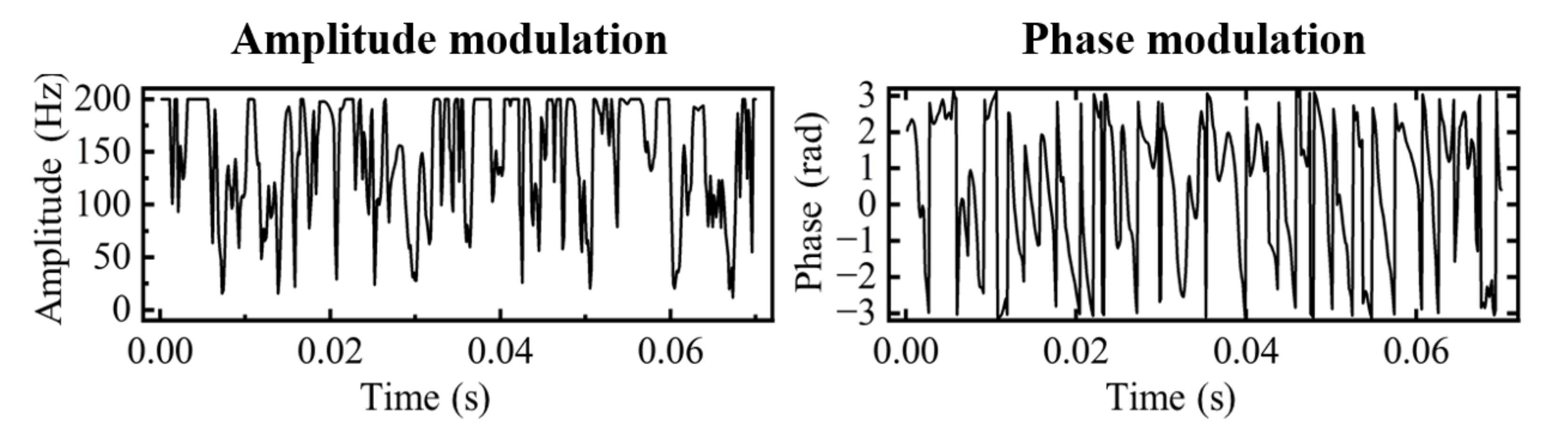
Figure 2. The
amplitude and phase modulation
of the "Optimal Control I" pulse.
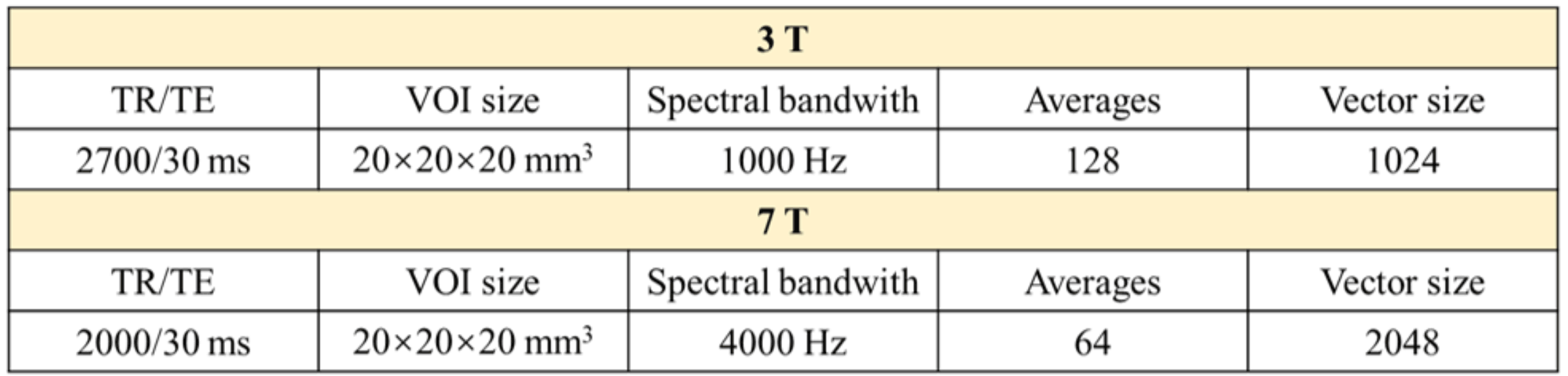
Table 1. Experimental parameters used in the experiments on the 3T
and 7T scanners.
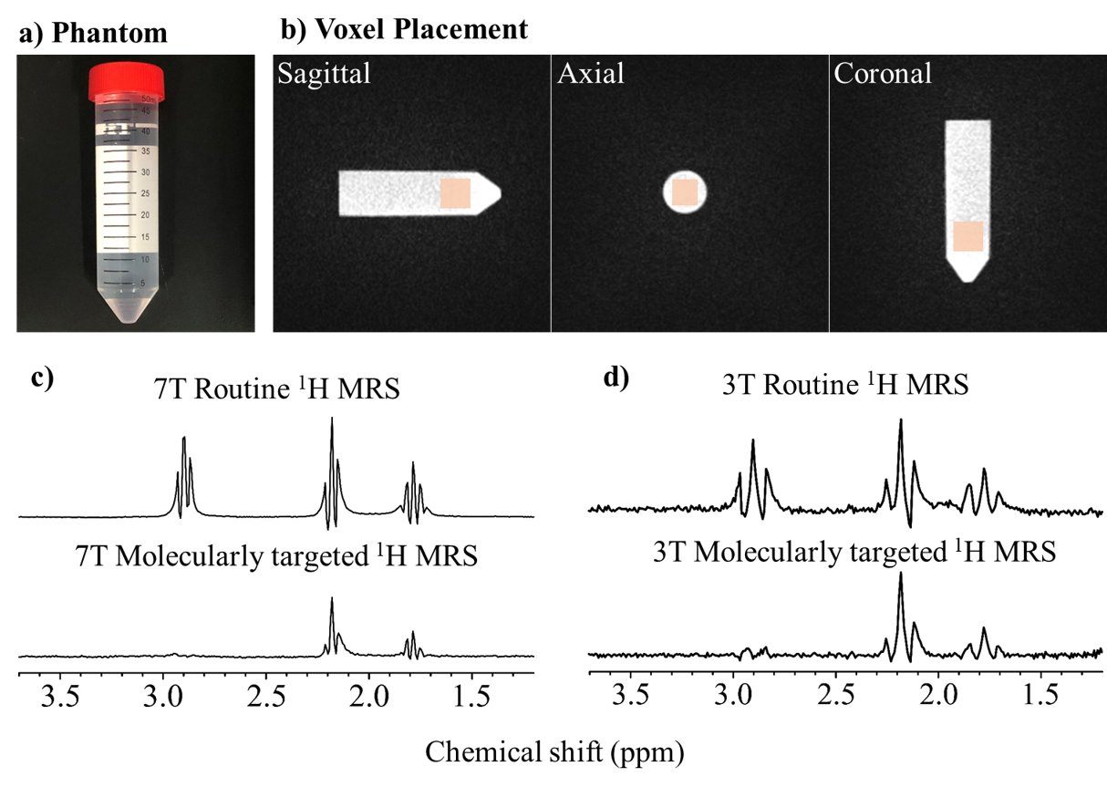
Figure 3. a) A photo of the phantom used in the experiments. The sample is a 50 ml centrifuge tube containing 50 ml of GABA solution in water. b) Localization of the magnetic resonance spectroscopy VOI (pink square) in the phantom. c) From top to bottom: The 7T routine 1H MR spectrum and the GABA-targeted spectrum. d) From top to bottom: The 3T routine 1H MR spectrum and the GABA-targeted spectrum.
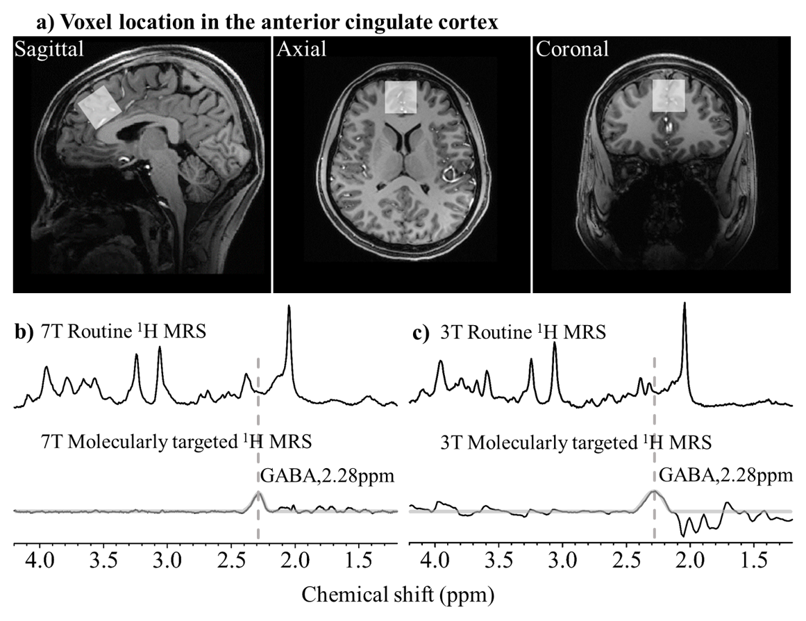
Figure 4. a) The VOI location in the anterior cingulate cortex (white square). b) From top to bottom: The 7T routine 1H MR spectrum and the GABA-targeted spectrum. c) From top to bottom: The 3T routine 1H MR spectrum and the GABA-targeted spectrum.
DOI: https://doi.org/10.58530/2023/2385