2369
Synthetic MRI derived relaxation mapping in evaluating cartilaginous degeneration caused by prolonged excessive exercise1Nanjing University of Chinese Medicine, Nanjing, China, 2GE Healthcare, MR Research China, Beijing, China, 3Jiangsu Province Hospital of Chinese Medicine, Nanjing, China
Synopsis
Keywords: Data Analysis, Osteoarthritis
The purpose of this study was to investigate whether prolonged overexertion can lead to cartilage degeneration through Synthetic MRI derived relaxation maps. In this study, 30 participants were recruited and measured with T1 and T2 mapping derived by Synthetic MRI. There was a statistically significant trend in cartilage T1 and T2 values in the long-term exercise group compared to the normal exercise group. Based on altered T1 and T2 relaxation properties of cartilage tissues, we conclude that chronic overexercise may lead to cartilage degeneration.Introduction
Knee osteoarthritis (KOA) is a disease based on degenerative pathological changes. KOA incidence is increasing year on year in China and globally in recent years[1]. The course of osteoarthritis of the knee is long, and the whole disease develops from light to heavy and from the tendon to the bone development rule. At the early and middle stage, KOA is mainly "tendon injury", that is, the appearance of joint pain, swelling, flexor disadvantage[2]. The theory of Prolonged walking injuring tendons is highly consistent with the pathogenesis of KOA. At present, the induction of mechanical factors in chronic degeneration of knee joints has been well demonstrated[3]. Cartilaginous degeneration is a typical pathological change of "tendon injury" and is the main cause of early clinical manifestation of KOA[4].MAGiC (Magnetic resonance image compilation), as a relatively novel synthetic MRI technique, is able to acquire T1, T2 and PD mapping simultaneous in a single scan[5]. With intrinsic properties of T1, T2 and PD, tissue can be specifically labeled and the corresponding biological or pathological changes can be monitored. Based on the promising findings reported in previous studies[6][7], MAGiC derived quantitative maps may also hold a potential in assessing cartilage degeneration after excessive exercise. However, this has not been studied so far.
Therefore, the purpose of this study was to investigate the alterations of cartilage tissue and determine their clinical significance in subjects with and without excessive exercise using MAGiC derived quantitative maps. T1 and T2 values in the medial condylar cartilage of the tibia platform and the epicondylar cartilage of the femur were compared to determine whether long-term exercise changed the cartilage tissue.
Materials and Methods
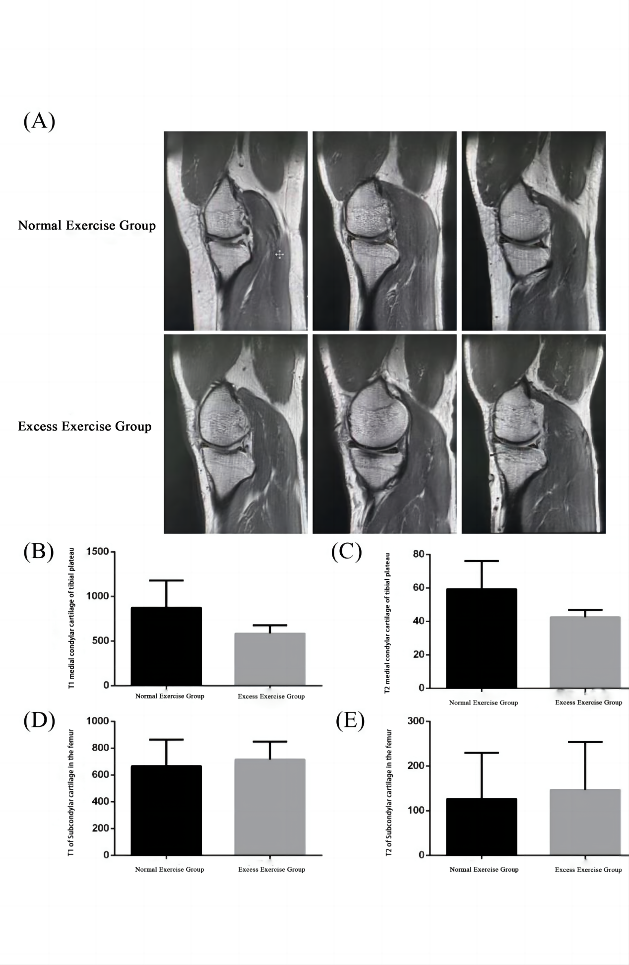
SubjectsA total of 15 male and 15 female participants were recruited for the study. The informed consent was obtained from each participant. All subjects were divided into two groups, including one marathon group (10 men, 5 women) and one normal exercise group (5 men, 10 women). The participants in the marathon group averaged two full marathon sessions a week.
MRI acquisition
All MRI experiments were performed on a 3T-scanner (SIGNA Architect, GE, USA) with an 8-channel flexible coil employed. Conventional T2WI and MAGiC for cartilage imaging were performed for each participants. For MAGiC, the corresponding scan parameters were defined as: field-of-view = 18cm x 18cm, matrix size = 320 x 256, slice thickness = 4mm, slice gap = 0.5mm, number of slice = 20, repetition time = 4621ms, echo time = 22.3/111.5ms, and scan time = 5 mins 14s.
Data analysis
All MAGiC data for cartilage were post-processed with a vendor-provided program (MAGIC, v.100.1.1). The corresponding T1, T2 mapping were obtained accordingly for each patient. Two experienced radiologists selected the areas with the most significant cartilaginous degeneration as regions of interest (ROI)s based on the T2WI images. The mean T1, T2 values of ROIs for each participant were used for statistic analysis.
Statistic analysis
All statistic analyses were performed in SPSS (version 23.0) and MedCalc software (version 15.2.2). Intra-class correlation coefficients analysis was applied to assess the inter-observer agreement of each parameter measurement over two radiologists. Differences in T1 and T2 between chronically excessive exercise and normal exercise groups were examined using both one-way analysis of variance (ANOVA) test and post-hoc independent sample t-tests. P < 0.05 was considered the threshold of statistic significance.
Results
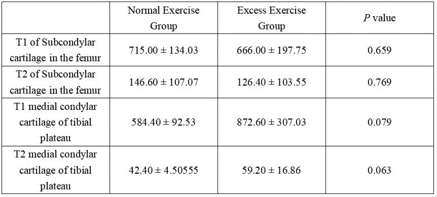
No significant difference of T1 and T2 was found in subcondylar cartilage of the femur between the marathon group and the normal exercise group(p=0.659, 0.769). However, there was a statistically significant trend of T1 and T2 (p=0.079, p=0.063) in medial condylar cartilage of tibial plateau between the marathon group and the normal exercise group, suggesting that excessive exercise may lead to potentially adverse changes in medial cartilage of the knee. (Figure1A-E, Table1).Discussion
In long-term clinical practice, we found higher levels of cartilage damage in subjects who experienced long-distance running or needed to walk for long periods of time. Meanwhile, studies have shown that people at risk of KOA are more likely to develop cartilaginous degeneration if they are hyperactive and bend their knees frequently [8].In this study, we evaluated knee cartilage in two groups of participants using quantitative T1 and T2 maps derived from synthetic MRI. Lower T1 and longer T2 values were shown after chronic overexercise than normal exercise. These may be explained that after chronic overexercise, the microstructures of cartilage tissue may be altered with larger porosity or edema[9], suggesting chronic overexercise caused cartilage damage.
Conclusion
In conclusion, our study demonstrated that quantitative T1 and T2 derived from synthetic MRI can effectively differentiate cartilage degeneration and may serve as reliable biomarkers for future clinical diagnosis of exercise-induced cartilage degeneration.Acknowledgements
No acknowledgement found.References
[1] Martel-Pelletier, J., Barr, A. J., Cicuttini, F. M., Conaghan, P. G., Cooper, C.,Goldring, M. B., et al. (2016). Osteoarthritis. Nat. Rev. Dis. Primers 2:16072
[2] Lv, Z., et al., Molecular Classification of Knee Osteoarthritis. Front Cell Dev Biol, 2021. 9: p. 725568.
[3] Khan, M.C.M., et al., The Influence of Running on Lower Limb Cartilage: A Systematic Review and Meta-analysis. Sports Med, 2022. 52(1): p. 55-74.
[4] Zeng, C.Y., et al., Benefits and Mechanisms of Exercise Training for Knee Osteoarthritis. Front Physiol, 2021. 12: p. 794062.
[5] Warntjes JB, Leinhard OD, West J, et al., Rapid magnetic resonance quantification on the brain: Optimization for clinical usage. Magn Reson Med.2008 Aug;60(2):320-9.
[6] Jiang Y, Yu L, Luo X, et al., Quantitative synthetic MRI for evaluation of the lumbar intervertebral disk degeneration in patients with chronic low back pain. Eur J Radiol. 2020 Mar;124:108858.
[7] Park S, Kwack K-S, Lee YJ, Gho S-M, Lee HY. Initial experience with synthetic MRI of the knee at 3T: comparison with conventional T1 weighted imaging and T2 mapping. Br J Radiol 2017; 90: 20170350.
[8] Hovis KK, Stehling C, Souza RB, et al. Physical activity is associated with magnetic resonance imaging-based knee cartilage T2 measurements in asymptomatic subjects with and those without osteoarthritis risk factors. Arthritis Rheum. 2011;63(8):2248-2256.
[9] Kotecha M, Klatt D, Magin RL. Monitoring cartilage tissue engineering using magnetic resonance spectroscopy, imaging, and elastography. Tissue Eng Part B Rev. 2013;19(6):470-484.
Figures