2368
Characterization of age- and gender-dependent differences in intervertebral disc strain using 3D-GRASP MRI under static mechanical loading1Radiology, NYU Grossman School of Medicine, New York, NY, United States
Synopsis
Keywords: MR Fingerprinting/Synthetic MR, Tissue Characterization, Intervertebral disc
The goal of this study was to assess the effect of age and gender in IVD characterization using a multiparameter MR Fingerprinting technique that can quantify T1, T2 and T1rho in a clinically feasible time. Seventeen healthy subjects were recruited. The goal of this study was to assess the effect of age and gender in IVD characterization using a multiparameter MR Fingerprinting technique that can quantify T1, T2 and T1rho in a clinically feasible time. This study suggests that the use of MR fingerprinting is a useful tool to gain insights to pathology resulting in IVD degeneration.INTRODUCTION
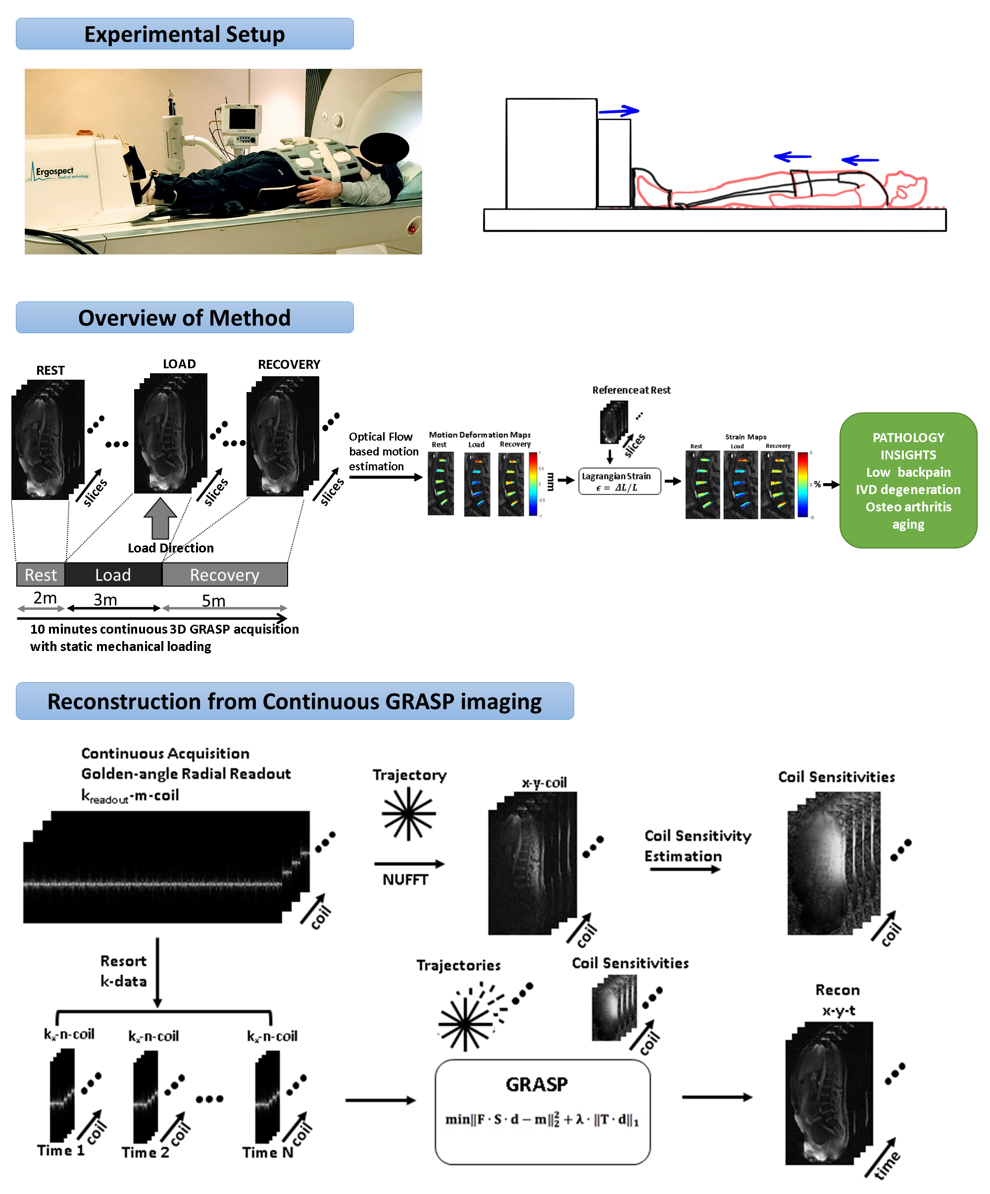
Chronic low back pain due to inter-vertebral disc (IVD) degeneration is a major cause of pain and disability worldwide1. The IVD is a fibrocartilage structure consisting of an inner gel-like core (nucleus pulposus) and a highly organized fibrous outer structure (annulus fibrosus) that together provide flexibility as well as compressive and tensile strength to the spine. It has traditionally been difficult to measure IVD strain in-vivo due to challenges in simultaneously loading the spine while acquiring image data with high spatial and temporal resolution. We have developed a MRI protocol using static mechanical loading and a continuous 3D-GRASP acquisition to quantify in-vivo IVD strain during loading and recovery2. Our study was performed to investigate age-dependent and gender-dependent differences in IVD strain of the lumbar spine measured using the 3D-GRASP MRI technique.METHODS
The study group consisted of 20 healthy subjects (10 females and 10 males; age=34.6±11.4 years; age range=22-56 years). All subjects signed written informed consent.Figure 1 shows the experimental setup. An MRI compatible ergometer (Ergospect, Innsbruck, Austria) was used to deliver simulated standing loads to the spine with a total force of 400 N (~50% body weight) during the MRI scan. The MRI protocol consisted of a rest-loading-recovery paradigm with a continuous 3D-GRASP acquisition performed on a 3T scanner (Prisma, Siemens Healthcare, Germany). The sagittal 3D-GRASP sequence consisted of a continuous MRI acquisition using a 3D golden angle stack-of-stars trajectory that acquired in-plane radial spokes (Kx-Ky) using the following parameters: repetition time=5ms, echo time=2.5ms, flip angle=12°, field of view=240x240mm2, slab thickness = 88 mm, matrix size=256x256x22, voxel resolution = 0.9x0.9x4mm3, receiver bandwidth=590Hz/pixel, radial spokes=4000, and total scan time=10min.
Raw data from the scanner was used for offline reconstruction as shown in Figure 1. The continuously acquired radial spokes were regrouped to form a sparse dataset consisting of 400 spokes per time frame giving a temporal resolution of one 3D dataset/min, which was used as input for compressed sensing (CS) reconstruction. An optical flow tracking algorithm3 was then used to determine the sub-voxel shifts in the IVD segments as a result of loading and recovery. The algorithm estimated voxel image intensity based velocity fields with the assumption that gray level intensities are preserved during displacement. Five IVD regions of interest (ROIs) of the lumbar spine were manually segmented (L1/L2, L2/L3, L3/L4, L4/L5, L5/S1) and 3D-motion deformation between each time-point and the reference frame were estimated. The Lagrangian strain (SL)4 for each ROI was calculated from the deformation fields and reference.
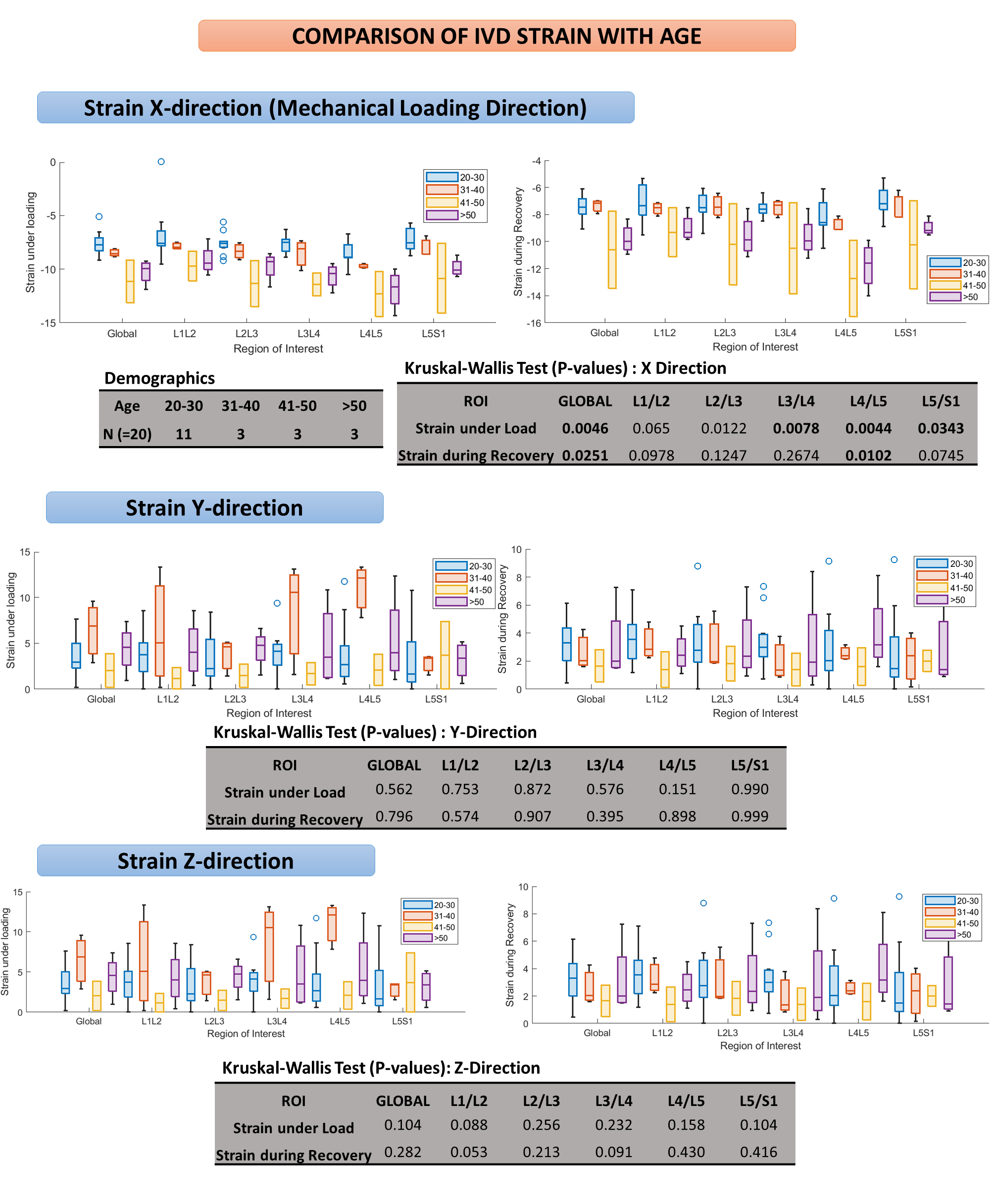
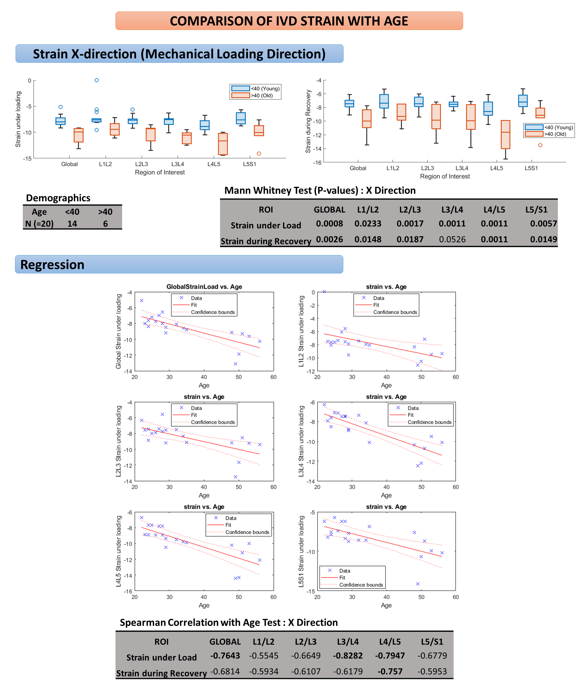
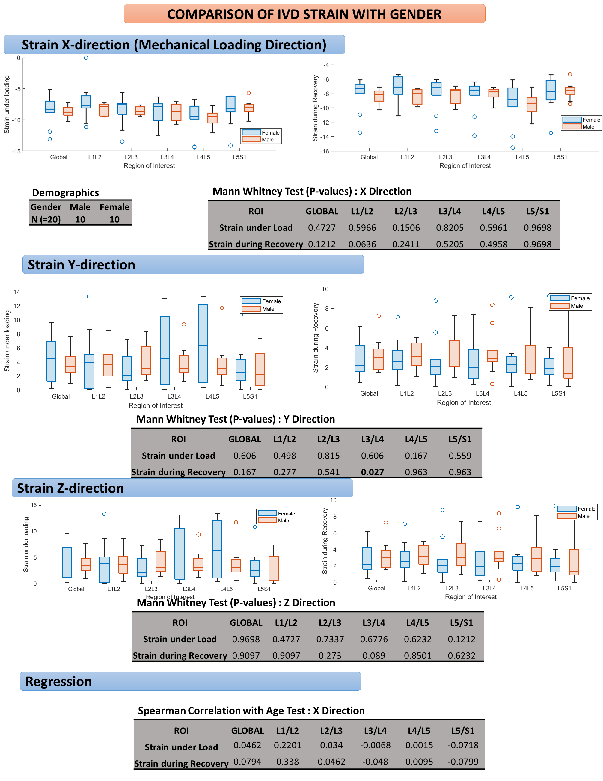
Kruskal-Wallis and Mann-Whitney tests were used to assess age-dependent differences in IVD strain for subjects separated into four age groups (20-30yrs;31-40yrs;41-50yrs;>50yrs) and two age groups (<40yrs;>40yrs), respectively. Spearman correlation coefficients were used to evaluate the association between age and IVD strain. Mann-Whitney tests were used to assess gender-dependent changes in IVD strain. Statistical significance was defined as P< 0.05.
RESULTS
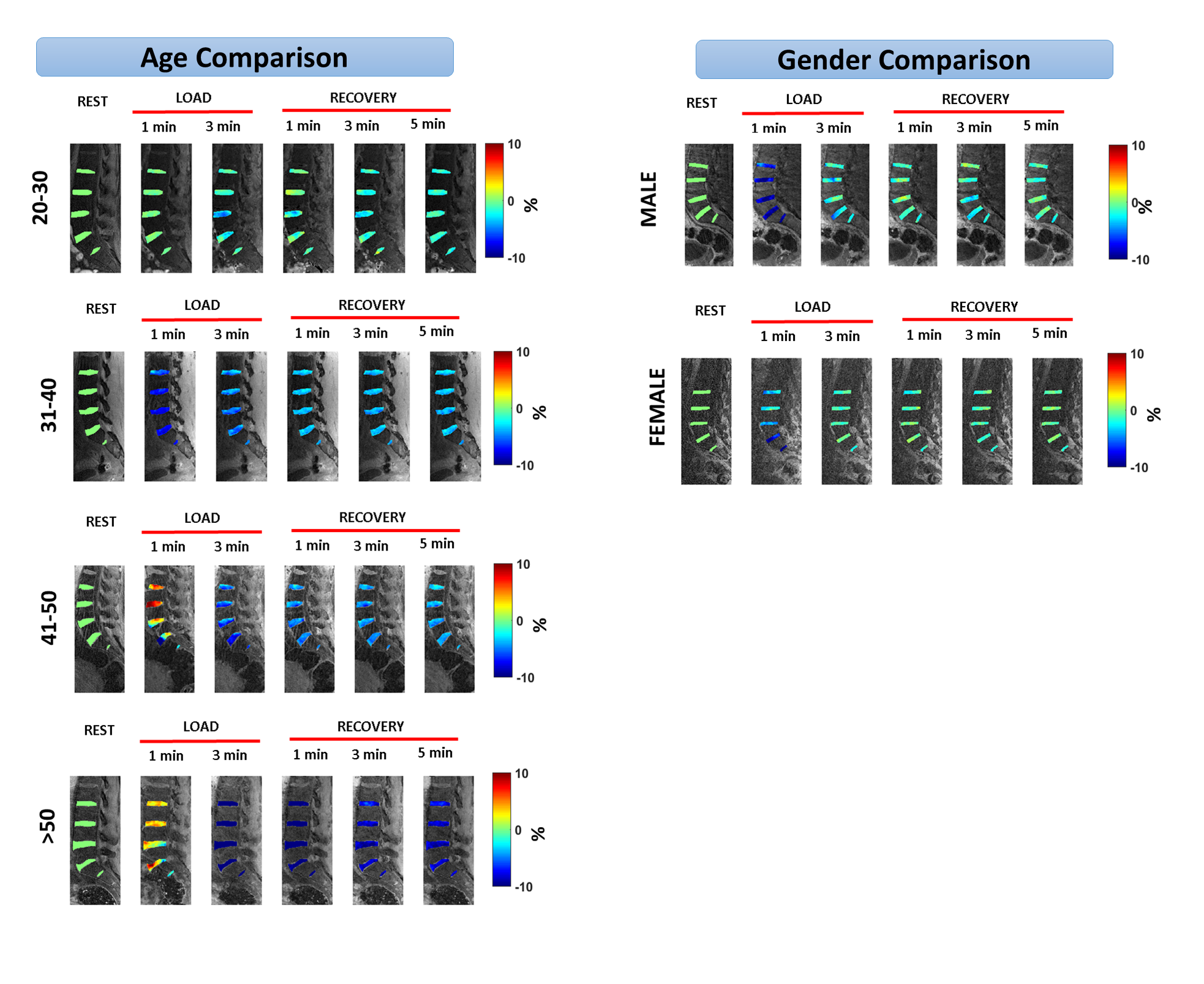
Figure 2 shows the results obtained from the in-vivo strain mapping, which demonstrates variations in strain in the individual IVDs of the lumbar spine from rest to loading to recovery. Figures 3 and 4 shows age-dependent differences of IVD strain during loading and recovery. There was a significant difference between age groups in global IVD strain in the X-direction during loading and recovery. A significant inverse correlation between age and global IVD strain was observed. There was no significant differences between age groups in IVD strain in the Y-direction or Z-direction. Figure 5 shows gender-related differences in IVD strain during loading and recovery. There was no significant differences between male and female subjects in global IVD strain in the X-direction, Y-direction, or Z-direction.DISCUSSION
Our study showed significant age-related differences but non-significant gender-related differences in IVD strain of the lumbar spine during static mechanical loading measured using the 3D-GRASP technique. Ex-vivo studies have documented evidence of asymptomatic IVD degeneration beginning as early as the third decade of life5. Early IVD degeneration is characterized by a decrease in the proteoglycan and water content of the nucleus pulposus5-7. The compositional changes of the nucleus pulposus result in increased stiffness and decreased compliance of the IVD8, which is responsible for the significant inverse correlation between age and IVD strain during loading observed in our study. The fact that significant inverse correlations between age and IVD strain were found in the X-direction but not in the Y-direction or Z-direction indicates that early IVD degeneration increases compressive stiffness to a greater extent than tensile stiffness during spine loading. Our study also documented significant age-dependent differences in IVD strain during recovery although the magnitude of the association was less than during loading.CONCLUSION
The results of our study demonstrate the significant role that aging plays in IVD strain changes of the lumbar spine during mechanical loading. Furthermore, our study demonstrates the use of a continuous 3D-GRASP MRI technique to characterize IVD strain of the lumbar spine during loading and recovery, which could be used in the future to investigate disease-related and treatment-related changes in the mechanical properties of the IVD in patients with back pain.Acknowledgements
This study was supported by NIH grants, R21-AR078357, R01-AR076328-01A1, R01-AR076985-01A1, and R01-AR078308-01A1 and was performed under the rubric of the Center of Advanced Imaging Innovation and Research (CAI2R), an NIBIB Biomedical Technology Resource Center (NIH P41-EB017183).References
1 Murray, C. J. et al. Disability-adjusted life years (DALYs) for 291 diseases and injuries in 21 regions, 1990-2010: a systematic analysis for the Global Burden of Disease Study 2010. Lancet 380, 2197-2223, doi:10.1016/S0140-6736(12)61689-4 (2012).
2 Menon, R. G., Zibetti, M. V. W., Pendola, M. & Regatte, R. R. Measurement of Three-Dimensional Internal Dynamic Strains in the Intervertebral Disc of the Lumbar Spine With Mechanical Loading and Golden-Angle Radial Sparse Parallel-Magnetic Resonance Imaging. J Magn Reson Imaging 54, 486-496, doi:10.1002/jmri.27591 (2021).
3 Zachiu, C., Denis de Senneville, B., Dmitriev, I. D., Moonen, C. T. W. & Ries, M. A framework for continuous target tracking during MR-guided high intensity focused ultrasound thermal ablations in the abdomen. J Ther Ultrasound 5, 27, doi:10.1186/s40349-017-0106-y (2017).
4 Chitiboi, T. & Axel, L. Magnetic resonance imaging of myocardial strain: A review of current approaches. J Magn Reson Imaging 46, 1263-1280, doi:10.1002/jmri.25718 (2017).
5 Singh, K., Masuda, K., Thonar, E. J., An, H. S. & Cs-Szabo, G. Age-related changes in the extracellular matrix of nucleus pulposus and anulus fibrosus of human intervertebral disc. Spine (Phila Pa 1976) 34, 10-16, doi:10.1097/BRS.0b013e31818e5ddd (2009).
6 Antoniou, J. et al. The human lumbar intervertebral disc: evidence for changes in the biosynthesis and denaturation of the extracellular matrix with growth, maturation, ageing, and degeneration. J Clin Invest 98, 996-1003, doi:10.1172/JCI118884 (1996).
7 Pearce, R. H., Grimmer, B. J. & Adams, M. E. Degeneration and the chemical composition of the human lumbar intervertebral disc. J Orthop Res 5, 198-205, doi:10.1002/jor.1100050206 (1987).
8 Inoue, N. & Espinoza Orias, A. A. Biomechanics of intervertebral disk degeneration. Orthop Clin North Am 42, 487-499, vii, doi:10.1016/j.ocl.2011.07.001 (2011).
Figures