2353
Synthetic MRI and FSE-PROPELLER duo diffusion-weighted imaging to differentiate malignant from benign head and neck tumors1the First Affiliated Hospital of Zhengzhou University, Zhengzhou, China, 2GE Healthcare, Shanghai, China
Synopsis
Keywords: MR Fingerprinting/Synthetic MR, Head & Neck/ENT
Accurate determination of the preoperative classification of histology of head and neck tumor remains a challenge. The aim of the study was to evaluate the feasibility and capability of synthetic MRI and stimulus and fast spin echo diffusion-weighted imaging (FSE-PROPELLER duo-DWI) for the differentiation of malignant from benign head and neck tumors. The results quantitively demonstrated the feasibility and also showed that T2 value is comparable to ADC value, and the combination of T2 and ADC values could provide improved diagnostic efficacy.Background and purpose
The histology of head and neck tumor is an important factor associated with the treatment and prognosis. However, accurate determination of the preoperative classification of histology of head and neck tumor remains a challenge. Diffusion-weighted imaging (DWI) can quantitatively evaluate the Brownian motion diffusion of water molecules in tissues at a cellular level expressed as apparent diffusion coefficient (ADC). Synthetic MRI based on a quantitative approach is used to estimate absolute physical properties, longitudinal and transverse relaxation times (T1 and T2) and proton density (PD) which are independent from the MRI scanners or scanning parameters at a given field strength. We aimed to evaluate the feasibility and capability of synthetic MRI and stimulus and FSE-PROPELLER duo-DWI with periodically rotated overlapping parallel lines with enhanced reconstruction for the differentiation of malignant from benign head and neck tumors and quantitively assess the diagnosis efficiency.Methods
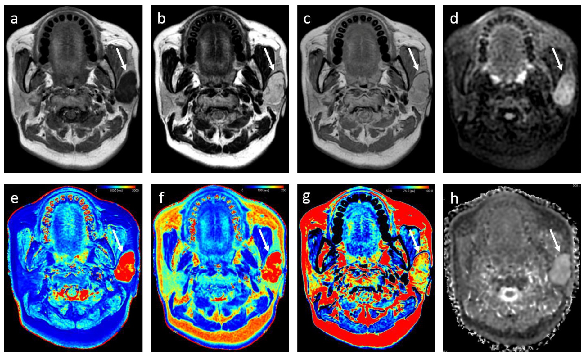
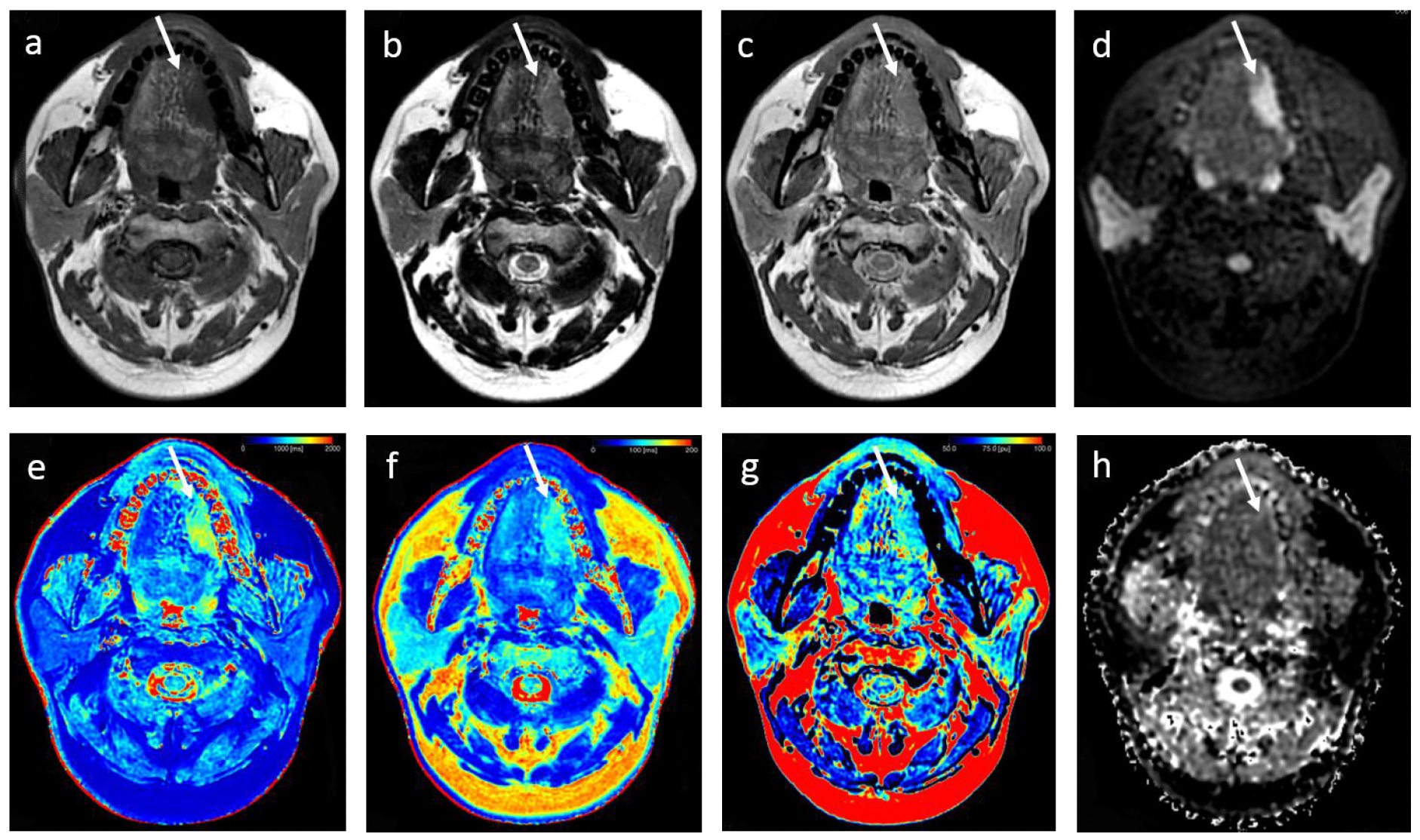
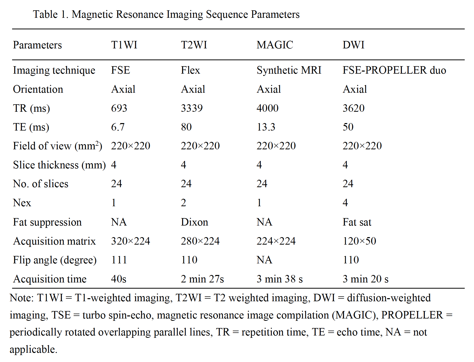
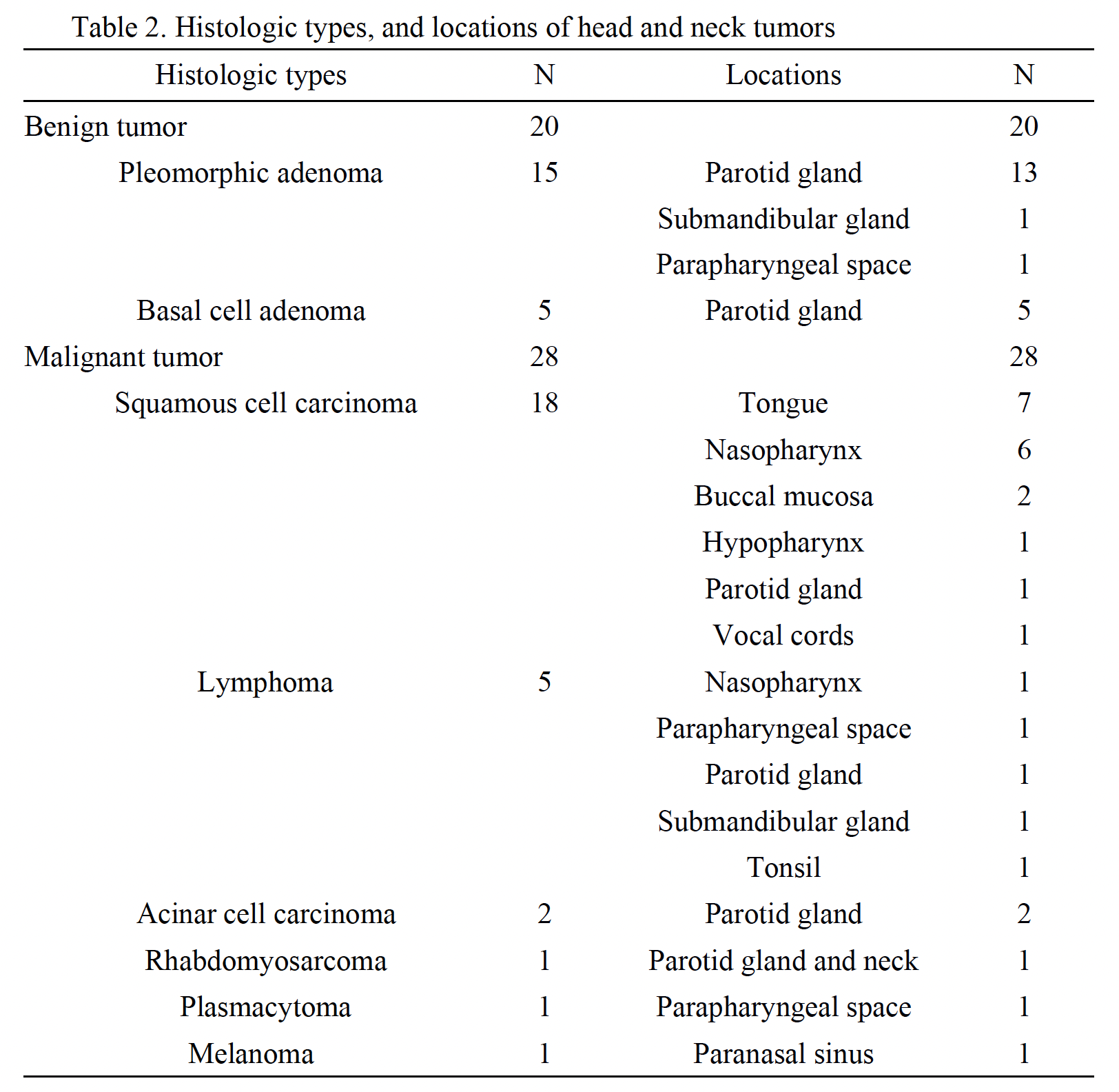
The study comprised 48 patients with pathologically confirmed head and neck tumors recruited from August 2022 to October 2022. The patients were divided into malignant (n = 28) and benign (n = 20) groups. All MRI acquisitions were performed on a 3T MR scanner (Premier, GE Healthcare, Milwaukee, WI, USA) in a supine position with a 21-channel head-neck coil. Synthetic MRI and FSE-PROPELLER duo-DWI (Detailed scan parameters refer to Table 1). T1, T2, and proton density (PD) values acquired on the synthetic MRI, and ADC values acquired on the FSE-PROPELLER duo-DWI. Then we used magnetic resonance image compilation (MAGIC) software to process the acquired data from synthetic MRI sequence. The two independent senior radiologists manually drew the regions of interest (ROIs) on synthetic T2WI to obtain the T1, T2, and PD values(Figure 1, Figure2). Postprocessing of FSE-PROPELLER duo-DWI was performed using the ADW 4.7 workstation. We then compared and analyzed the ability to differentiate benign from malignant head and neck tumors. The diagnostic value of the combined T2 and ADC values was based on the logistic regression analysis. We conducted the receiver operating characteristic (ROC) curve and the area under the curve (AUC), sensitivity, specificity, positive predictive value (PPV), and negative predictive value (NPV) were further calculated to ascertain diagnostic performance.Results
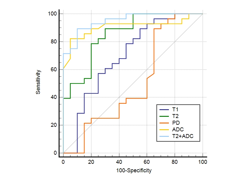
The T1, T2 and ADC values of benign tumors were significantly higher than those of the malignant tumors (1741.13 ± 662.64 ms vs 1390.06 ± 241.09 ms, 157.43 ± 72.23 ms vs 97.64 ± 14.91 ms, 2.03±0.31 × 10⁻³ mm2/s vs 1.46 ± 0.37 × 10⁻³ mm2/s, all P < 0.05). No significant differences were found in PD values between the two groups (t’ = -0.125, P = 0.901) (Table 2). ROC analysis showed that in differentiating malignant from benign head and neck tumors, T2+ADC (cut off value, > 0.55; AUC, 0.950) showed optimal diagnostic performance, which was better than that of T1 (cut off value, ≤ 1675.84 ms; AUC, 0.698) , T2 (cut off value, ≤ 113.24ms; AUC, 0.855) and PD (cut off value, > 80.67 ms; AUC, 0.568) (p = 0.0030, 0.0464, and p < 0.0001, respectively), but the difference in AUC between ADC (cut off value, ≤ 1.60 × 10⁻³ mm2/s; AUC, 0.906) and T2+ADC or T2 did not reach signifcance (p = 0.2648 and 0.4604, respectively)(Figure 3).Conclusion
In summary, the quantitative T1, T2, and PD values obtained by synthetic MRI and ADC value obtained by FSE-PROPELLER duo-DWI were helpful for discriminating malignant from benign head and neck tumors. The overall diagnostic performance of synthetic MRI was inferior to FSE-PROPELLER duo-DWI. However, T2 value is comparable to ADC value, and the combination of synthetic MRI and FSE-PROPELLER duo-DWI could provide improved diagnostic efficacy. In the future, larger population and multi-center studies are needed to affirm the results of this study.Acknowledgements
We are particularly grateful to all the people who have given us help on our article.References
1. Law BKH, King AD, Ai QY, Poon DMC, Chen W, Bhatia KS, et al. Head and neck tumors: Amide proton transfer MRI. Radiology. 2018;288:782–90.
2. Payabvash SMD. Quantitative diffusion magnetic resonance imaging in head and neck tumors. Quant Imaging Med Surg. 2018;8:1052–65.
3. Liu YJ, Lee YH, Chang HC, Chung HW, Wang CW, Juan CH, et al. Imaging quality of PROPELLER diffusion-weighted MR imaging and its diagnostic performance in distinguishing pleomorphic adenomas from Warthin tumors of the parotid gland. NMR Biomed. 2020;33:1–11.
4. Granberg T, Uppman M, Hashim F, Cananau C, Nordin LE, Shams S, et al. Clinical feasibility of synthetic MRI in multiple sclerosis: A diagnostic and volumetric validation study. Am J Neuroradiol. 2016;37:1023–9.
5. Meng T, He H, Liu H, Lv X, Huang C, Zhong L, et al. Investigation of the feasibility of synthetic MRI in the differential diagnosis of non-keratinising nasopharyngeal carcinoma and benign hyperplasia using different contoured methods for delineation of the region of interest. Clin Radiol. 2021;76:238.e9-238.e15.
Figures