2347
Assessing the development of brain's structural connectivity in the perinatal stage with diffusion MRI1Harvard Medical School & Boston Children's Hospital, Boston, MA, United States
Synopsis
Keywords: Neonatal, Data Analysis
This study analyzed the development of brain's structural connectivity during the perinatal period using the diffusion MRI data of 263 subjects from the dHCP dataset. We used anatomically constrained probabilistic tractography to reconstruct structural connectomes. We computed connectivity metrics to assess network integration and segregation and tested for the effects of postmenstrual age (PMA), premature birth, and gender. Results showed increasing network integration and segregation with PMA in the range 29 to 45 weeks. Premature birth and male gender were associated with significantly lower rates of increase in global efficiency.Introduction
The human brain undergoes significant and rapid developments from the mid-fetal stage to the neonatal period. Highly structured and organized brain networks emerge and develop in this period. Tightly-orchestrated and complex processes that give rise to these networks may be prone to adverse genetic or environmental factors such as premature birth. To study normal and abnormal brain development, diffusion-weighted magnetic resonance imaging (dMRI) can be used to perform quantitative structural connectivity analysis. However, studying the structural connectivity of the perinatal brain has been especially difficult due to the challenges of scanning subjects early in life. In this work, we used the data from 263 subjects in the developing Human Connectome Project (dHCP) [1] to compute important metrics of structural connectivity. We analyzed the development of these metrics with age and the impacts of gender and premature birth.Methods
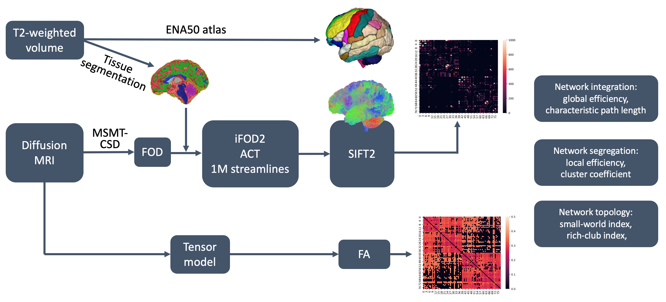
Data processing: Figure 1 depicts the main steps in our data processing pipeline. The dMRI data were denoised, followed by B1 field inhomogeneity correction. Subsequently, all dMRI and anatomical data (i.e., T2 images and tissue segmentations) were resampled to isotropic resolution of 1 mm. Multi-tissue constrained spherical deconvolution was used to estimate the fiber orientation distributions (FODs) [2]. Anatomically constrained probabilistic tractography was applied on the computed FOD and the tissue segmentation map to generate 1 million valid streamlines. Maximum angle between successive streamline tracing steps was set to 30 degrees, and a FOD amplitude threshold of 0.01 was used as the stopping criterion. SIFT2 algorithm [3] was used to compute cross-sectional area multipliers to enhance the biological plausibility of the tractogram. A research fellow and a research assistant visually inspected the tractography quality and approved 263 subjects for further analysis. The neonatal brain gray matter parcellation from the Edinburgh Neonatal Atlas (ENA50) [4] was used to define the connectome nodes. Since it is recommended to use some measure of tissue microstructure integrity to weight the connections, network edge strengths were defined to be the mean FA along the streamlines. Graph theoretical analysis was performed using GRETNA software [5]. We quantified network integration in terms of global efficiency and characteristic path length, and network segregation in terms of local efficiency and clustering coefficient.Statistical analysis: General linear models (GLMs) were used to assess the correlation between structural connectivity measures and PMA. In order to test for the effects of preterm birth and gender, term/preterm birth and gender were added as the main effects with age and group as covariates. If the interaction effect was significant, rates of age-dependent trend in network measures would be significantly different between groups.
Results and Discussion
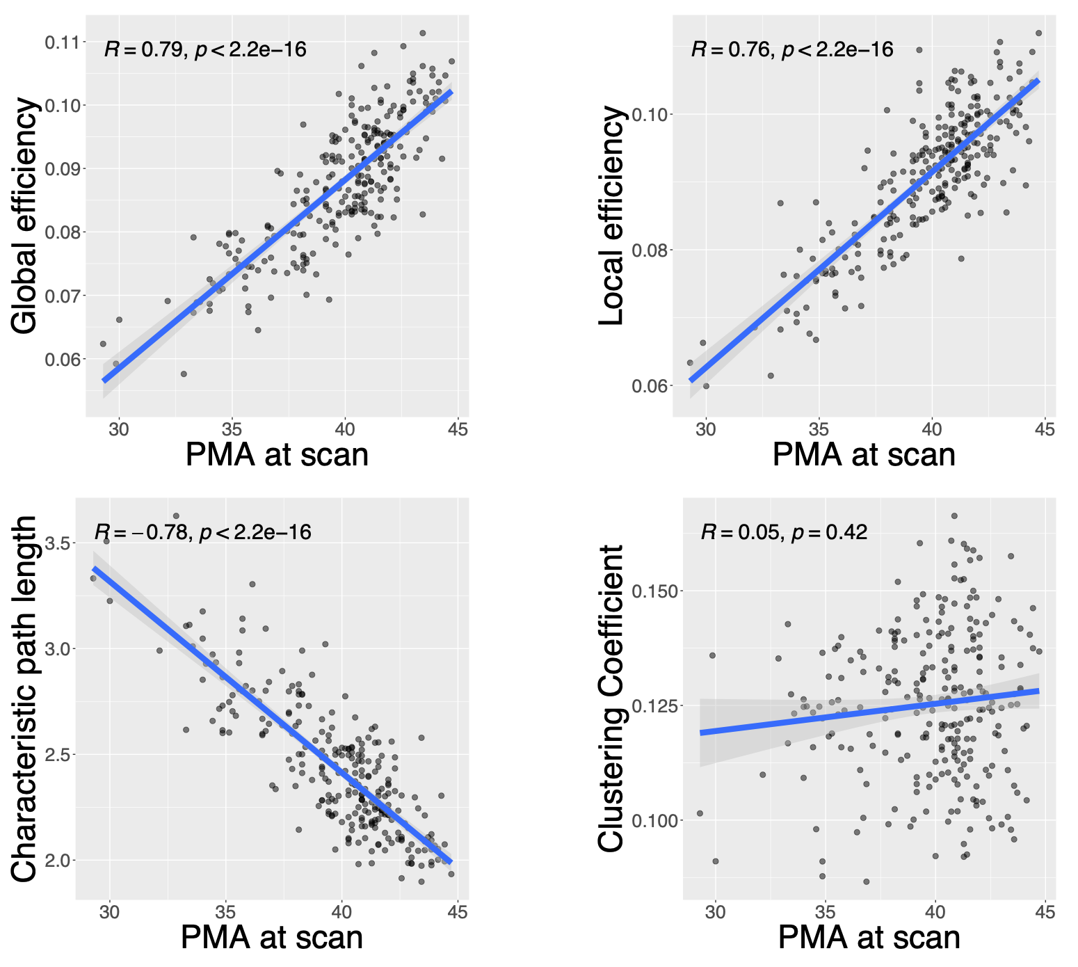
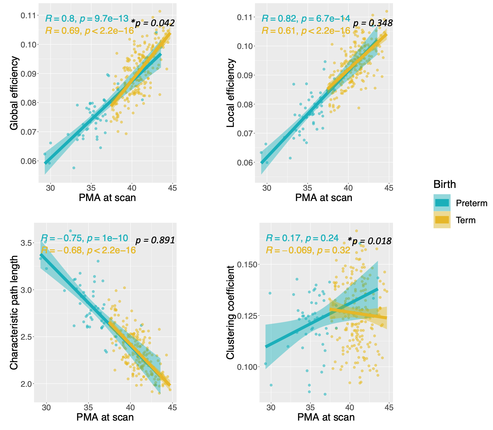
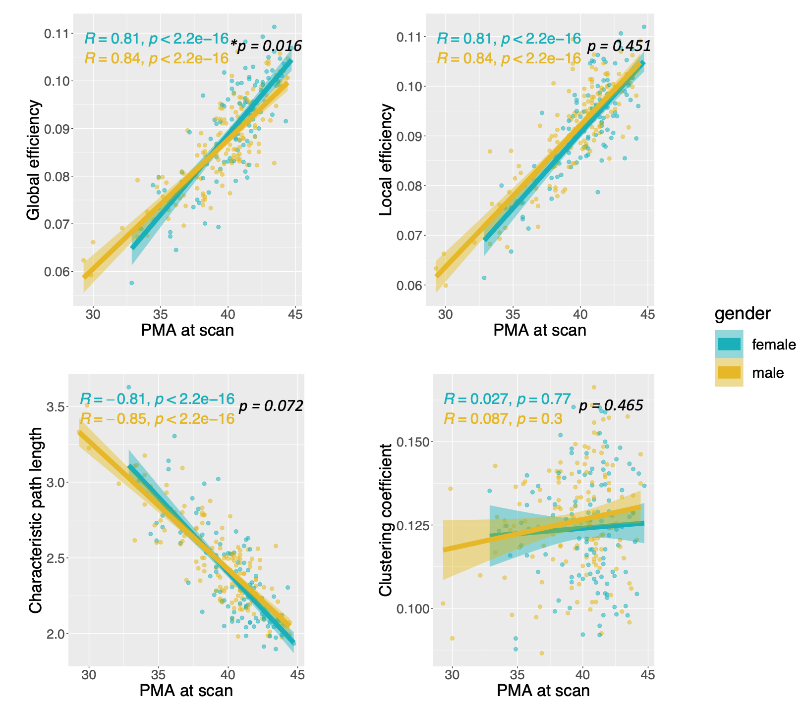
As shown in Figure 2, analysis of the pool of all 263 subjects showed that PMA at scan was positively correlated with global efficiency (r = 0.79, p < 0.001) and local efficiency (r = 0.76, p < 0.001), while negatively correlated with characteristic path length (r = -0.78, p < 0.001). The correlation with clustering coefficient was insignificant (p= 0.42). Overall, these results indicate a significant increase in both network integration and network segregation.As shown in Figures 3 and 4, there were no significant effects for gender and preterm birth on local efficiency and characteristic path length. The rate of increase in global efficiency was significantly lower in preterm-born subjects compared with term-born subjects (p = 0.042). Rate of increase in global efficiency was also significantly lower in males compared with females (p = 0.016). Additionally, we observed a different trend in the clustering coefficient between term and preterm subjects, which was statistically significant (p = 0.018). Surprisingly, while there was a clear increase in the clustering coefficient for preterm-born subjects, there was a slight decrease for term subjects.
Conclusion
In this study, we assessed the integration and segregation of structural brain networks in the perinatal stage and explored the impact of premature birth and gender. Using the high-quality data available through the dHCP project, we were able to include a much larger number of subjects than in most prior works on this difficult age group.We observed a significant increase in network integration with PMA. This suggests a rapid increase in the brain's ability to integrate information from distributed brain regions, and increased ease and efficiency of communication between different brain regions. We also observed a significant increase in network segregation with PMA. This suggests that the brain's ability to support specialized information processing in interconnected sub-regions grows rapidly in this stage.
We also observed significant effects for premature birth and gender. Premature birth was associated with a slower increase in global efficiency, but surprisingly a faster increase in clustering coefficient. The latter observation may benefit from more detailed analysis and interpretation. Rate of increase in global efficiency was also higher in females than in males. While females had a lower global efficiency at 35 weeks PMA, they had a higher global efficiency by 45 weeks PMA.
Acknowledgements
References
1. Bastiani, Matteo, Jesper LR Andersson, Lucilio Cordero-Grande, Maria Murgasova, Jana Hutter, Anthony N. Price, Antonios Makropoulos et al. "Automated processing pipeline for neonatal diffusion MRI in the developing Human Connectome Project." Neuroimage 185 (2019): 750-763.
2. Jeurissen, Ben, Jacques-Donald Tournier, Thijs Dhollander, Alan Connelly, and Jan Sijbers. "Multi-tissue constrained spherical deconvolution for improved analysis of multi-shell diffusion MRI data." NeuroImage 103 (2014): 411-426.
3. Smith, Robert E., Jacques-Donald Tournier, Fernando Calamante, and Alan Connelly. "SIFT2: Enabling dense quantitative assessment of brain white matter connectivity using streamlines tractography." Neuroimage 119 (2015): 338-351.
4. Blesa, Manuel, Paola Galdi, Gemma Sullivan, Emily N. Wheater, David Q. Stoye, Gillian J. Lamb, Alan J. Quigley, Michael J. Thrippleton, Mark E. Bastin, and James P. Boardman. "Peak width of skeletonized water diffusion MRI in the neonatal brain." Frontiers in Neurology 11 (2020): 235.
5. Wang, Jinhui, Xindi Wang, Mingrui Xia, Xuhong Liao, Alan Evans, and Yong He. "GRETNA: a graph theoretical network analysis toolbox for imaging connectomics." Frontiers in human neuroscience 9 (2015): 386.
Figures