2338
Volumetric MRI analysis of occipital gyrus in fetuses with isolated ventriculomegaly1Department of Diagnostic Radiology, Guangzhou Women and Children's Medical Center, Guangzhou Medical University,Guangdong Provincial Clinical Research Center for Child Health, 510623, China, Guangzhou, China
Synopsis
Keywords: Fetal, Brain, Fetal MRI; Isolated ventriculomegaly
The lateral ventricle is closely related to the occipital lobe anatomically. We found changes in cortical volume and white matter volume in isolated fetuses ventriculomegaly. This study will address whether occipital and occipital gyrus volumes differ in IVM and investigate the developmental pattern of the occipital lobe with increasing gestational age investigate. This study will help to provide the research basis for the study the neurodevelopment in IVM fetuses.Introduction
Isolated ventriculomegaly (IVM) is the most common abnormality of the fetal central nervous system, and some fetuses have neurological retardation after birth1. In IVM, the occipital horn of the lateral ventricle is the most significant area to dilate2. However, there is a lack of research on the occipital gyrus of IVM fetus. The purpose of this study was to illustrate the changes of occipital gyrus volume in IVM fetuses by manually segmenting and calculating the volume of occipital brain region, and to reveal the trend of occipital gyrus changes in IVM fetuses with gestational age.Methods
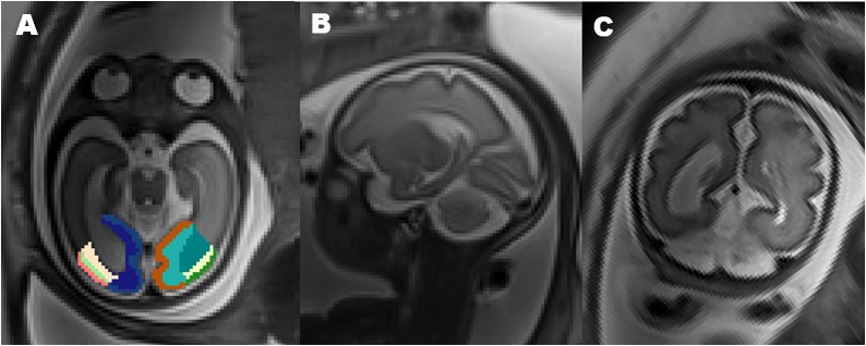
The protocol to perform the present study was reviewed and approved by the local Internal Review Board. We used in T2-weighted resonance imaging of 44 fetus (22 controls and 22 with IVM; gestation ages ranging from 26 to 35 GW). Normal fetal controls were from a non-CNS abnormality on MRI. Inclusion criteria for the IVM group consisted of a singleton pregnancy and IVM (at least 1 lateral ventricle with a width of 10–15 mm without additional CNS abnormalities). These subjects are examined by a 3.0 T scanner (Siemens Skyra, Germany). Data acquisition includes Multiple stacks of slice half-Fourier spin echo (HASTE) T2W slice images (slice thickness = 3 mm, no gap) were planned with respect to the fetal brain. Anatomical areas of each slice of the fetal brain were outlined freehand on the axial images using ITA-SNAP 3.8, the coronal and sagittal planes were used for reference to improve accuracy3 (Figure 1). White matter and cortical matter volumes in the occipital gyrus (include superior occipital gyrus, mild occipital gyrus, inferior occipital gyrus, cuneus, lingual gyrus) of each cerebral hemisphere were manually outlined on T2 using the Developing Brain Region Annotation with Expectation-Maximization (Draw-EM) atlas as template4. We respectively calculate each slice of the volume of gray matter, white matter in the occipital gyrus and the volume of gray matter, white matter in the occipital lobe. Finally, we used SPSS 26.0 (SPSS, Chicago, IL, USA) to analyze all data differences between the IVM group and the control group, and to statistically analyze the correlation between each index and gestational age.Results
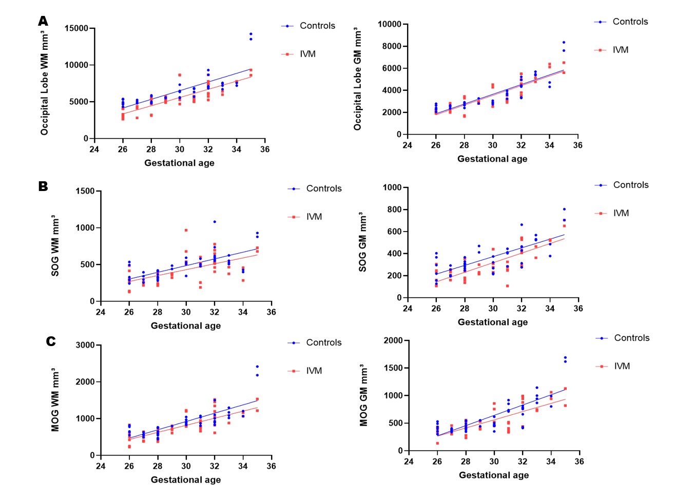
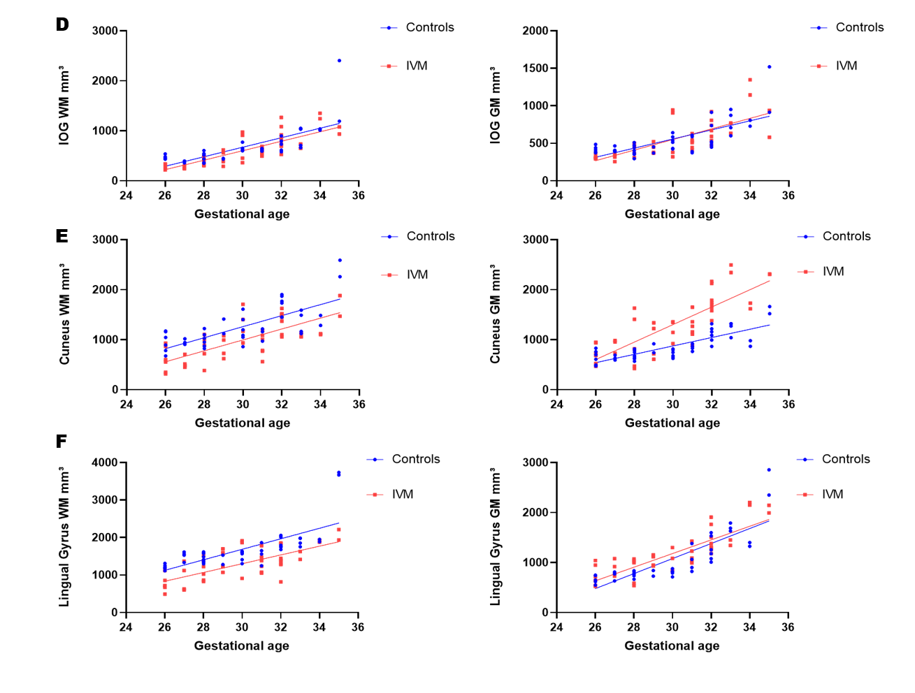
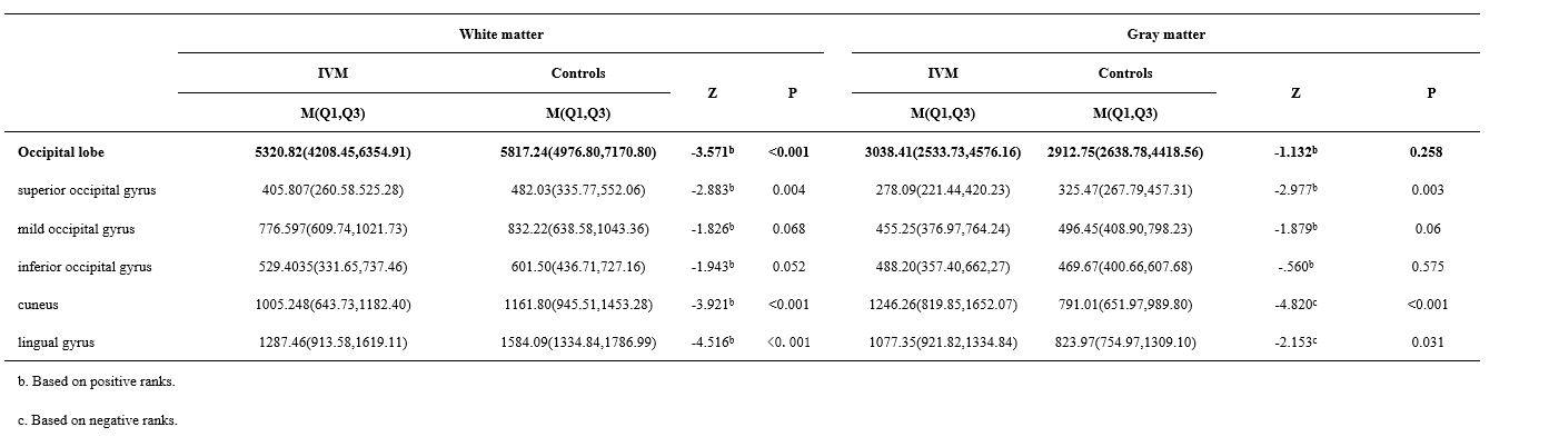
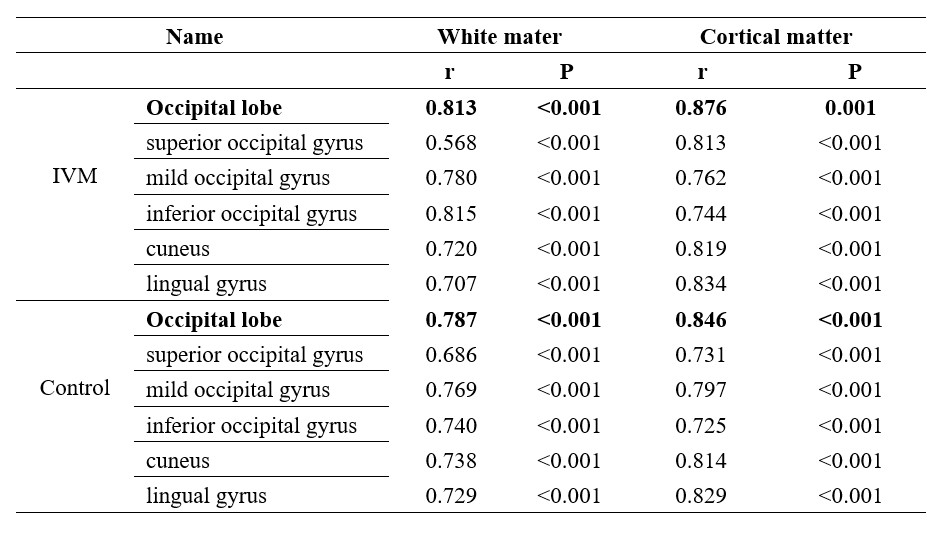
The volume characteristics of occipital lobe and occipital gyrus were further calculated. Fetuses with IVM had significantly decreased occipital white matter and the total occipital lobe volume than controls (P < .0.05; Table 1), while there were no differences in gray matter volume (P > 0.05; Table 1). Fetuses with IVM had significantly larger cuneus cortical and lingual gyrus cortical matter volume than controls (P < .005; Table 1). However, fetuses with IVM had significantly decreased cuneus white matter and lingual white matter volume than controls (P < .001; Table 1); and decreased superior occipital gyrus cortical matter and white matter volume (P <0.05; Table 1). We further found that the volume of superior occipital gyrus was moderately correlated with the increase of gestational age in IVM fetuses, while the volume of gray and white matter in other gyrus were highly correlated with the increase of gestational age (P <0.05, r <0.6; P <0.05, r >0.6 Table 2, Fig. 2,3). IVM fetal occipital lobe volume was smaller than normal group, but the growth rate was not significantly different (Table1,2). Superior occipital gyrus smaller volume and volume growth with the gestational age was not significant. (P <0.05 Table 1; r < 0.6, P <0.05 Table 2).Discussion
This study found that isolated ventricular ventriculomegaly was associated with changes in white and gray matter volume in several gyri tissues in the occipital lobe of the fetal brain compared with normal fetuses. As far as we know, this study is the first to examine regional gyrus volume changes in utero IVM fetuses. We speculated the occipital lobe gyrus volume change was due to two reasons, first, the mechanical expansion of the lateral ventricle increased the surface area which may result in a larger number of progenitor neurons, leading to an increase in the volume of gray matter in the adjacent occipital gyrus, the second is the expanded lateral ventricle occipital horn compressions the occipital white matter, resulting in its volume reduction5,6, the change of superior occipital gyrus may affect the higher level visual association processes7. The occipital lobe was closely related to visual and language development8,9. Therefore, we believe the structural and functional links between the volume changes of occipital gyrus and other brain regions in fetuses with IVM are of interest.Conclusion
Compared with normal fetuses, IVM fetuses show volume changes in the superior occipital gyrus, cuneus and lingual gyrus, the volume of occipital lobe of IVM fetus is small, but increases with gestational age. This may be related to neurodevelopmental delays after birth.Acknowledgements
This study was supported by Science and Technology Projects in Guangzhou. (202201020630,202201020627)References
1.Pagani G, Thilaganathan B, Prefumo F. Neurodevelopmental outcome in isolated mild fetal ventriculomegaly: systematic review and meta-analysis. Ultrasound Obstet Gynecol. 2014 Sep;44(3):254-60.
2.Pisapia JM, Rozycki M, Akbari H, Bakas S, Thawani JP, Moldenhauer JS, Storm PB, Zarnow DM, Davatzikos C, Heuer GG. Correlations of atrial diameter and frontooccipital horn ratio with ventricle size in fetal ventriculomegaly. J Neurosurg Pediatr. 2017 Mar;19(3):300-306.
3.Jarvis D, Akram R, Mandefield L, Paddock M, Armitage P, Griffiths PD. Quantification of total fetal brain volume using 3D MR imaging data acquired in utero. Prenat Diagn. 2016 Dec;36(13):1225-1232.
4.Makropoulos A, Aljabar P, Wright R, Hüning B, Merchant N, Arichi T, Tusor N, Hajnal JV, Edwards AD, Counsell SJ, Rueckert D. Regional growth and atlasing of the developing human brain. Neuroimage. 2016 Jan 15;125:456-478. doi: 10.1016/j.neuroimage.2015.10.047.
5.Hahner N, Benkarim OM, Aertsen M, Perez-Cruz M, Piella G, Sanroma G, Bargallo N, Deprest J, Gonzalez Ballester MA, Gratacos E, Eixarch E. Global and Regional Changes in Cortical Development Assessed by MRI in Fetuses with Isolated Nonsevere Ventriculomegaly Correlate with Neonatal Neurobehavior. AJNR Am J Neuroradiol. 2019 Sep;40(9):1567-1574.
6.Vasung L, Rollins CK, Zhang J, Velasco-Annis C, Yang E, Lin I, Sutin J, Warfield SK, Soul J, Estroff J, Connolly S, Barnewolt C, Gholipour A, Feldman HA, Grant PE. Abnormal development of transient fetal zones in mild isolated fetal ventriculomegaly. Cereb Cortex. 2022 Mar 29:bhac125.
7.Uddén J, Snijders TM, Fisher SE, Hagoort P. A common variant of the CNTNAP2 gene is associated with structural variation in the left superior occipital gyrus. Brain Lang. 2017 Sep;172:16-21.
8.Zhang H, Liu J, Zhang Q. Neural representations for the generation of inventive conceptions inspired by adaptive feature optimization of biological species. Cortex. 2014 Jan;50:162-73.
9.Espinosa JS, Stryker MP. Development and plasticity of the primary visual cortex. Neuron. 2012 Jul 26;75(2):230-49.
Figures