2306
Improved visualization of T1-weighted carotid artery plaque imaging using time-efficient spiral spin-echo DIXON with Compressed SENSE1Division of Radiology, Miyazaki University Hospital, Miyazaki, Japan, 2Department of Radiology, Faculty of Medicine, University of Miyazaki, Miyazaki, Japan, 3Philips Japan, Tokyo, Japan
Synopsis
Keywords: Blood vessels, Vessels
In MR carotid plaque imaging, black-blood imaging can diagnose plaque components and predict plaque hardness with a high degree of accuracy. In this study, we proposed T1-weighted black-blood carotid plaque images using non-ECG gated time-efficient Spiral spin-echo (SE) with Compressed SENSE (CS-Spiral SE) and compared with conventional ECG-gated DIR-TSE. We have demonstrated that the non-gated CS-Spiral SE could provide improved visualization in carotid plaque imaging with superior contrast to DIR-TSE with sufficient fat and blood signal suppression within a shorter scan time. It has a great potential to help more accurate assessment and characterization of the carotid atherosclerotic plaque.Introduction
In MR carotid plaque imaging, black-blood imaging can diagnose plaque components and predict plaque hardness with a high degree of accuracy1. Recently, Spin-echo (SE) sequence with Periodically Rotated Overlapping ParallEL Lines with Enhanced Reconstruction (PROPELLER) has been adopted for MR carotid plaque imaging instead of conventional electrocardiogram (ECG)-gated double inversion recovery Cartesian-TSE (DIR-TSE)2-5. PROPELLER is a more suitable method than DIR-TSE for obtaining high-contrast multisection images while keeping minimal motion artifacts because: (1) by combining presaturation-pulses similar to other conventional sequences, flow signal within the lumen can be suppressed, (2) motion-related misregistration can be readily corrected by the PROPELLER technique, and (3) it can be used for image acquisition with constant TR in any patient because the ECG-gating method is no longer needed2. However, SE PROPELLER basically requires quite long scan time, e.g., literature indicated the scan time was around 7 minutes5.Spiral MRI6, on the other hand, provides several advantages over both Cartesian and PROPELLER MRI. First, spiral MRI is faster, because of longer readout durations by using a spiral-out readout, which enable a concurrent decrease in scan time while simultaneously increasing image SNR. Recent improvements allow applying spiral MRI in the clinical fields7,8, such as image reconstruction using a conjugate-gradient algorithm for joint off-resonance deblurring and Dixon-based water/fat separation9, which can intrinsically improve the B0 eddy current induced image blurring in addition to generate robust water and fat only images. Furthermore, Spiral SE inherently has a reduced sensitivity to both in-plane and through-plane flow-induced artifacts6. Moreover, Spiral MRI can be combined with compressed sensitivity encoding (Compressed SENSE10) for further accelerating the acquisition time while maintaining its SNR efficiency.
In this study, we compared T1-weighted black-blood carotid plaque images of non-gated CS-Spiral SE and conventional DIR-TSE to demonstrate the clinical feasibility of CS-Spiral SE T1-weighted carotid plaque imaging.
Methods
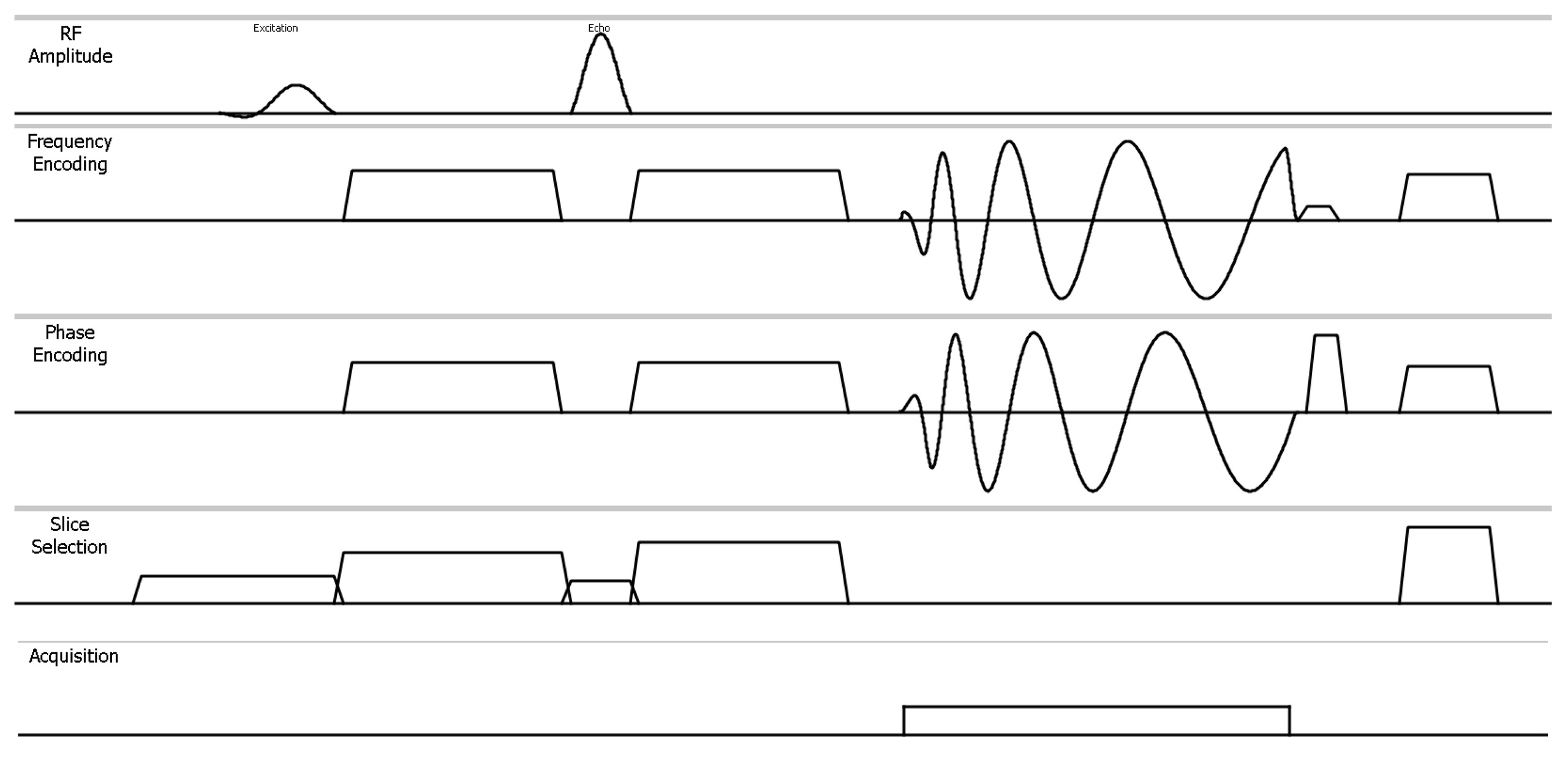
A sequence diagram of CS-Spiral SE is shown in Figure 1. Spiral SE inherently has a reduced sensitivity to both in-plane and through-plane flow artifacts. A set of additional large crusher gradients along the all encoding directions around the 180° refocusing pulse of the SE further reduce through-plane flow artifacts even though post contrast-enhancement scan7. The overall reduced sensitivity of Spiral SE to flow-induced errors eliminates the need for additional spatial saturation bands. Removal of these saturation bands in Spiral SE also reduces magnetization transfer effects and further improves the tissue contrast in addition to constant TR.In this study, we combined the Spiral sampling and CS reconstruction to further accelerate the Spiral SE. The sampling scheme of Spiral MRI is inherently suited for the CS reconstruction. In Spiral MRI, the sampling density decreases towards the outer parts of k-space providing a natural form of variable density, while the non-Cartesian sampling also promotes incoherence. Scan time reduction in CS-Spiral is achieved naturally by reducing the overall number of spiral arms acquired while still creating a smooth incoherent sampling pattern.
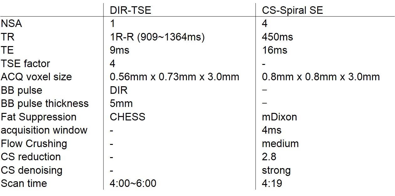
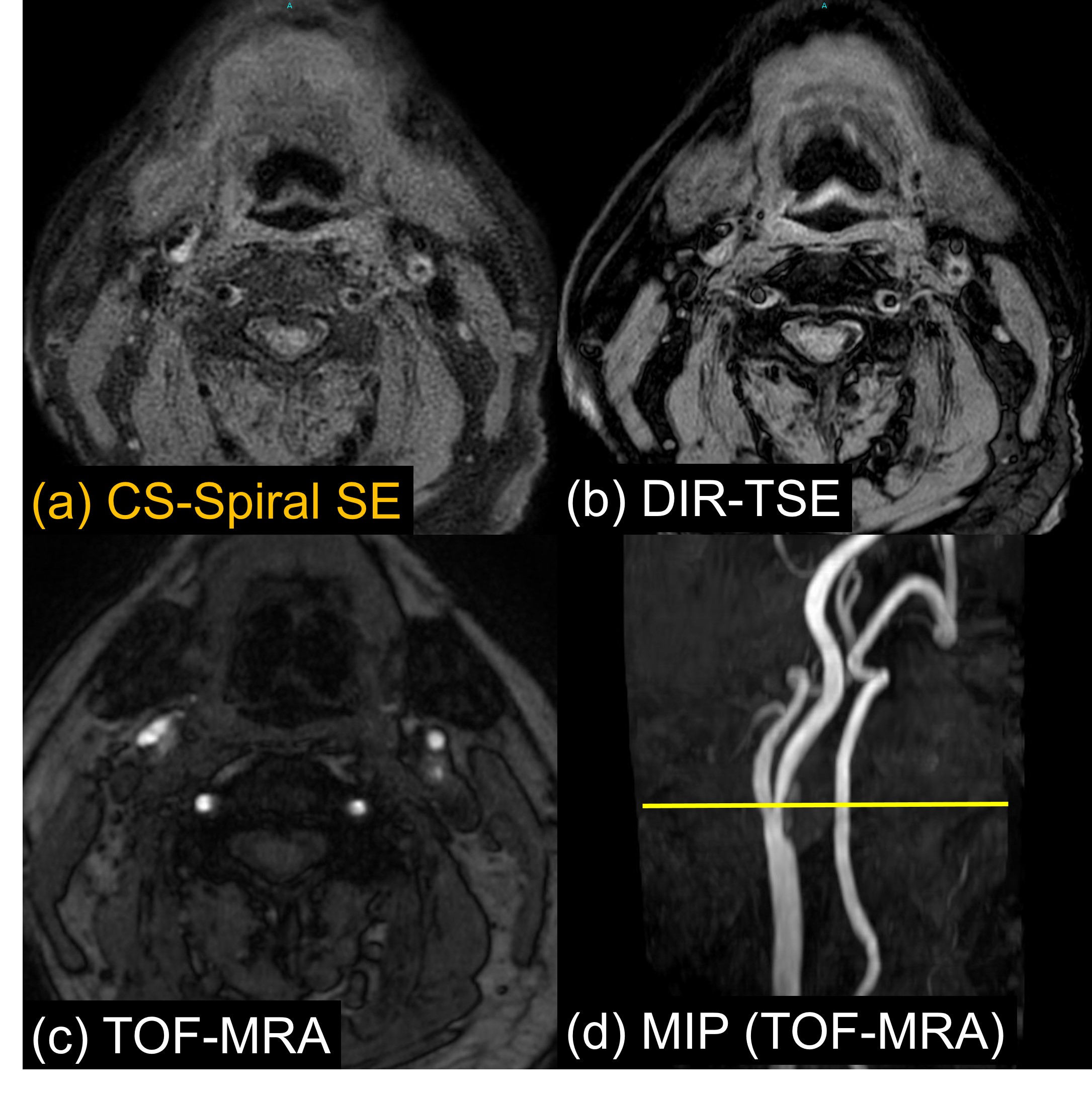
A total of 10 patients underwent carotid plaque imaging using Ingenia 3.0T CX (Philips, Best, the Netherlands), and all obtained images were evaluated as follows. Signal intensities of carotid artery plaque/sternocleidomastoid muscle and carotid artery plaque/vessel lumen were measured manually on non-gated CS-Spiral SE and DIR-TSE images, respectively. A contrast ratio (CR) was calculated from each signal intensity. In addition, visual assessment was performed by two radiologists using a 5-point scale for carotid plaque and vessel wall11. CR was statistically analyzed using a paired two-group t-test, and visual assessment was the Wilcoxon signed-rank test.
Imaging parameters of CS-Spiral SE and conventional DIR-TSE we used in this study are summarized in Table 1.
Results and Discussion
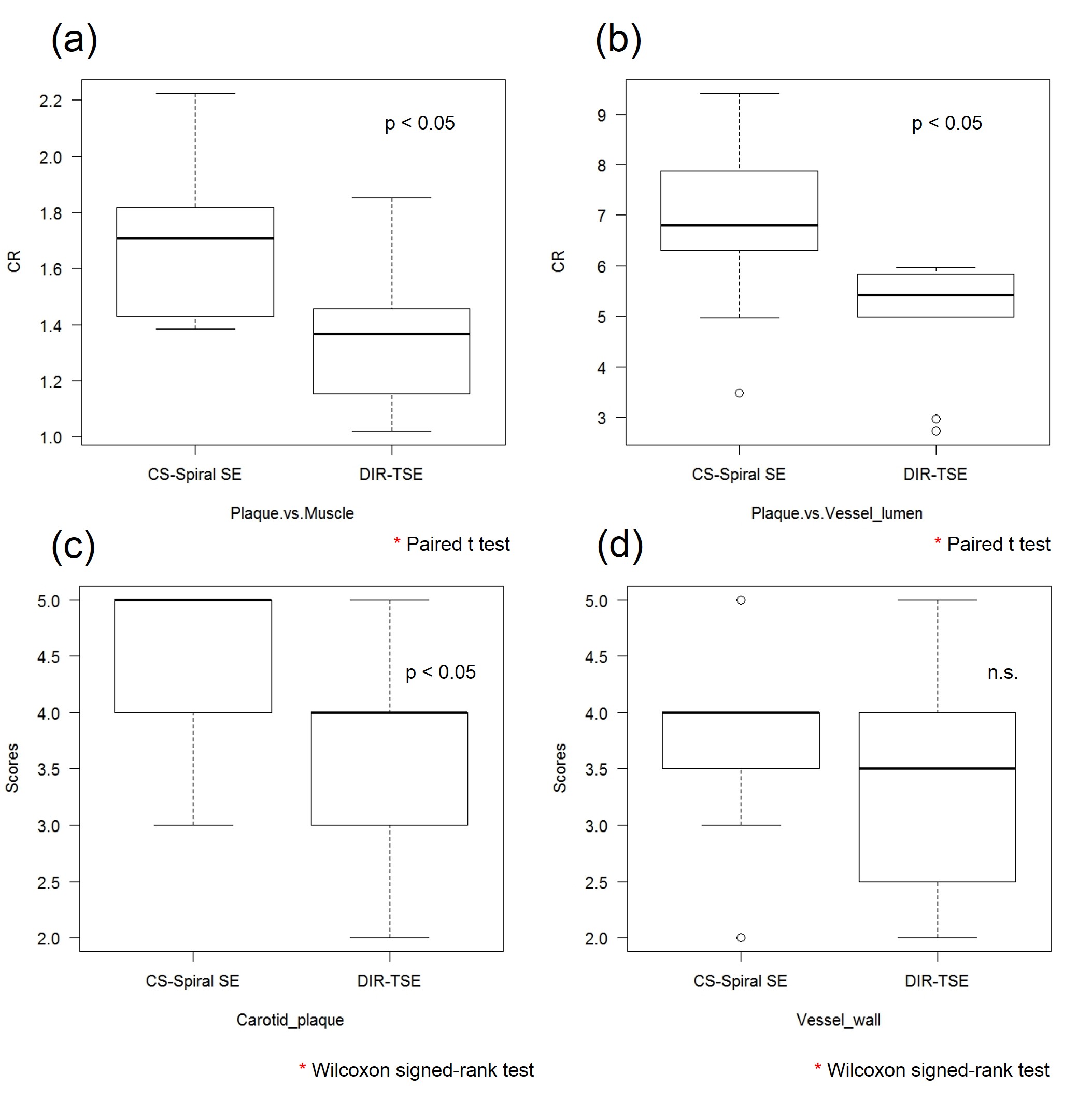
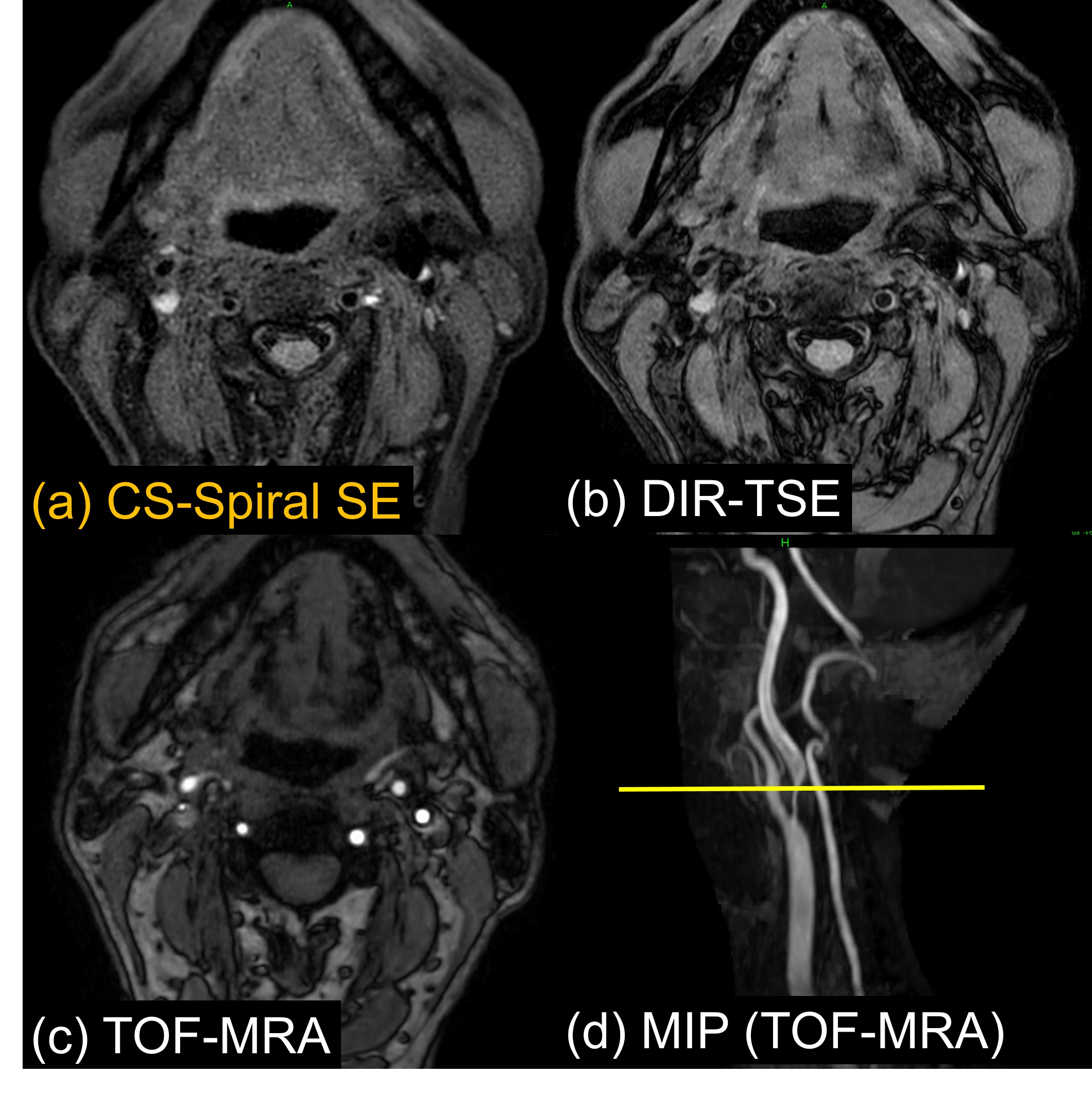
CS-Spiral SE showed significantly higher CR of both carotid artery plaque/sternocleidomastoid muscle and carotid artery plaque/vessel lumen compared to those of DIR-TSE (Fig. 2a,b). CS-Spiral SE also indicated significantly higher scores of visual evaluation of carotid artery plaques (Fig. 2c,d). Although the blood vessel wall showed a slightly higher value, there was no significant difference. Representative clinical images of CS-Spiral SE and DIR-TSE in patients with atherosclerotic plaques are shown in Figure 3 and Figure 4.Conclusion
We have demonstrated the feasibility of a new sequence for carotid plaque imaging using CS-Spiral SE technique. The non-gated CS-Spiral SE could provide improved visualization in carotid artery plaque imaging with superior contrast to conventional DIR-TSE with sufficient fat and blood signal suppression within a shorter scan time. This sequence has a great potential to help more accurate assessment and characterization of the carotid atherosclerotic plaque.Acknowledgements
No acknowledgement found.References
1. Yoshida K, et al. Characterization of carotid atherosclerosis and detection of soft plaque with use of black-blood MR imaging. AJNR Am J Neuroradiol 2008;29:868–874.
2. Narumi S, et al. Prediction of carotid plaque characteristics using non-gated MR imaging: correlation with endarterectomy specimens. AJNR Am J Neurorad 2013;34:191–197.
3. Narumi S, et al. Altered carotid plaque signal among different repetition times on T1-weighted magnetic resonance plaque imaging with self-navigated radial-scan technique. Neuroradiology 2010;52:285–290.
4. Yoneyama M, et al. Hyperecho PROPELLER-MRI: Application to rapid high-resolution motion-insensitive T2 -weighted black-blood imaging of the carotid arterial vessel wall and plaque. J Magn Reson Imaging. 2017 Feb;45(2):515-524. doi: 10.1002/jmri.25377.
5. Saito A, et al. Carotid plaque signal differences among four kinds of T1-weighted magnetic resonance imaging techniques: a histopathological correlation study. Neuroradiology. 2012 Nov;54(11):1187-94.
6. Pipe JG, et al. Spiral trajectory design: a flexible numerical algorithm and base analytical equations. Magn Reson Med 2014;71(1):278-285.
7. Li Z, et al. A Spiral Spin-Echo MR Imaging Technique for Improved Flow Artifact Suppression in T1-Weighted Postcontrast Brain Imaging: A Comparison with Cartesian Turbo Spin-Echo. AJNR Am J Neuroradiol 2015.
8. Ooi MB, et al. Spiral T1 Spin-Echo for Routine Postcontrast Brain MRI Exams: A Multicenter Multireader Clinical Evaluation. AJNR Am J Neuroradiol. 2020 Feb;41(2):238-245. doi: 10.3174/ajnr.A6409.
9. Wang D, et al. Joint water-fat separation and deblurring for spiral imaging. Magn Reson Med 2018 Jun;79(6):3218-3228. doi: 10.1002/mrm.26950.
10. Geerts-Ossevoort L, et al. Compressed SENSE Speed done right. Every time. The Netherlands: Philips Healthcare; 2018 Jan. Report No: 4522 991 31821.https://www.philips.de/content/dam/b2bhc/de/resourcecatalog/landingpages/ingeniaelition/White_Paper_Compressed_SENSE-opt.pdf
11. Okuchi S, et al. Visualization of carotid vessel wall and atherosclerotic plaque: T1-SPACE vs. compressed sensing T1-SPACE. Eur Radiol. 2019 Aug;29(8):4114-4122.
Figures