2297
Comparison of Arterial Pulsatility of Cerebral Perforating Arteries between Dual-VENC and Single-VENC Phase-Contrast MRI at 7T
Jianing Tang1,2, Elizabeth Joe3, Helena Chui3, and Lirong Yan2,3
1Biomedical Engineering, Northwestern University, Evanston, IL, United States, 2Radiology, Northwestern University, Evanston, IL, United States, 3Department of Neurology, University of Southern California, Los Angeles, CA, United States
1Biomedical Engineering, Northwestern University, Evanston, IL, United States, 2Radiology, Northwestern University, Evanston, IL, United States, 3Department of Neurology, University of Southern California, Los Angeles, CA, United States
Synopsis
Keywords: Blood vessels, High-Field MRI
Dysfunction of cerebral perforating arteries is a major pathology of small vessel disease. Previous study has demonstrated the pulsatility of cerebral perforating arteries can be measured by 7T high-resolution PC-MRI. In this study, we systematically compared the performance of PC-MRI with single and dual-VENC on pulsatility index (PI) measurements of cerebral perforating arteries. Our results showed that dual-VENC provides more reliable PI measurements than single VENC. PI measurements were insensitive to the temporal resolution of PC-MRI acquisition but showed greater variations with single VENC. The pilot study suggests that PI of cerebral perforating arteries was significantly higher in aged participants.Introduction
Cerebral perforating arteries play an essential role in maintaining the metabolic activities of deep brain nuclei1. Dysfunction of perforating arteries is associated with lacunar infarcts, basal ganglia hemorrhages, and deep microbleeds, which increases the risks of neurovascular disease and cognitive decline2. Small vessel disease (SVD) is represented by pathological changes in cerebral perforating arteries. Therefore, direct assessment of the pulsatility of perforating arteries can offer valuable insight into the role of vascular dysfunction in the pathogenesis of small vessel disease. Imaging of small arteries is beneficial from 7T with high SNR. Recent 7T studies have demonstrated the feasibility of detecting flow hemodynamics of cerebral perforating arteries using submillimeter-resolute phase-contrast MRI (PC-MRI) with both single VENC and dual VENC, separately3,4. However, the performance of single VENC and dual-VENC PC-MRI on LSA pulsatility measurements has not been fully studied. The goal of this study is to compare the pulsatility index (PI) of LSAs calculated from dual VENC and single VENC PC-MRI in terms of the PI reproducibility, PI dependency on temporal resolution and aging effects.
Methods
MRI experiments:All experiments were performed on a 7T MAGNETOM Terra MRI system (Siemens Healthcare, Erlangen, Germany) using an 8-transmit/32-receive head coil (Nova Medical, Wilmington, MA). Retrospectively gated single-slice 2D PC-MRI sequences with VENCs of 20 cm/s and 40 cm/s were acquired on 31 participants (age: 51.8 ± 21.5). The other image parameters included: FOV = 180x200 mm2, slice thickness = 2 mm, voxel size = 0.2x0.2x2.0 mm, flip angle = 58°, TE/TR= 9.72/95ms, 20 phases across a cardiac cycle. A 3D high-resolution TOF was performed on each subject prior to PC-MRI scans to locate the perforating arteries (Figure 1a). Test-retest PC-MRI scans were performed on 8 participants to evaluate the reproducibility of PI measurement. PC-MRI scans with temporal resolutions of 63ms, 95ms, and 127ms were performed on 9 participants to test the effects of temporal resolution on the sensitivity of perforator detection and the reliability of PI measurement.
Image processing and statistical analysis:
Two separate ROI masks were generated for single VENC and dual VENC, respectively. Background phase correction and phase unwrapping were done before the velocity and PI were calculated. In dual VENC image processing, high VENC phase maps were used to unaliase the phase wrapping in low VENC phase maps to ensure proper velocity profile. Kolmogorov-Smirnov test was performed on PI from both single and dual VENC to determine whether each dataset follows Gaussian distribution. A Wilson Rank-Sum test was applied to the two PI datasets according to the normality test result. One-way repeated measurement ANOVA test and post-hoc pair-wise rank-sum test were conducted to study the effect of temporal resolution on PI measurements. The participants were further separated into young and older groups to study the aging effect on LSA pulsatility.
Results
There was no significant difference in the PI values measured using PC-MRI with single VENC of 40cm/s (PI=0.70+/-0.24) and dual VENCs of 20cm/s and 40cm/s (PI=0.63+/- 0.19), as shown in Figure 2. The repeatability analysis on test-retest PI measurements was carried out using Bland–Altman plots (Figure 3). Both single- and dual-VENC methods show significant correlation between two PI measurements (r = 0.91, 0.98, p<0.001, respectively). The 95% confidence intervals were [-0.34 0.16] and [-0.11 0.06] for single- and Dual-VENC PI measurements, respectively. The test-retest results indicate that dual-VENC offers much better test-retest reproducibility for PI measurement with a higher correlation coefficient and smaller standard deviation. Figure 4 shows the comparison of PI values at three temporal resolutions in PC-MRI acquisitions. No significant difference was detected in PI values among all three temporal resolutions with both single-VENC and dual-VENC (p = 0.15, 0.18). However, the PI measurements with single VENC showed overall greater variations at three temporal resolutions, compared to those with dual VENC. In dual-VENC, PI measured at the temporal resolution of 95ms exhibited the lowest variations, compared to those with the other two temporal resolutions. Figure 5 shows the PI comparison between young and aged groups. PIs measured by both single-VENC and dual VENC show significantly higher PI values in the aged group than those in the young group (p<0.001), suggesting the LSA pulsatility increases with age.Discussion & Conclusion
In this study, we have systematically compared the performance of PC-MRI with single VENC and dual VENCs on the LSA pulsatility measurement. LSA pulsatility has been successfully assessed using PC-MRI with both single- and dual-VENC. Compared to single-VENC, dual-VENC shows more reliable PI measurements with better reproducibility and reduced variations measured at different temporal resolutions. Elevated PI is observed in aged adults compared to young adults, suggesting LSA PI could be a sensitive vascular marker for aging and small vessel disease. Future studies will evaluate the association of LSA pulsatility with cognitive functions, to further demonstrate the role of LSA dysfunction in the pathophysiology of aging and small vessel disease.Acknowledgements
This work is supported by grants from NIH R01NS118019, RF1AG072490, and BrightFocus Foundation A20201411S.References
1. Hakim AM. Small Vessel Disease. Front Neurol. 2019;10:1020. Published 2019 Sep 24. doi:10.3389/fneur.2019.01020Fyfe, I.
2. Global small vessel disease brain changes predict cognitive decline. Nat Rev Neurol 16, 2–3 (2020). Bouvy WH, Geurts LJ, Kuijf HJ, et al. Assessment of blood flow velocity and pulsatility in cerebral perforating arteries with 7-T quantitative flow MRI. NMR Biomed. 2016;29(9):1295-1304. doi:10.1002/nbm.3306
3. Tang J, Ma SJ, Yan L, Assessment of arterial pulsatility of cerebral perforating arteries using 7T high resolution dual-VENC phase-contrast MRI. In: Proc 28th Annual Meeting ISMRM, London 2022.
Figures
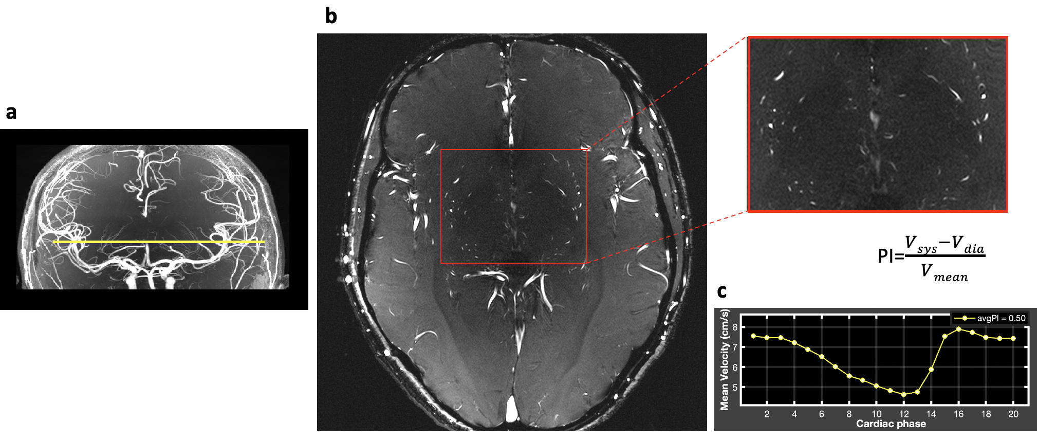
Figure 1. a. TOF image. Yellow line indicates the imaging position of PC-MRI. b. PC-MRI structural image. Red box indicates the basal ganglia region where the LSAs locate. c. The resulting velocity profile and PI calculation of LSAs.
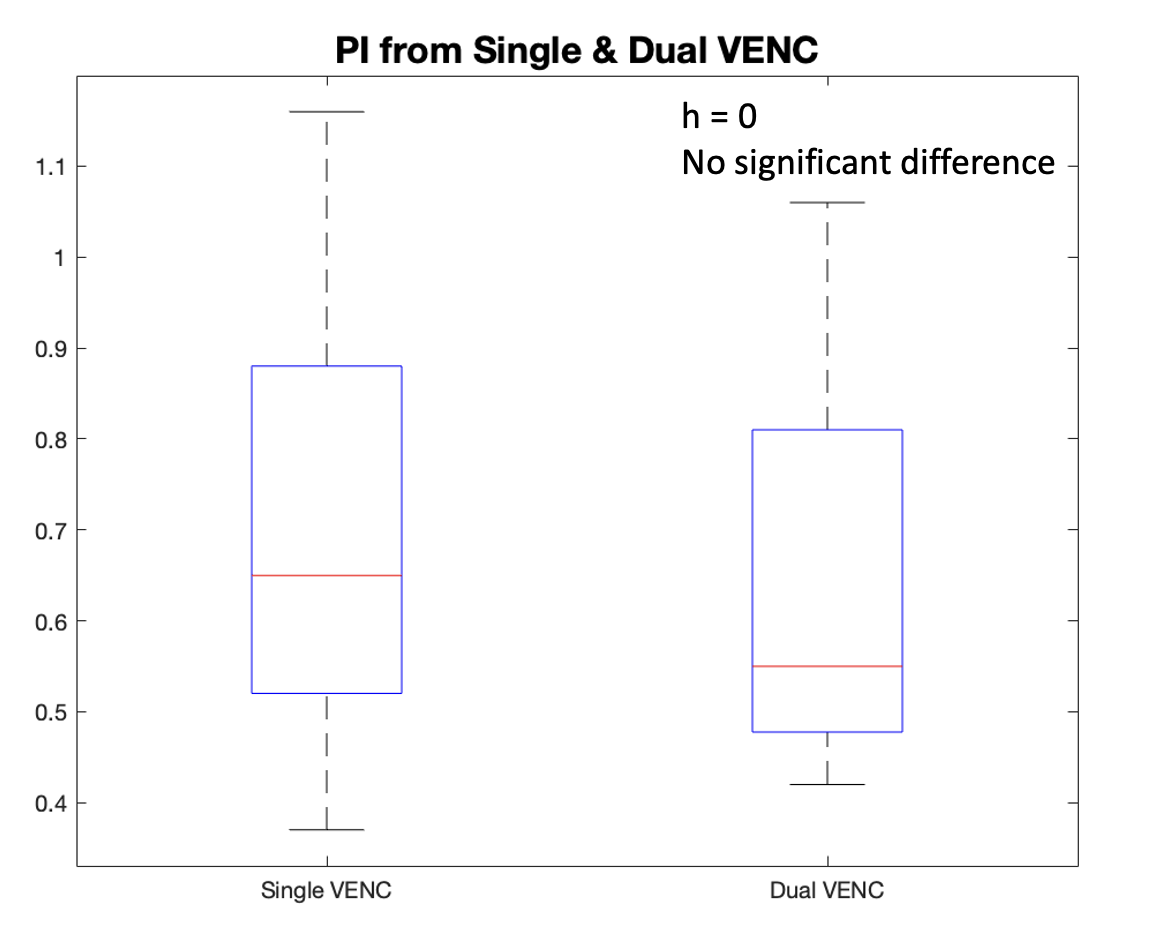
Figure 2. Boxplot of PI values measured using PC-MRI with a single VENC and dual-VENC. No significant difference in PI values was found between single and dual-VENC.
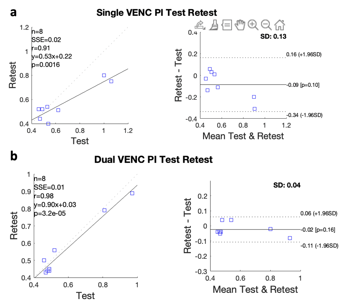
Figure 3. a. Bland-Altman Plot of PI test-retest PI measurements using a single VENC (a) and dual-VENC (b).
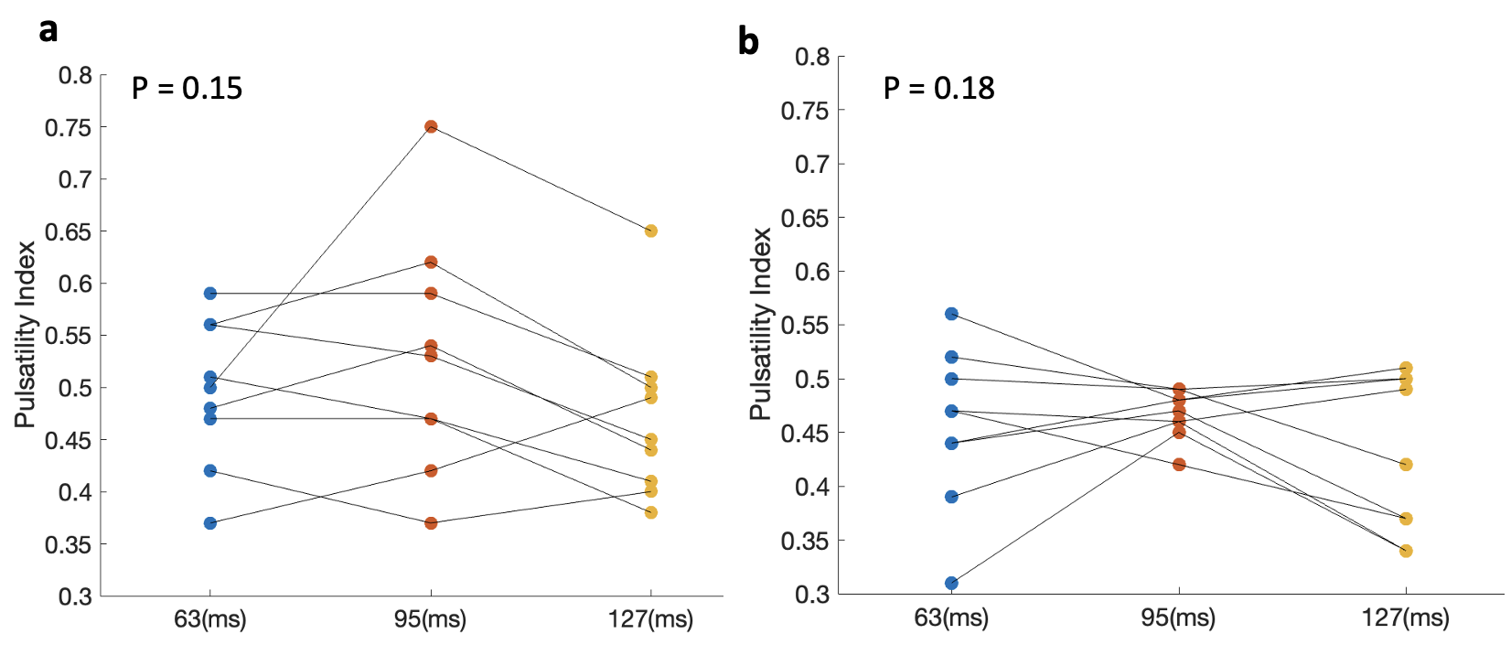
Figure 4. PI comparison at three temporal resolutions using a single VENC (a) and dual VENCs (b).
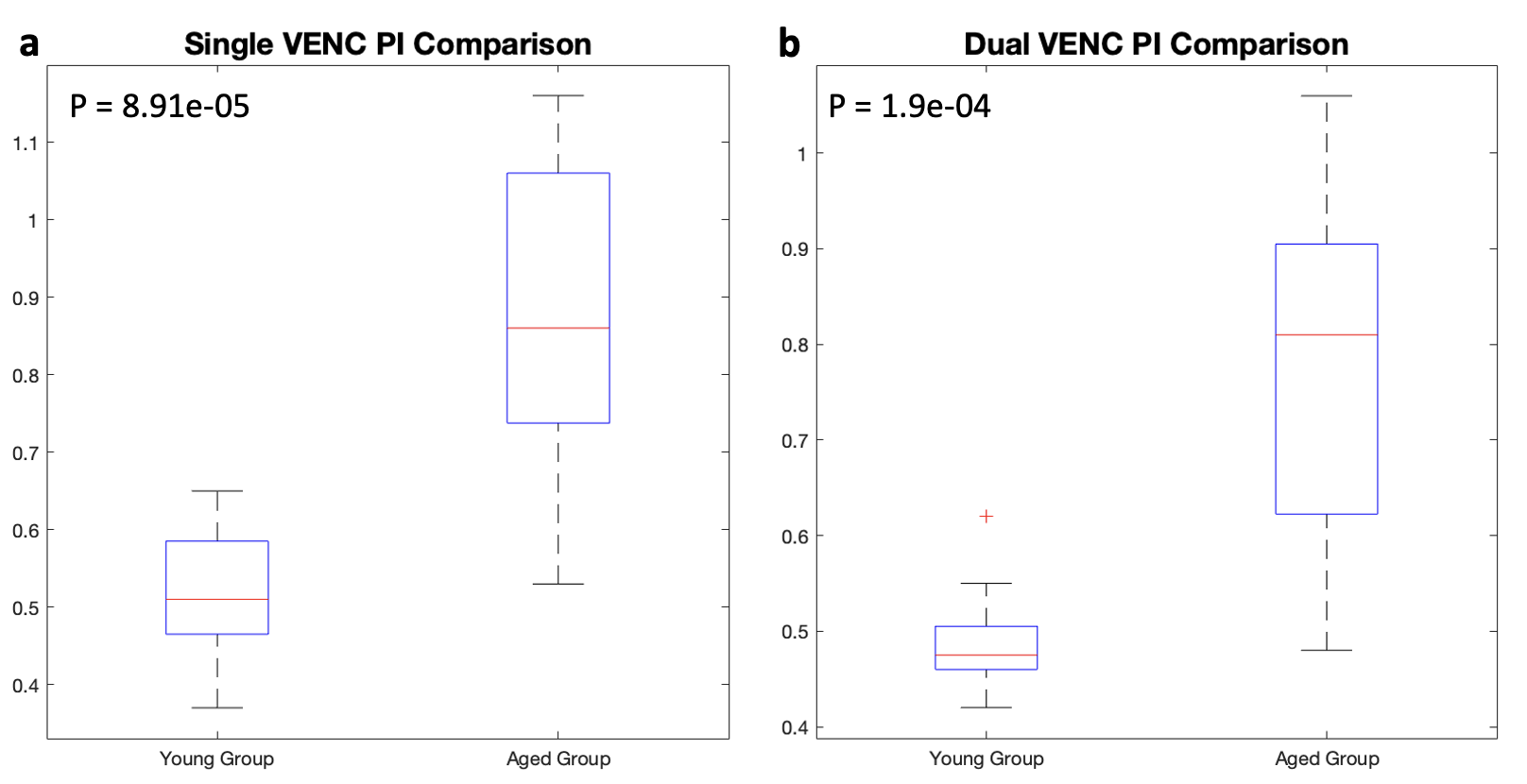
Figure 5. PI comparison between young and aged participants from single-VENC (a) and dual-VENC (b) measures.
DOI: https://doi.org/10.58530/2023/2297