2294
Dobutamine-induced Stress Altered Cerebral Blood Flow in Healthy Adults: a 3D Pseudocontinuous Arterial Spin Labeling Study1Beijing Friendship Hospital, Capital Medical University, Beijing, China, 2School of Biological Science and Medical Engineering, Beihang University, Beijing, China, 3National Space Science Center, Chinese Academy of Sciences, Beijing, China, 4Philips Healthcare, Beijing, China
Synopsis
Keywords: Blood vessels, Arterial spin labelling, Dobutamine Stress
It is not fully known whether dobutamine commonly clinically used in echocardiography and short-term treatment of congestive heart failure has effect on brain microcirculatory behavior. In this study, we sought to investigate the effect of dobutamine on cerebral haemodynamics in healthy volunteers. The results showed that dobutamine-induced stress significantly decreased the CBF in the anterior circulation, mainly in frontal lobe. Furthermore, volunteers with high BMI, large BA diameter and low SBP during dobutamine stress test are more likely to have stress-induced decrease of CBF.Introduction
Dobutamine is a widely used sympathomimetic drug in clinical practice for cardiac stress testing to non-invasively detect myocardial ischemia and coronary artery disease[1-3]. Clinically, dobutamine was suggested to be associated with an increase in cerebral perfusion in septic patients [4]. Inconsistently, dobutamine was reported to cause a decrease of cerebral blood flow (CBF) from the data of five patients, which was estimated by the transcranial color Doppler imaging (TCDI) technique [5]. The contrasting results of these studies reflect dobutamine's complex mechanisms of action. There is a chance that methodologic heterogeneity could cause inconsistent results, especially in patients with cerebrovascular diseases [6-8]. Therefore, the effect of dobutamine on CBF of either patients or healthy subjects is still unclear and probably multifactorial, involving CO, blood pressure, morphology of cerebral blood vessel. Moreover, alterations of microcirculatory behavior in brain of healthy subjects after dobutamine infusion may occur even with normal systemic haemodynamics and could contribute to hypoperfusion or hyperperfusion. Consequently, we aimed to explore CBF alterations in healthy subjects after undergoing dobutamine infusion by using the 3D pseudocontinuous arterial spin labeling(3D-pcASL) sequence.Methods
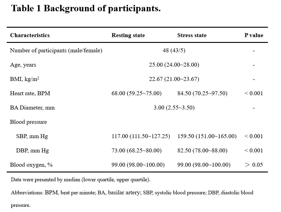
Forty-eight healthy volunteers, aged 23 to 34 years, free of cardiovascular and cerebrovascular disease, underwent magnetic resonance imaging (MRI) to obtain the CBF maps by using 3D ASL before and during the dobutamine stress test. In addition, cerebrovascular morphology was obtained based on 3D time-off-light (3D-TOF) magnetic resonance angiography (MRA) sequence. Electrocardiogram, heart rate, blood pressure and blood oxygen were simultaneously recorded before, during the dobutamine injection and in recovery (not conducted during MRI scan). Basilar artery (BA) were evaluated and measured by two radiologists with extensive experience in neuroimaging based on the MRA images. Wilcoxon signed-rank tests were used to calculate the differences in demographic and clinical characteristics between the two time points with the statistical threshold set at 0.05. Group differences in CBF between the two time points were tested using a paired t-test with age as a covariate. For multiple comparison correction, we used a False Discovery Rates [FDR]-corrected method (voxel level P < 0.001, pixel level P< 0.05; cluster size>719). Each cluster with significant difference was saved and used as a mask for the purpose of the subsequent region of interest (ROI)-based analyses. Then, the CBF values of the ROIs in the forty eight subjects were extracted with the xjView toolbox. A logistic regression analysis was used to identify predictors of CBF responses during dobutamine stress. The variables considered were age, BMI, heart rate, systolic blood pressure, and diastolic blood pressure in resting state and BA diameter.Results
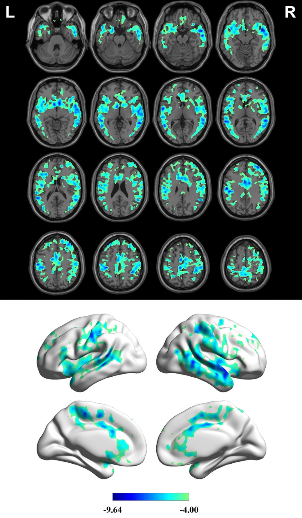
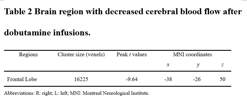
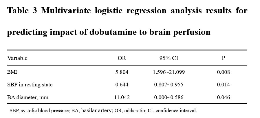
Heart rate (HR), systolic blood pressure (SBP), diastolic blood pressure (DBP) significant increased after dobutamine infusion (Table 1). Blood oxygen did not differ significantly between these two states. Compared with the CBF in rest state, the measurements in stress state showed decreased CBF in anterior circulation, mainly in the frontal Lobe (FDR-corrected; voxel level P < 0.001, pixel level P< 0.05; cluster size =16225) (Table 2, Figure 1). Logistic regression results showed that BMI, SBP in rest state and BA diameter were significantly associated with the change of CBF in frontal Lobe (Table 3).Discussion
In this study, we used 3D pseudocontinuous arterial spin labeling to probe the effect of dobutamine on CBF of the whole brain. We have demonstrated that dobutamine-induced stress decreased the CBF in anterior circulation, mainly in frontal lobe. The CBF in all brain regions was within the normal range during dobutamine infusion, however, there was a significant decrease in CBF in anterior circulation during dobutamine stress. Consistently, dobutamine was reported to cause a decrease of CBF from the data of five patients estimated by TCDI technique [5]. Previous study have suggested that ICA blood flow significantly decreased during heavy exercise, which implicates CBF of anterior circulation may decrease following the decrease of ICA blood flow [9]. However, dobutamine was suggested to be associated with an increase in cerebral perfusion in septic patients estimated by TCDI [10]. One explanation may due to the different dose of dobutamine, which was higher in our study than in this study. Another explanation was that patients with circulatory diseases are likely to have disrupted cerebral autoregulation, whereas the effects of dobutamine agents may be stronger. Furthermore, administration of phenylephrine, a clinically used vasopressor like dobutamine, was associated with a trend towards a greater reduction in frontal lobe oxygenation in diabetics [11, 12]. These results support that frontal lobe is more vulnerable to vasopressors. In comparison, the CBF of posterior circulation, supplied by the vertebral arteries that combine to form the basilar artery, has no significant change. Similarly, the blood flow velocity waveform of the vertebral artery were not altered by dobutamine in a dog model [13]. These results suggest that posterior circulation is less susceptible to dobutamine compared with anterior circulation.Conclusion
Dobutamine-induced stress significantly decreased the CBF in the anterior circulation. Furthermore, volunteers with high BMI, low SBP and larger BA diameter during dobutamine stress test are more likely to have stress-induced decrease of CBF. These findings suggest that more attention should be paid to blood pressure, BMI and cerebrovascular morphology of patients in dobutamine administration.Acknowledgements
Not applicable.References
1.Cortigiani L, Sorbo S, Miccoli M, Scali MC, Simioniuc A, Morrone D, Bovenzi F, Marzilli M, Dini FL. Prognostic value of cardiac power output to left ventricular mass in patients with left ventricular dysfunction and dobutamine stress echo negative by wall motion criteria. Eur Heart J Cardiovasc Imaging 2017, 18(2): 153-158.
2.Biswas S, Malik AH, Bandyopadhyay D, Gupta R, Goel A, Briasoulis A, Fonarow GC, Lanier GM, Naidu SS. Meta-analysis Comparing the Efficacy of Dobutamine Versus Milrinone in Acute Decompensated Heart Failure and Cardiogenic Shock. Curr Probl Cardiol 2022: 101245.
3.Jubran A, Schnittger I, Tremmel J, Pargaonkar V, Rogers I, Becker HC, Yang S, Mastrodicasa D, Willemink M, Fleischmann D, Nieman K. Computed Tomographic Angiography-Based Fractional Flow Reserve Compared With Catheter-Based Dobutamine-Stress Diastolic Fractional Flow Reserve in Symptomatic Patients With a Myocardial Bridge and No Obstructive Coronary Artery Disease. Circ Cardiovasc Imaging 2020, 13(2): e009576.
4.Berre J, De Backer D, Moraine J-J, Melot C, Kahn RJ, Vincent J-LJCcm. Dobutamine increases cerebral blood flow velocity and jugular bulb hemoglobin saturation in septic patients. Critical care medicine 1997, 25(3): 392-398.
5.Donati A, Coltrinari R, Loggi S, Pallotto R, Ruzzi M, Scarcella M, Valentini I, Pelaia P. Influence of dobutamine and norepinephrine and cerebral blood flow: preliminary data. Critical Care 2004, 8(1): 1-1.
6. Burley CV, Francis ST, Whittaker AC, Mullinger KJ, Lucas S. Measuring resting cerebral haemodynamics using MRI arterial spin labelling and transcranial Doppler ultrasound: comparison in younger and older adults. Brain and behavior 2021, 11(7): e02126.
7.Clyde BL, Resnick DK, Yonas H, Smith HA, Kaufmann AM. The Relationship of Blood Velocity As Measured by Transcranial Doppler Ultrasonography to Cerebral Blood Flow Determined by Stable Xenon Computed Tomographic As Studies after Aneurysmal Subarachnoid Hemorrhage. Neurosurgery 1996, 38(5): 896-905.
8.Mahoney L, Shah G, Crook D, Rojas-Anaya H, Rabe H. A Literature Review of the Pharmacokinetics and Pharmacodynamics of Dobutamine in Neonates. Pediatr Cardiol 2016, 37(1): 14-23.
9.Sato K, Ogoh S, Hirasawa A, Oue A, Sadamoto T. The distribution of blood flow in the carotid and vertebral arteries during dynamic exercise in humans. J Physiol 2011, 589(Pt 11): 2847-2856.
10.Berre J, De Backer D, Moraine JJ, Melot C, Kahn RJ, Vincent JL. Dobutamine increases cerebral blood flow velocity and jugular bulb hemoglobin saturation in septic patients. Crit Care Med 1997, 25(3): 392-398.
11.Brassard P, Pelletier C, Martin M, Gagne N, Poirier P, Ainslie PN, Caouette M, Bussieres JS. Influence of norepinephrine and phenylephrine on frontal lobe oxygenation during cardiopulmonary bypass in patients with diabetes. J Cardiothorac Vasc Anesth 2014, 28(3): 608-617. 12. Nissen P, Brassard P, Jørgensen TB, Secher NH. Phenylephrine but not ephedrine reduces frontal lobe oxygenation following anesthesia-induced hypotension. Neurocritical care 2010, 12(1): 17-23. 13. Van Bel F, Steendijk P, Teitel DF, de Winter JP, Van der Velde ET, Baan J. Cerebral blood flow velocity: the influence of myocardial contractility on the velocity waveform of brain supplying arteries. Ultrasound in medicine & biology 1992, 18(5): 441-449.
Figures