2289
Using R2* map to evaluate the changes of deep brain nuclei in patients with Parkinson 's disease1the First Affiliated Hospital of Dalian Medical University, Dalian, China, 2Department of Radiology, the First Affiliated Hospital of Dalian Medical University, Dalian, China, 3Clinical and Technical Support, Philips Healthcare, Beijing, Beijing, China
Synopsis
Keywords: Parkinson's Disease, Relaxometry, R2*,deep brain nuclei
R2* is highly sensitive not only to tissue iron level, but also to the microscopic iron distribution. In this study, we first characterize the distribution of R2* within the deep brain nuclei in Parkinson's disease patients and health controls, especially the lateral difference. Secondly, we also evaluate the clinical usefulness of regional R2* for the differential diagnosis between PD and HC group. The results showed that there was no lateral difference in R2* values in the HC and PD groups. The R2* value of the right thalamus in the HC group was statistically higher than that in the PD group.Introduction
Parkinson's disease (PD) is a major progressive neurodegenerative disease associated with the loss of neuromelanin-containing dopaminergic neurons of the substantia nigra (SN)[1]. Proton transverse relaxation rate (R2* ) is positively correlated with the content of paramagnetic substances such as iron and deoxyhemoglobin, which can quantitatively evaluate the change of tissue oxygen content. It may also provide different tissue information, such as the association with α-synuclein. Previous studies have shown that abnormal deposition of intracranial iron is one of the pathogenesis of PD and the deposition occurs mostly in the brainstem and basal ganglia[2]. In this study, we first characterize the distribution of iron-related signal (R2*) within the deep brain nuclei in PD patients and health controls (HC), especially the lateral difference. Secondly, we also evaluate the clinical usefulness of regional R2* for the differential diagnosis between PD group and HC group.Methods
Twenty PDs and twenty HCs were prospectively recruited and all were right-handed. Informed consent was acquired from each subject. Participants were scanned using a 3.0T MR scanner (Ingenia CX , Philips Healthcare, Best, the Netherlands) with a 32-channel head coil to acquire the MR sequence of strategically acquired gradient echo (STAGE) and R2* images were obtained after post-processing. The bilateral caudate nuclei (CN), putamen (PUT), globus pallidus (GP), thalamus (THU), red nuclei (RN), substantia nigra (SN) and dentate nuclei (DN) of PDs and HCs were delineated on R2* images by signal processing in nuclear magnetic resonance (SPIN) software. ROI was repeatedly measured three times at the same slice and then their average was calculated. Independent sample t test (normal distribution) or Mann-Whitney U test (non-normal distribution) was used to compare the R2* values of bilateral nuclei in each group and between the two groups. Data processing was performed by two researchers with more than 3 years of experience in image processing. And the intra-class correlation coefficient (ICC) was used to test the consistency of the data.Results
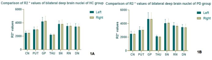
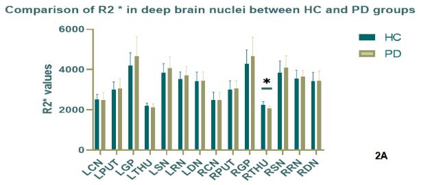
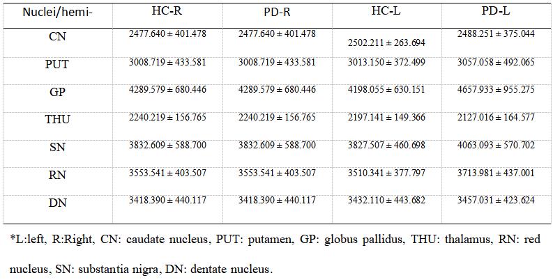
The results showed that there was no lateral difference in R2* values in the HC and PD groups (Figure 1). The R2* value of the right thalamus in the HC group was significantly higher than that in the PD group (Figure 2). The data consistency of two observers was good (intraclass correlation coefficient >0.75). The R2* values of the deep brain nuclei in the HC and PD groups are shown in the Table 1.Discussion
The loss of dopamine neurons in the substantia nigra is the main pathological change of Parkinson's disease. The abnormal deposition of iron is also considered to be one of the pathogenesis of Parkinson's disease, and mainly occurs in the substantia nigra pars compacta [2]. In this study, the R2* value of bilateral substantia nigra and red nucleus in PDs was lower than that in HCs, that is, the abnormal iron deposition in substantia nigra and red nucleus in PDs was higher than that in HCs, but the difference was not statistically significant. The reason may be due to the small sample size of this experiment, which needs to be further expanded. The R2* value of the right thalamus in the PDs was significantly lower than that in the HCs, which may be related to the increase of iron deposition in PDs, which induced oxidative stress and caused vascular injury. Patients with a longer course of PD will have decreased cerebral blood flow perfusion and decreased accumulation of deoxyhemoglobin values, resulting in decreased R2* [3, 4]. Previous studies have shown that abnormal iron deposition is mainly in substantia nigra, red nucleus, globus pallidus and putamen, while the pathological iron deposition in thalamus and caudate nucleus is not obvious [5], and the pattern of abnormal iron deposition is greatly affected by the age of onset [6]. Therefore, exploring the relationship between R2* and deep brain nuclei is helpful for the prevention and treatment of PD.Conclusion
The results showed that there was no lateral difference in R2* values in the HC and PD groups. The R2* value of the right thalamus in the HC group was statistically higher than that in the PD group. To explore the relationship between R2* and deep brain nuclei is helpful for the prevention and treatment of PD.Acknowledgements
Thanks to all colleagues involved in this study.References
[1] Lees AJ, Hardy J, Revesz T. Parkinson's disease. Lancet. 2009;373:2055–2066.
[2] Liu Z, Shen HC, Lian TH, et al. Iron deposition in substantia nigra: abnormal iron metabolism, neuroinflammatory mechanism and clinical relevance. Sci Rep. 2017, 7(1):14973.
[3] Kapitán M, Ferrando R, Diéguez E, et al. Cambios de la perfusión cerebral en la enfermedad de Parkinson: relación con la duración de los síntomas [Regional cerebral blood flow changes in Parkinson's disease: correlation with disease duration]. Rev Esp Med Nucl. 2009, 28(3):114-20.
[4] Seiler A, Deichmann R, Nöth U, et al.Oxygenation-Sensitive Magnetic Resonance Imaging in Acute Ischemic Stroke Using T2'/R2' Mapping: Influence of Relative Cerebral Blood Volume. Stroke. 2017, 48(6):1671-1674.
[5]Shu Hongge, Qi Jianpin, ZhuWenzhen, et al. The Quantitative Study of Brain lron Deposition in Parkinson's Disease [J]. Journal of huazhong university of science and technology (health sciences) ,2009,38(04):527-530.
[6]Seiler A, Deichmann R, Nöth U, et al. Oxygenation-Sensitive Magnetic Resonance Imaging in Acute Ischemic Stroke Using T2'/R2' Mapping: Influence of Relative Cerebral Blood Volume. Stroke. 2017, 48(6):1671-1674.
Figures