2278
CEST and MISL detect regional neuropathologies in a Parkinson’s disease mouse model at 3T
Joseph H. C. Lai1, Jianpan Huang1, Yang Liu1,2, Se Weon Park1,2, Jiadi Xu3, and Kannie W. Y. Chan1,2,3,4,5
1Department of Biomedical Engineering, City University of Hong Kong, Hong Kong, Hong Kong, 2Hong Kong Centre for Cerebro-Cardiovascular Health Engineering (COCHE), Hong Kong, Hong Kong, 3Russell H. Morgan Department of Radiology and Radiological Science, The Johns Hopkins University School of Medicine, Baltimore, MD, United States, 4City University of Hong Kong Shenzhen Research Institute, Shenzhen, China, 5Tung Biomedical Sciences Centre, City University of Hong Kong, Hong Kong, Hong Kong
1Department of Biomedical Engineering, City University of Hong Kong, Hong Kong, Hong Kong, 2Hong Kong Centre for Cerebro-Cardiovascular Health Engineering (COCHE), Hong Kong, Hong Kong, 3Russell H. Morgan Department of Radiology and Radiological Science, The Johns Hopkins University School of Medicine, Baltimore, MD, United States, 4City University of Hong Kong Shenzhen Research Institute, Shenzhen, China, 5Tung Biomedical Sciences Centre, City University of Hong Kong, Hong Kong, Hong Kong
Synopsis
Keywords: Parkinson's Disease, CEST & MT
Reveal alterations in molecules and CSF-tissue water exchange could enable interactive interventions in PD. Here we applied CEST and MISL to study the changes in a PD model at 3T. We observed significant changes (P<0.05) at 2.6ppm on day 28 and at -3.5ppm on day 7, which could indicate a substantial decrease of dopamine and lipid-related pathology at the substantia nigra, respectively. Along the nigrostriatal pathway, we observed a gradual MISL drop at the ventricles, which was significantly lower at day 28 (P<0.05) and might imply the CSF-tissue-related pathologies. Our findings could serve as potential biomarkers for early PD detection.INTRODUCTION
Parkinson's disease (PD) is an age-related neurodegenerative disease, with key pathologies including the loss of dopaminergic neurons at the substantia nigra pars compacta (1,2) and the deposition of unwanted Lewy bodies in the brain (3). Although levodopa has been well-established for alleviating PD symptoms at the clinical level (3,4), no single treatment approach can cure or reverse the disease status. One of the key hurdles comes from the ambiguity of PD pathology at the molecular level.With the advances in PD-related research, different animal models have been developed to study the etiologies and reveal subtle molecular changes (5). Recent studies have shown that aquaporin 4 (AQP4) may play a role in PD (6,7). As such, we proposed to study the molecular changes of an MPTP-induced PD mouse model with Chemical Exchange Saturation Transfer (CEST) MRI. CEST is an MRI technique that can image molecular changes up to the millimolar range (8). The recently developed Magnetization transfer Indirect Spin Labeling (MISL) protocol can quantify cerebrospinal fluid (CSF) water exchange with the surrounding macromolecules in the brain and AQP4-related diseases (9). Since the MPTP model is well established for preclinical PD research (1,5,10,11), we will use this model to analyze the underlying molecular changes. We anticipate this can provide valuable information for early diagnosis and treatment planning.
METHODS
A set of dopamine phantoms was prepared to study the CEST properties. In brief, 1 mg/mL of dopamine hydrochloride (Sigma) was dissolved in PBS with different pH.For in vivo study, 3 C57BL/6 male mice (8-10 weeks) received 1-methyl-4-phenyl-1,2,3,6-tetrahydropyridine (MPTP, purity ≥ 98%, Sigma) via intraperitoneal injection (18 mg/kg per dose, diluted in saline) four times in 24 hours with an interval of at least 2 hours (1,5,10,11). The control group received the same volume of saline for the same period.
The phantom and the mice were scanned by a horizontal bore 3T Bruker BioSpec animal scanner (Bruker, Germany) with a 40-mm volume transceiver coil. The CEST sequence was referenced to our previously reported protocol (12). In addition, a MISL sequence was acquired referencing to the protocol from Li et al. (9). In brief, TR=5000ms, TE=250ms, B1=2.0μT, and tsat=3s. We used -10ppm as the labeled MT signal and 300ppm as the control image. The MISL images were calculated by subtracting the labeled MT signal from the control, followed by normalizing the control image. The MRI readout and other parameters were identical to the in vivo CEST sequence. The scan time of the MISL acquisition was about 4 min. The PD group was longitudinally imaged before, 7 days, and 28 days after injection. For the control mice, the scan was only performed on day 7.
One-way ANOVA analyzed longitudinal comparisons, and the comparisons with control were analyzed by Student’s t-test, all by GraphPad Prism 8.0.1.
RESULTS & DISCUSSION
The exchangeable protons of dopamine generated CEST contrast at 1.2ppm and 2.6ppm (Figure 1A and B). The peaks were sensitive to acidic pH (Figure 1C). We further studied the B1 dependence of the two peaks (Figure 1D-F). Since 2.6ppm is further away from water, we used it to monitor the dopamine change in vivo.Our in vivo study analyzed the longitudinal CEST change at the substantia nigra with three different offsets, i.e. 2.6ppm, 3.5ppm for amide protons, and -3.5ppm for aliphatic protons (Figure 2). As expected, the signal at 2.6ppm decreased by 18.4% on day 28 (Figure 2B, P<0.05). Signals at 3.5ppm (Figure 2C) and -3.5ppm (Figure 2D) changed over time, yet the changes were insignificant. We compared the PD mice with the control on day 7 since a stabilized loss of dopaminergic neurons was reported at this timepoint (10). Interestingly, a significant signal increase, around 12.9%, was observed at -3.5ppm (Figure 2G, P<0.05).
We further studied the CSF-tissue water exchange along the nigrostriatal pathway. Two consecutive slices anterior to the substantia nigra, covering the third and lateral ventricles, were imaged by MISL (Figure 3). Figure 3B showed that the MISL signal gradually decreased after MPTP intoxication (-13.7% on day 7) and reached a minimum on day 28 (-21.1%, P<0.05). We did not observe a significant difference in the MISL on day 7 (Figure 3C). The slice 3mm anterior to the substantia nigra also showed a decrease in MISL signal longitudinally (Figure 3E), and day 7 showed a 10.6% reduction compare to the control (Figure 3F, -10.6%). Representative MISL maps from the 1.5mm anterior slice were shown in Figure 3G.
Although we observed some trends in regional CEST and MISL signals, conclusions could not be drawn due to limited number of animals. A large cohort of animal study together with validation by histology is underway to further study the changes in these imaging parameters and the underlying neuropathology.
To summarize, this study demonstrated the detectability of dopamine at 1.2ppm and 2.6ppm. CEST detects changes at 2.6ppm, 3.5ppm, and -3.5ppm in the PD model's both regionally and longitudinally. A significant decrease at 2.6ppm was observed on day 28 after disease induction (P<0.05). Moreover, MISL indicated a significant decrease in MISL on day 28 (P<0.05), which could indicate CSF-tissue-related pathologies. Our findings could provide valuable information for early PD detection.
Acknowledgements
Authors would like to acknowledge the funding supports from Research Grants Council (11102218, PDFS2122-1S01, 11200422, RFS2223-1S02, C1134-20G); City University of Hong Kong (7005433, 7005626, 9239070, 9609307, 9610560); National Natural Science Foundation China (81871409); Tung Biomedical Sciences Centre; Hong Kong Centre for Cerebro-cardiovascular Health Engineering.References
- Meredith GE, Rademacher DJ. MPTP mouse models of Parkinson’s disease: An update. J Parkinsons Dis 2011;1:19–33 doi: 10.3233/JPD-2011-11023.
- Braak H, Ghebremedhin E, Rüb U, Bratzke H, del Tredici K. Stages in the development of Parkinson’s disease-related pathology. Cell Tissue Res 2004;318:121–134 doi: 10.1007/s00441-004-0956-9.
- Jankovic J, Tan EK. Parkinson’s disease: Etiopathogenesis and treatment. J Neurol Neurosurg Psychiatry 2020;91:795–808 doi: 10.1136/jnnp-2019-322338.
- de Bie RMA, Clarke CE, Espay AJ, Fox SH, Lang AE. Initiation of pharmacological therapy in Parkinson’s disease: when, why, and how. Lancet Neurol 2020;19:452–461 doi: 10.1016/S1474-4422(20)30036-3.
- Prasad EM, Hung SY. Behavioral tests in neurotoxin-induced animal models of parkinson’s disease. Antioxidants 2020;9:1–50 doi: 10.3390/antiox9101007.
- Nagelhus EA, Ottersen OP. Physiological Roles of Aquaporin-4 in Brain. Physiol Rev 2013;93:1543–1562 doi: 10.1152/physrev.00011.2013.-Aquaporin-4.
- Tamtaji OR, Behnam M, Pourattar MA, Jafarpour H, Asemi Z. Aquaporin 4: A key player in Parkinson’s disease. J Cell Physiol 2019;234:21471–21478 doi: 10.1002/jcp.28871.
- van Zijl PCM, Yadav NN. Chemical exchange saturation transfer (CEST): What is in a name and what isn’t? Magn Reson Med 2011;65:927–948 doi: 10.1002/mrm.22761.
- Li AM, Chen L, Liu H, Li Y, Duan W, Xu J. Age-dependent cerebrospinal fluid-tissue water exchange detected by magnetization transfer indirect spin labeling MRI. Magn Reson Med 2022;87:2287–2298 doi: 10.1002/mrm.29137.
- Jackson-Lewis V, Przedborski S. Protocol for the MPTP mouse model of Parkinson’s disease. Nat Protoc 2007;2:141–151 doi: 10.1038/nprot.2006.342.
- Bagga P, Crescenzi R, Krishnamoorthy G, et al. Mapping the alterations in glutamate with GluCEST MRI in a mouse model of dopamine deficiency. J Neurochem 2016;139:432–439 doi: 10.1111/jnc.13771.
- Lai JHC, Liu J, Huang J, et al. CEST MRI of temporal changes of hematoma in Intracerebral Hemorrhage ( ICH ) mouse at 3T. International Society for Magnetic Resonance in Medicine 2021:2014–2016 doi: 10.1002/mrm.22891.5.
Figures
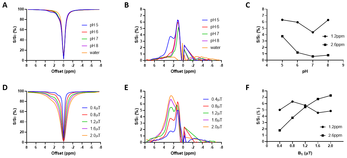
Figure 1. (A) Z-spectra and (B) Lorentzian difference
(LD) spectra of the dopamine phantoms at different pH show that dopamine is
detectable at 1.2ppm and 2.6ppm. The pH dependence at the two offsets when B1 =
0.8μT are summarized in (C). (D) Z-spectra and (E)
LD spectra of dopamine at pH 5 show that 2.6ppm is more sensitive to the change
of B1. The B1 dependence at the two offsets are
summarized in (F).
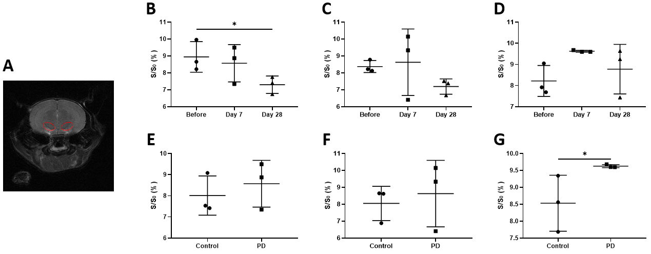
Figure 2. (A) Red ellipses in the T2 reference
image indicated the ROI of substantia nigra. CEST analysis was performed at (B
& E) 2.6ppm, (C & F) 3.5ppm, and (D & G) -3.5ppm. (B – D) were
longitudinal analyses of paired mice, while (E – G) studied the difference
between PD and control mice at day 7 (*P<0.05, n=3). Values are means ±
SD.
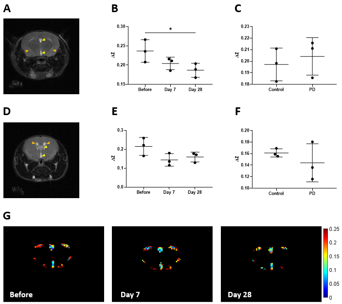
Figure 3. A T2 reference image showed the brain slice at
(A) 1.5mm anterior to substantia nigra, with corresponding longitudinal MISL
result in (B) and comparison with control at day 7 in (C). A T2 reference image
showed the brain slice at (D) 3mm anterior to SN, with corresponding longitudinal
MISL result in (E) and comparison with control at day 7 in (F). Yellow arrows
indicated the third ventricle, while orange arrows indicated the lateral ventricles
(*P<0.05, n=3). Values are means ± SD. Representative MISL maps at different
timepoints from the 1.5mm anterior slice are gathered in (G).
DOI: https://doi.org/10.58530/2023/2278