2268
Bone marrow fat composition assessed by fast and simple 1D MRS combined with intermolecular double-quantum coherence on 3.0 T
Jianfeng Bao1, Xiao Wang2, Yuchuan Zhuang3, Liangjie Lin4, Yong Zhang2, and Jingliang Cheng2
1Department of Magnetic Resonance Imaging, The First Affiliated Hospital of Zhengzhou University, zhengzhou, China, 2Department of Magnetic Resonance Imaging, The First Affiliated Hospital of Zhengzhou University, Zhengzhou, China, 3Department of Electronic Science, University of Rochester, ROCHESTER, NY, United States, 4Philips Healthcare, Beijing, China
1Department of Magnetic Resonance Imaging, The First Affiliated Hospital of Zhengzhou University, zhengzhou, China, 2Department of Magnetic Resonance Imaging, The First Affiliated Hospital of Zhengzhou University, Zhengzhou, China, 3Department of Electronic Science, University of Rochester, ROCHESTER, NY, United States, 4Philips Healthcare, Beijing, China
Synopsis
Keywords: Bone, Fat, intermolecular double-quantum coherence,fat composition,magnetic resonance spectroscopy
Unsaturated fatty acids in the bone marrow may play a critical role in bone metabolism, however, these peaks cannot be well resolved by conventional MRS techniques due to local intensive B0 inhomogeneities. We introduced intermolecular double-quantum coherence to enhance the spectrum resolution on clinical 3.0 T within one minute and more unsaturated peaks can be identified.Unsaturated
fatty acids in the bone marrow may play a critical role in bone metabolism,
however, these peaks cannot be well resolved by conventional MRS techniques due
to local intensive B0 inhomogeneities. We introduced intermolecular
double-quantum coherence to enhance the spectrum resolution on clinical 3.0 T
within one minute and more unsaturated peaks can be identified.
Purpose
Growing evidence suggests that bone marrow fat is more than simply filling tissues in bone cavities, and different fatty acids, as shown in Figure 1a, can regulate bone metabolism in several signaling pathways. However, it is difficult to resolve for the conventional MRS method. Because the bone marrow fat exists in a complex microenvironment, which has solid bone, liquid water, and semi-solid and semi-liquid fat. The resulting natural local B0 field inhomogeneity is the big problem to obtain high-resolution MRS. To overcome the line boarding caused by off-resonance effects, more recently, our group demonstrated the intermolecular double-quantum coherence (iDQC) technique with 2D spectrum MRS. Here, we want to show that the time-saving method,1D acquisition, can enhance the resolution on 3.0 T as well, which may be more acceptable for the clinal scan.Method
As shown in Figure 1b, the localized iDQC signal can be obtained with the pulse sequence modified based on the PRESS. The red pulse is a 90°was gauss-shaped (12 ms), which was used to selectively excite the fat methylene peak at 1.3 ppm to generate the dipole field. A pair of coherence selection gradients (CSGs) with an area ratio of 1:2 was applied along the Z direction to select iDQC signals.TR = 5s, t1 = 20~52 ms. The acquisition time was 50 ms with 1104 points. The amplitude for CSGs was 20 mT/m. The durations for the first and the second CSG were 3 ms and 6 ms, respectively. The data was processed using a customized Matlab program. The scan was performed on the lumbar vertebra on a 62-year-old Chinese female.Results
Compared with the PRESS result shown in Figure 1c, only two main broad peaks, more additional fatty acids peaks are well resolved with the localized iDQC method, including allylic methylene (2.0 ppm), methylene (2.2 ppm), diallylic methylene (2.7 ppm), and olefinic (5.3 ppm). Figure 1d shows the representative experimental results with the different colors corresponding to various fatty acids. Figure 2 shows the spectrum characterizes varies over evolution time. It should be noted that the methylene peaks at 2.0 and 2.2 ppm, marked by the red dashed box, are important to calculate the monounsaturated/polyunsaturated fatty acids (MOFA/PUFA).Conclusions
The proposed fast 1D and simple localized iDQC method shows good resistant properties against the B0 inhomogeneity caused by the trabecular bone. With well-resolved fatty acid peaks, this method may hold a potential to explore the pathological changes in bone-related diseases in the future.Acknowledgements
Acknowledgment: This work was supported by the National Natural Science Foundation of China under grant number 81601470.References
No reference found.Figures
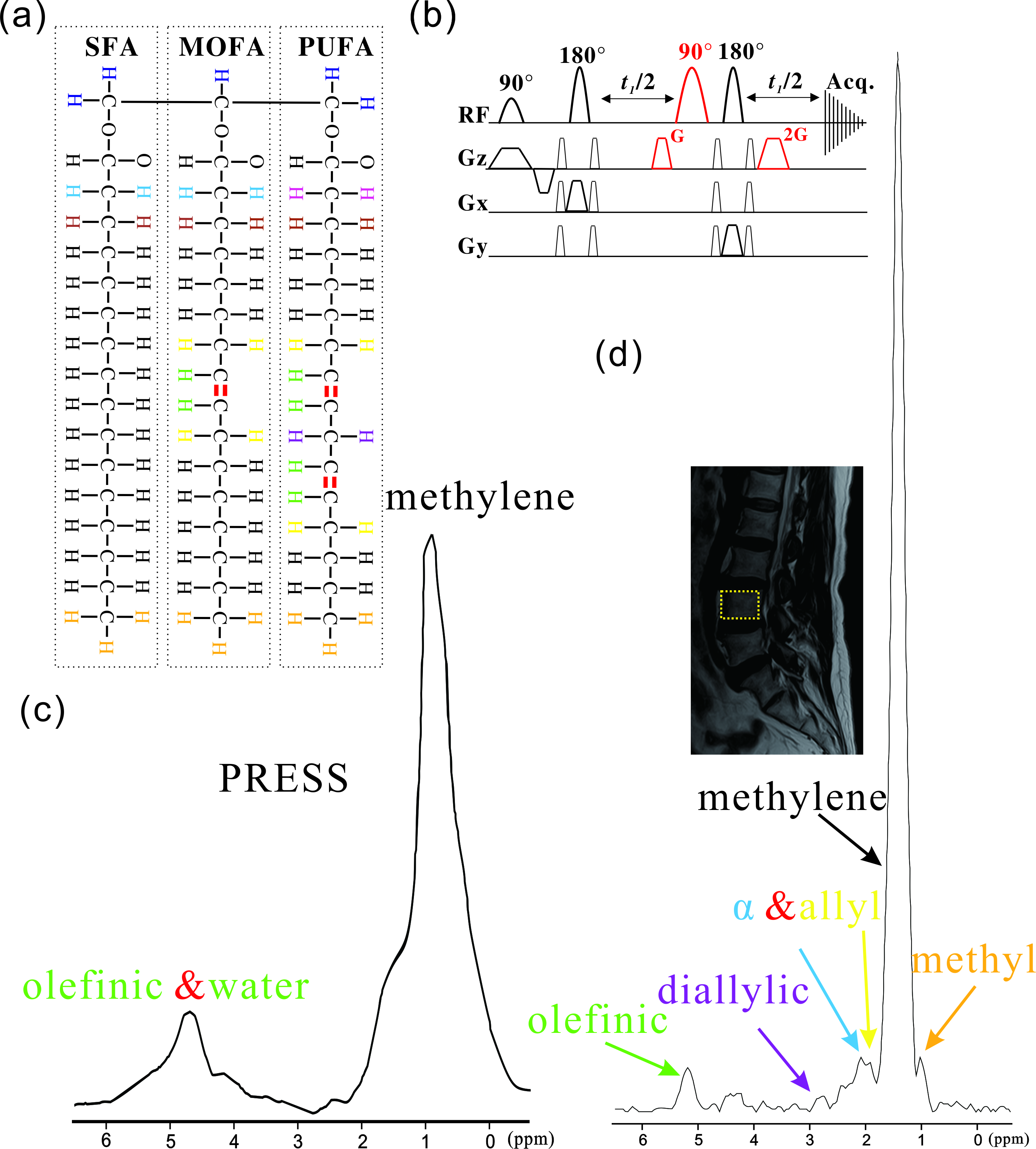
Figure 1 (a) there kinds of
fatty acids and different colors represent protons in different chemical
environments;(b) 1D localized iDQC pulse sequence used in this work, the black
color is PRESS module for localizing and the red color is for generating iDQC
signals; (c) PRESS results and (D) 1D localized iDQC result.
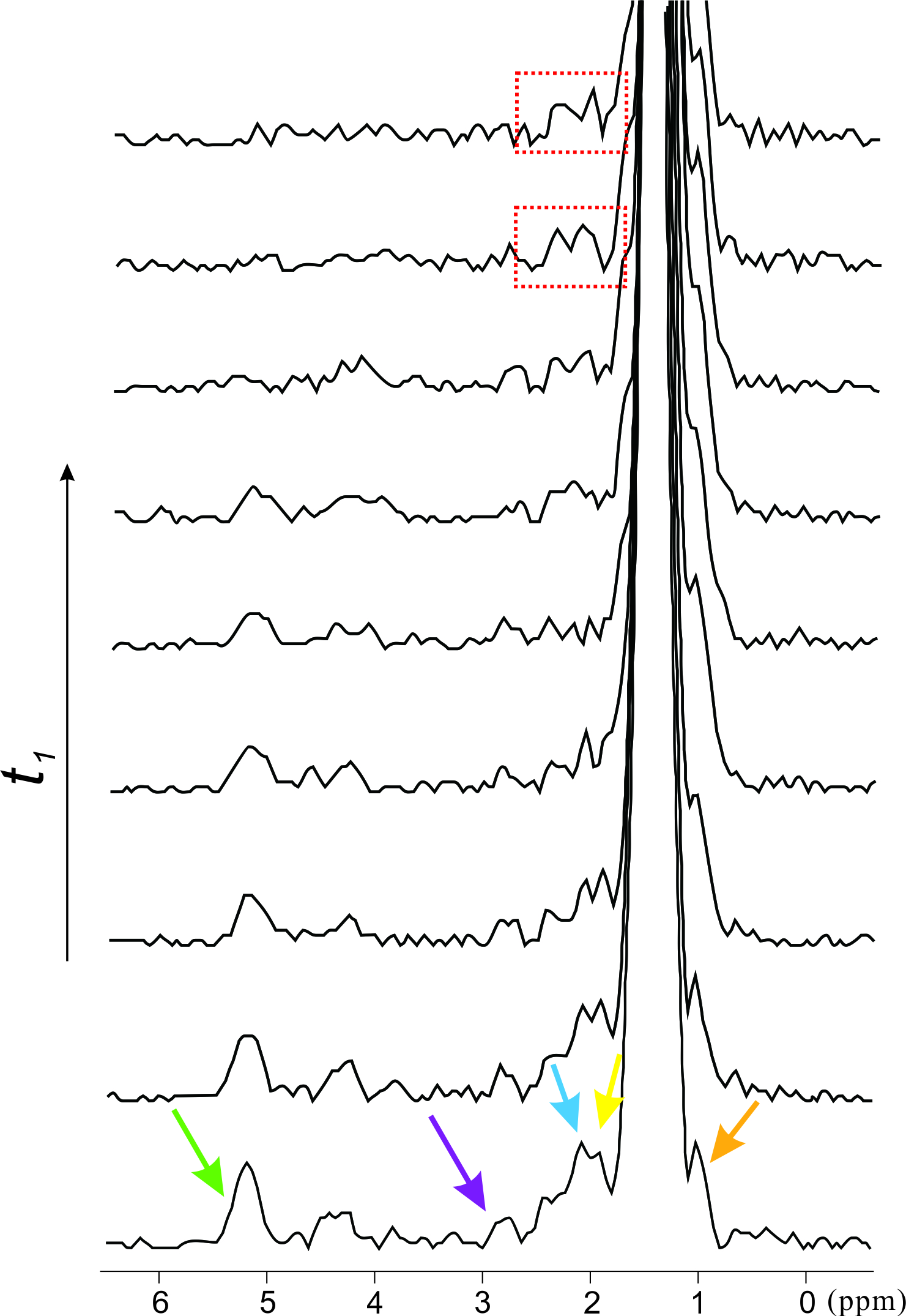
Figure 2 1D iDQC 1H
MR spectra with increasing t1. The allylic methylene (2.0 ppm) and
methylene (2.2 ppm) are prominent with a relatively long t1.
DOI: https://doi.org/10.58530/2023/2268