2264
Radiomics based on conventional MRI sequences to differentiate inert fibrogenic tumors and invasive fibrogenic tumors of trunk and limbs
Peng Gao1, Weisheng Zhang1, and Hexin Feng2
1Dalian Medical University, Dalian, China, 2China Medical University, Shenyang, China
1Dalian Medical University, Dalian, China, 2China Medical University, Shenyang, China
Synopsis
Keywords: Muscle, Tumor
区分不同亚型的纤维化肿瘤仍然具有挑战性,但具有重要的临床意义。本研究的目的是评估基于常规 MRI 序列的影像组学在区分惰性纤维化肿瘤和躯干和四肢浸润性纤维化肿瘤的价值。预测模型基于 T1 图像的逻辑回归在区分惰性纤维瘤和侵袭性纤维瘤方面具有最好的预测效率,对 T1 和 T2 预测模型贡献最大的特征分别是 idn 和最小轴长。病变的大小和深度对判断肿瘤的性质有一定的参考价值。Introduction
Incomplete treatment caused by inaccurate preoperative assessment can not only lead to frequent local recurrence, but also lead to distant metastasis, which seriously affects the quality of life and shortens the life span of patients1. In the process of differential diagnosis of benign and malignant soft tissue tumors, especially those with homologous but heterogeneous soft tissue tumors, even doctors with rich experience in bone and muscle imaging diagnosis often apply traditional imaging. It is difficult to give an accurate and convincing diagnosis, but the application of radiomics can conduct deeper mining of traditional medical imaging images, so it is possible to distinguish benign and malignant soft tissue tumors.Methods
The clinical and magnetic resonance imaging data of 89 patients with fibrogenic tumors (FTS) confirmed by postoperative pathology were analyzed retrospectively. Three-dimensional region of interest (3DROI) was delineated layer by layer on tumor lesions in T1 and T2 sequences of magnetic resonance imaging(MRI) of 89 patients by using 3D Slicer, and 107 histological features of 7 categories were extracted and the features with statistical significance between groups were reduced by Lasso regression and 10% cross-validation, and the correlation between high-performance features was verified. Finally, patients were divided into training group and verification group according to the ratio of 7: 3. logistic regression, random forest and support vector machine were used to build prediction models for the training sets, and verification set was used to verify the diagnostic accuracy of the models.Results
In the clinical features and routine imaging features of inert group and invasive group, the size and depth of lesions were statistically significant. The statistical features of T1 sequence included correlation difference, high correlation and high gray scale, idn, kurtosis and elongation, while the statistical features of T2 sequence included elongation, skewness, minimum axial length and maximum two-dimensional diameter. The logistic regression based on T1 image has the best performance. The areas under ROC curve of training set and verification set are 0.827 and 0.813, and the accuracy, specificity and sensitivity are 0.774, 0, 722, 0.846 and 0.852, 0.929 and 0.769, respectively. In addition, from the random forest model, it can be concluded that idn and minimum axis length are the characteristics that contribute the most to the prediction models of T1 and T2. The prediction model based on linear kernel function in support vector machine is relatively more stable and accurate.Discussion
This study is based on the inert and invasive fiber source of the trunk and extremities in imageomics of conventional MRI sequence images.The results showed that the training group of logistic regression model based on T1WI image features AUC (specificity and sensitivity) of the two groups were 0.827 (84.6%, 72.2%) and 0.813 respectively(76.9%, 92.9%). The prediction accuracy of the confusion matrix is 77.4% and 85.1% respectively, both at a high level. It is reported that the differential diagnosis of lipoma and well differentiated liposarcoma lesions was carried out by using imageomics. The results showed that the AUC of the imageomics model based on T1WI image features was 0.832, which was significantly higher than the AUC values (0.74, 0.72, 0.61) diagnosed by three radiologists. The diagnostic efficacy of the radiologic model was similar to our best model. It is also reported that the classification accuracy of the support vector machine model based on the characteristics of T1WI images to identify benign and malignant soft tissue tumors is as high as 93%3, which is also better than the 90% classification accuracy of radiologists in the control group. To sum up, the imageomics model based on T1WI image features is feasible and reliable in the field of soft tissue tumor differential diagnosis, and its diagnostic efficacy is better than that of radiologists to a certain extent.conclusion
The prediction model of logistic regression based on T1 image has the best prediction efficiency for distinguishing inert fibrous tumors from invasive fibrous tumors, and the features that contribute the most to T1 and T2 prediction models are idn and minimum axial length, respectively. The size and depth of lesions have certain reference value for judging the nature of tumors.Acknowledgements
No acknowledgement found.References
1.VOS M, STARMANS M, TIMBERGEN M, et al. Radiomics approach to distinguish between well differentiated liposarcomas and lipomas on MRI. 2019, 106(13): 1800- 1809. JUNTU J, SIJBERS J, DE 2.BACKER S, et al. Machine learning study of several classifiers trained with texture analysis features to differentiate benign from malignant soft-tissue tumors in T1-MRI images [J]. 2010, 31(3): 680-689. XU W, 3.HAO D, HOU F, et al. Soft Tissue Sarcoma: Preoperative MRI-Based Radiomics and Machine Learning May Be Accurate Predictors of Histopathologic Grade. 2020, 215(4): 963-969.Figures
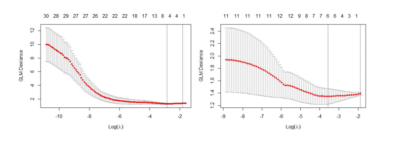
Figure 1: The features are dimensionalized by Lasso method.
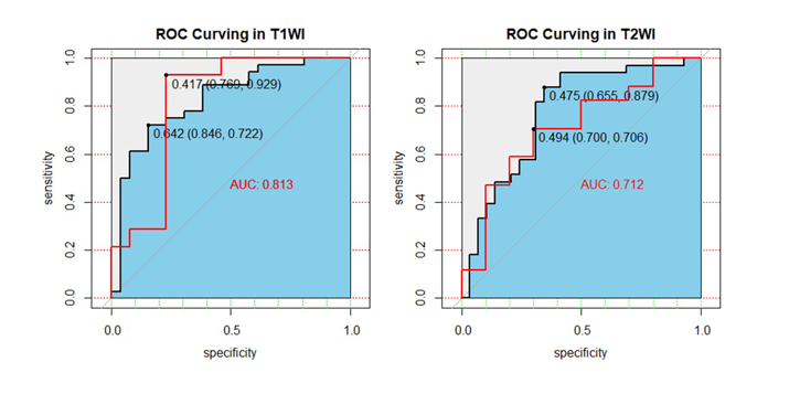
Figure 2: ROC curve of logistic
regression model

Figure 3: contribute degree of features
to T1 and T2 prediction models
DOI: https://doi.org/10.58530/2023/2264