2257
Whole-tumor histogram analysis of DWI and DCE-MRI for soft tissue sarcoma: Correlation with HIF-1alpha expression1Huashan hospital Fudan university, Shang hai, China, 2MR Collaborations, Siemens Healthcare Ltd., Shang hai, China, 3Radiology, Huashan hospital Fudan university, Shang hai, China
Synopsis
Keywords: MSK, Diffusion/other diffusion imaging techniques
In this study, we initially revealed the feasibility of conventional MRI features and whole-tumor histogram features of diffusion-weighted imaging (DWI) and dynamic contrast-enhanced MRI (DCE-MRI) parameters in assessing hypoxia-inducible factor 1-alpha (HIF-1α) expression of soft tissue sarcomas (STS). On this basis, we performed a short-term survival analysis of the study population. The findings suggest that MRI morphological features and histogram features of quantitative parameters help predict higher HIF-1α expression and may significantly predict prognosis in patients with STS.Introduction
STSs are highly heterogeneous malignant tumors originating from mesenchymal components [1]. Due to the high mortality and recurrence rates, scholars have preferred to evaluate the relationship between the internal microenvironmental state with patient clinical outcomes [2]. Several studies have reported that the hypoxic microenvironment is essential for adverse events such as high recurrence and high radiotherapy resistance in the sarcoma population [3]. Among them, activation of HIF-1α is a crucial process mediating the adaptive cellular response to the hypoxic microenvironment. Currently, it is shown that high HIF-1α expression in STS is associated with higher mortality [4]. Thus, assessing the hypoxic level and monitoring HIF-1α expression in STS helps determine prognosis and treatment response.Purpose
To investigate the correlation of histogram metrics from DWI and DCE-MRI parameters with HIF-1alpha expression in soft tissue sarcoma STS.Methods
We enrolled 71 patients with STS who underwent 3.0 T MRI, including conventional MRI, DWI, and DCE-MRI sequences. Location, maximum tumor diameter, envelope, T2-weighted tumor heterogeneity, peritumoral edema, peritumoral enhancement, necrosis, tail-like pattern, bone invasion, and vessel/nerve invasion and/or encasement were determined using conventional MRI images. The whole-tumor histogram metrics were calculated on the apparent diffusion coefficient (ADC), Ktrans, Kep, and Ve maps. Independent samples t-test and one-way ANOVA were used for testing the differences between normally distributed categorical data with HIF-1alpha expression. Pearson and spearman correlations and multiple linear regression analyses were performed to determine the correlations between histogram metrics and HIF-1alpha expression. Survival curves were plotted using the Kaplan-Meier method.Results
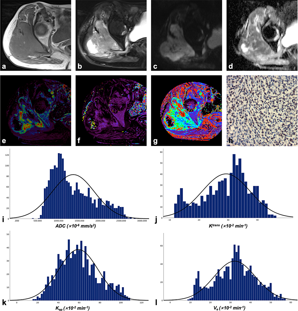
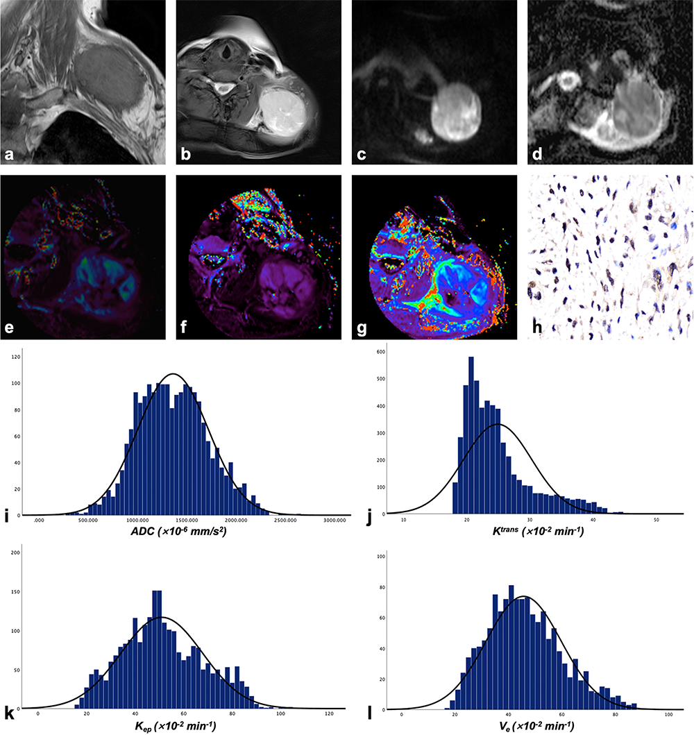
Seventy-one patients were included in the final analysis, including 40 males (mean age, 58 years) and 31 females (mean age, 61 years). Regarding conventional MRI features, only highly heterogeneous on T2-weighted images (55.6±19.9% vs. 45.4±20.5%, p=0.041) and more than 50% necrotic area (57.3±20.4% vs. 43.9±19.7%, p=0.002) were prone to indicate STS with higher HIF-1alpha expression. Histogram metrics obtained from ADC (mean, median, 10th, and 25th percentile values), Ktrans (mean, median, 75th, and 90th percentile values), and Kep (90th percentile values) were significantly correlated with HIF-1alpha expression. Multiple linear regression analysis demonstrated that more than 50% necrosis, ADCskewness, Ktrans90th, and grade III were independently associated with HIF-1alpha expression (standardized β coefficient for more than 50% necrosis=0.074, p=0.011; for ADCskewness=-0.332, p=0.003; for Ktrans90th=0.203, p=0.027; for grade III=0.314, p=0.004) Fig.1 and 2 show typical histogram distributions of DWI and DCE-MRI maps in the presence of lower- and higher-HIF-1alpha expression cases.After radical resection, all patients were managed with individualized treatment, including local radiotherapy, adjuvant chemotherapy, or targeted therapy according to histological type and genetic test results. Participants were followed up for 6–36 months. There were ten metastatic relapses, 16 local relapses, and 18 deaths. The 3-year survival probabilities of the series were 0.61 for disease-free survival and 0.73 for overall survival.
Discussions
This study used whole-tumor histogram analysis to reveal that DWI and DCE-MRI parameters correlated with HIF-1alpha expression in STS. The univariate results showed that the higher T2WI tumor heterogeneity and necrosis over 50% had significantly higher HIF-1alpha expression. Multifactorial analysis showed that necrosis over 50%, ADCskewness, Ktrans90th, and FNCLCC grade III identified HIF-1alpha expression as the most promising predictors. This result indicates the potential of conventional MRI combined with DWI and DCE-MRI analysis as imaging biomarkers for predicting the oxygenation level of STS. In addition, HIF-1alpha expression above 75% may be associated with poorer OS and MFS.Conclusion
DWI and DCE-MRI histogram parameters were significantly correlated with HIF-1alpha expression in STS.Acknowledgements
No acknowledgement found.References
1. Clark MA, Fisher C, Judson I, Thomas JM. Soft-tissue sarcomas in adults. N Engl J Med 2005;353:701-11.
2. Merry E, Thway K, Jones RL, Huang PH. Predictive and prognostic transcriptomic biomarkers in soft tissue sarcomas. NPJ Precis Oncol 2021;5:17.
3. Zhang M, Qiu Q, Li Z, Sachdeva M, Min H, Cardona DM, DeLaney TF, Han T, Ma Y, Luo L, Ilkayeva OR, Lui K, Nichols AG, Newgard CB, Kastan MB, Rathmell JC, Dewhirst MW, Kirsch DG. HIF-1 Alpha Regulates the Response of Primary Sarcomas to Radiation Therapy through a Cell Autonomous Mechanism. Radiat Res 2015;183:594-609.
4. Nystrom H, Jonsson M, Werner-Hartman L, Nilbert M, Carneiro A. Hypoxia-inducible factor 1alpha predicts recurrence in high-grade soft tissue sarcoma of extremities and trunk wall. J Clin Pathol 2017;70:879-85.
Figures