2242
Application value of restriction spectrum imaging in differentiation of hepatocellular carcinoma and intrahepatic cholangiocarcinoma1Zhengzhou University People’s Hospital & Henan Provincial People’s Hospital, Henan, China, 2Henan University People’s Hospital & Henan Provincial People’s Hospital, Henan, China, 3Xinxiang Medical University & Henan Provincial People's Hospital, Henan, China, 4MR Collaboration, Central Research Institute, United Imaging Healthcare, Shanghai, China, 5Department of Medical Imaging, Zhengzhou University People’s Hospital & Henan Provincial People’s Hospital, Zhengzhou, China
Synopsis
Keywords: Liver, Cancer
Diffusion-weighted imaging (DWI) can be used to evaluate the degree of tumor cell proliferation and tumor necrosis. The diffusion motion of water molecules is assumed to be Gaussian distribution, which has some limitations. In recent years, some advanced models have emerged that can provide more parameters to explore complex biological behaviors. Restricted spectral imaging (RSI) is one of them. However, the application of this model in liver disease is rare. The purpose of this study was to explore the potential value of the RSI model in the preoperative non-invasive differential diagnosis of hepatocellular carcinoma (HCC) and intrahepatic cholangiocarcinoma (ICC).Synopsis
Restriction spectrum imaging (RSI) is an advanced magnetic resonance technology to detect water diffusion through multiple b values and different directions. RSI model can reveal the biological behavior of tumor tissues, provide additional microenvironmental information, and be more helpful to detect the structure and changes in tumor cells. This study aims to explore the value of the whole-tumor histogram metrics derived from RSI in differentiating hepatocellular carcinoma (HCC) and intrahepatic cholangiocarcinoma (ICC). Our result showed that RSI model can improve the diagnostic accuracy of differentiating HCC and ICC. The RSI model has great potential in non-invasive evaluation of HCC and ICC.Introduction
Hepatocellular carcinoma (HCC) and intrahepatic cholangiocarcinoma (ICC) are the most common primary liver cancers[1]. Different pathologic types determine different treatments and prognosis. Needle biopsy is the gold standard for determining the type of pathology, but it is invasive and carries the risk of tumor spread or bleeding. Diffusion-weighted imaging (DWI) can noninvasively detect the microstructure of tissues and has been widely used in the characterization, detection and prognosis of tumors[2]. RSI is a novel diffusion model, which separates the dispersion of water molecules into several microscopic tissue compartments to further explore complex biological behavior[3]. However, hardly has the RSI been applied for abdominal imaging. The objective of this study was to explore the potential value of RSI model in distinguishing HCC from ICC.Material and Methods
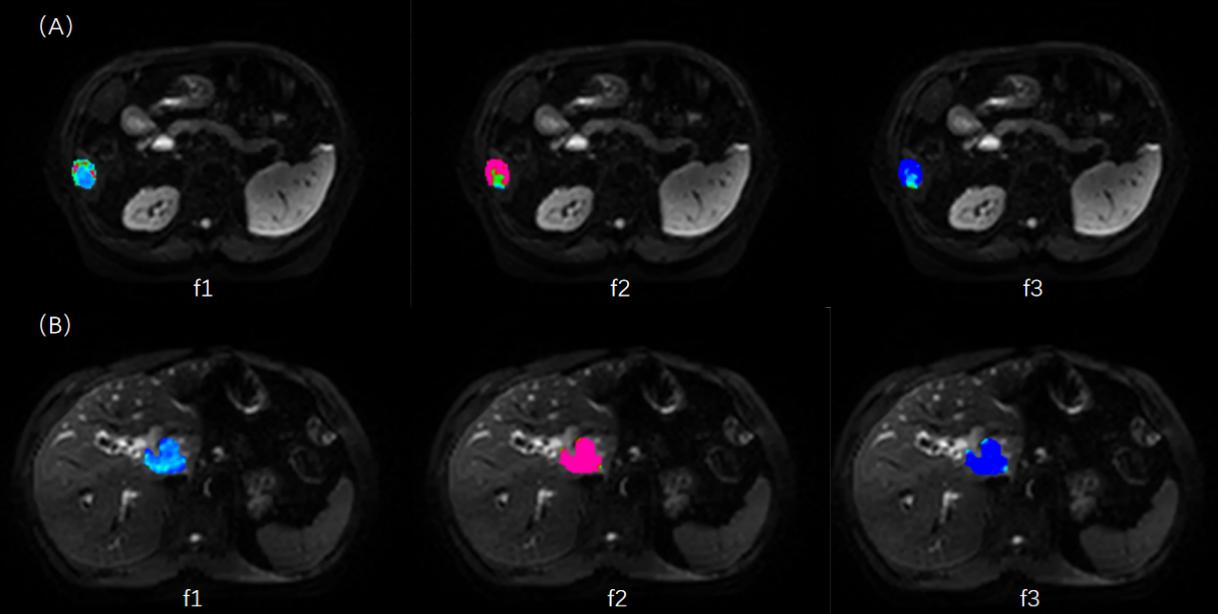
A total of 27 patients were enrolled in the study, including 15 patients with HCC and 12 patients with ICC. All the RSI images were acquired using a 3.0T MR scanner (uMR 790, United Imaging Healthcare, Shanghai, China) with 13 b values (b values: 0,25,50,100,150,200,400,600,800,1000,1500,2000,and 3000 s/mm2). Parametric maps were calculated by previously reported fitting methods (Figure 1). The independent-sample t-test and Mann-Whitney U test were used to compare the difference of each parameter value between the two groups. Binary logistic regression was used to combine parameters. Receiver operating characteristic (ROC) analysis was performed to assess the diagnostic performance, and the differences were assessed using DeLong analysis.Results
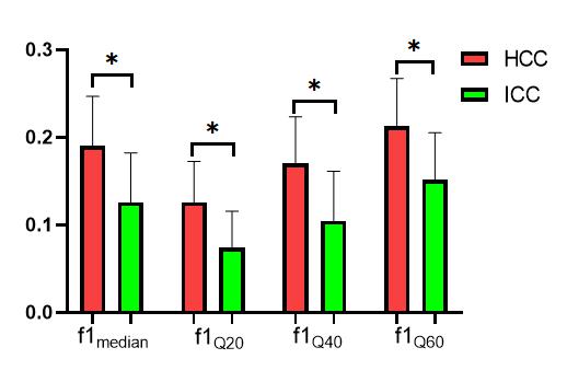
The f1median, f1Q20, f1Q40, f1Q60 were higher and ADCmean was lower in HCC than in the ICC (P = 0.001, 0.004, 0.001, 0.001 and 0.016, respectively, Figure 2). The ROC analysis showed that f1median, f1Q20, f1Q40, f1Q60 and ADCmean all had a positive effect on differentiating HCC and ICC, and the AUCs were 0.811, 0.817, 0.817, 0.822 and 0.744, respectively (all P < 0.05, Figure 3). There was no significant difference in AUC values among the parameters. The combination of RSI-derived parameters and ADC had the strongest predictive potential (AUC = 0.872; sensitivity, 93.33%; specificity, 83.33%; P < 0.001 ).Discussion
In this study, we demonstrated that the RSI model can effectively distinguish HCC from ICC. The parameter f1 represents the restricted signal of the intracellular water compartment[4,5]. ADC reflects the diffusion rate of water molecules and is negatively correlated with the proliferation status of tumor cells[6]. Compared with ICC, HCC has a higher density of tumor cells, smaller intercellular space, and a higher degree of water diffusion movement restriction. The center part of ICC is composed of a large number of loose fibrous tissues, and the tumor cells are mostly located in the periphery and can be arranged into adenoids, which promotes the diffusion movement of water molecules[7]. Therefore, the parameters f1 in the HCC group are significantly higher and ADCmean is significantly lower than those in the ICC group.Conclusion
The RSI-derived parameters may serve as noninvasive and quantitative imaging markers, which can effectively improve the diagnostic performance of HCC and ICC.Acknowledgements
No acknowledgementsReferences
1. Lee YT, Wang JJ, Luu M, et al. Comparison of Clinical Features and Outcomes Between Intrahepatic Cholangiocarcinoma and Hepatocellular Carcinoma in the United States. Hepatology. 2021;74(5):2622-2632.
2. Martinez-Heras E, Grussu F, Prados F, et al. Diffusion-Weighted Imaging: Recent Advances and Applications. Semin Ultrasound CT MR. 2021 Oct;42(5):490-506.
3. Krishnan AP, Karunamuni R, Leyden KM, et al. Restriction Spectrum Imaging Improves Risk Stratification in Patients with Glioblastoma. AJNR Am J Neuroradiol. 2017;38(5):882-889.
4. Qin Y, Tang C, Hu Q, et al. Quantitative Assessment of Restriction Spectrum MR Imaging for the Diagnosis of Breast Cancer and Association With Prognostic Factors. J MAGN RESON IMAGING. 2022 Oct 7.
5. Besser AH, Fang LK, Tong MW, et al. Tri-Compartmental Restriction Spectrum Imaging Breast Model Distinguishes Malignant Lesions from Benign Lesions and Healthy Tissue on Diffusion-Weighted Imaging. Cancers (Basel). 2022 ;14(13):3200.
6. Jiang J, Fu Y, Hu X, et al. The value of diffusion-weighted imaging based on monoexponential and biexponential models for the diagnosis of benign and malignant lung nodules and masses. Br J Radiol. 2020;93(1110):20190400.
7. Zou X, Luo Y, Li Z, et al. Volumetric Apparent Diffusion Coefficient Histogram Analysis in Differentiating Intrahepatic Mass-Forming Cholangiocarcinoma From Hepatocellular Carcinoma. J MAGN RESON IMAGING. 2019;49(4):975-83.
Figures