2239
Preoperative evaluation of liver regeneration following hepatectomy in hepatocellular carcinoma using magnetic resonance elastography1West China Hospital, Sichuan University, Cheng Du, China, 2MR Research China,GE Healthcare, Bei Jing, China, 3Sanya People’s Hospital, San Ya, China
Synopsis
Keywords: Liver, Elastography, liver regeneration, elasticity imaging techniques, hepatocellular carcinoma, hepatectomy
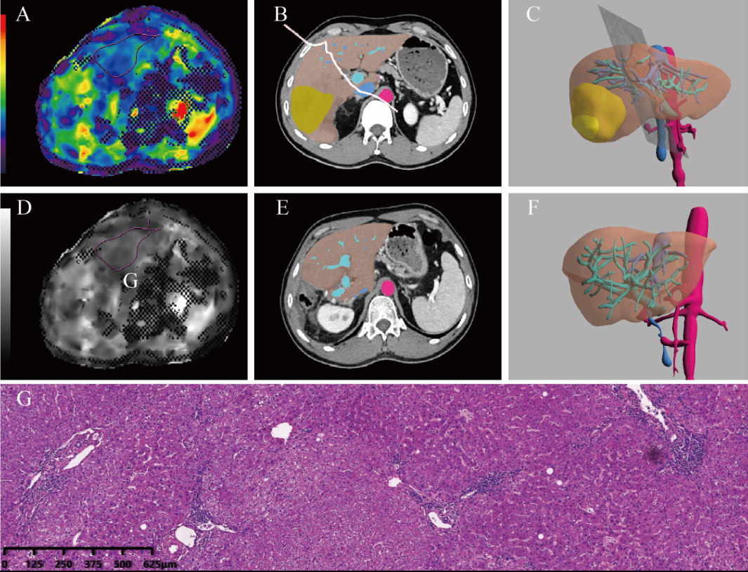
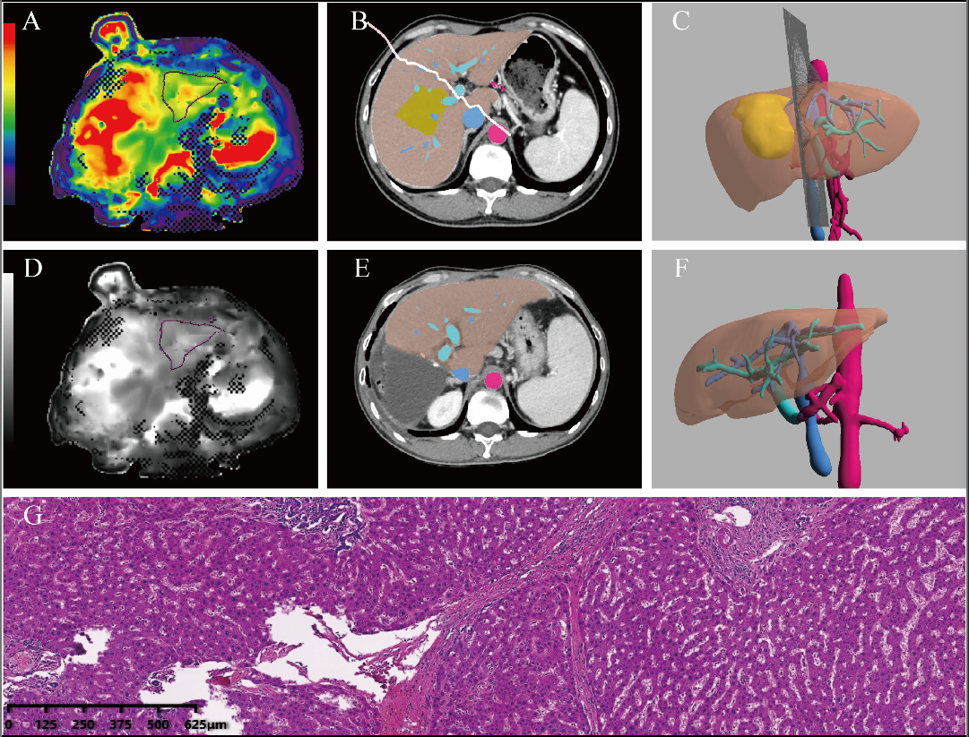
The liver stiffness (LS) value based on MRE may be a useful preoperative predictor of liver regeneration in patients with HCC. The LS derived from MRE shows a significant negative correlation with fibrosis, an important predictor of liver regeneration. In the low and high parenchymal hepatic resection rate (PHRR) groups, there was no significant relationship between regeneration index (RI) and LS values of MRE. However, a negative relationship was shown between LS values and RI in the intermediate PHRR group.Background
For patients with hepatocellular carcinoma (HCC) undergoing hepatectomy, insufficient remnant liver regenerative capacity can lead to liver failure. The aim of this study was to evaluate the potential role of magnetic resonance elastography (MRE) for the preoperative prediction of liver regeneration in patients with HCC after partial hepatectomy (PH).Methods
A total of 54 patients with HCC undergoing MRE prior to PH were retrospectively included. The total functional liver, volume of preoperative future liver remnant (LVpre), and volume of postoperative liver remnant (LVpost), respectively, were measured, and the regeneration index (RI) and parenchymal hepatic resection rate (PHRR) were manually calculated. Univariate and multivariate logistic regression analyses were conducted to identify factors associated with a high RI, and receiver operating characteristic (ROC) curves were employed to evaluate the diagnostic performance of the liver stiffness (LS) values. Patients were classified into three subgroups based on the value of PHRR: low PHRR (<30%), intermediate PHRR (30–50%), and high PHRR (>50%). Subsequently, Spearman correlation analysis was used to investigate the relationship between LS values and RI in the subgroups.Results
Multivariable analysis revealed a low LS value was associated with greater odds of a high RI [odds ratio (OR), 0.049; 95% confidence interval (CI): 0.002 to 0.980]. An optimal cutoff value of 3.30 kPa was used to divide all patients into a low RI group and a high RI group with an area under the curve (AUC) value of 0.882 (95% CI: 0.767 to 0.996). A significant negative relationship between RI and LS values (r=−0.799; P<0.001) was observed in the intermediate PHRR subgroup.Conclusions
The LS values based on MRE may serve as a potential preoperative predictor of liver regeneration for patients with HCC undergoing PH.Acknowledgements
Funding: This work was supported by the Science and Technology Support Program of Sichuan Province (Nos. 2021YFS0144, 2021YFS0021, and 2020YFS0121), China Postdoctoral Science Foundation (No. 2021M692289), and Post-Doctor Research Project, West China Hospital, Sichuan University (No. 2020HXBH130).References
1. Golse N, Bucur PO, Adam R, Castaing D, Sa Cunha A, Vibert E. New paradigms in post-hepatectomy liver failure. J Gastrointest Surg 2013;17:593-605.
2. Park CC, Nguyen P, Hernandez C, Bettencourt R, Ramirez K, Fortney L, Hooker J, Sy E, Savides MT, Alquiraish MH, Valasek MA, Rizo E, Richards L, Brenner D, Sirlin CB, Loomba R. Magnetic Resonance Elastography vs Transient Elastography in Detection of Fibrosis and Noninvasive Measurement of Steatosis in Patients With Biopsy-Proven Nonalcoholic Fatty Liver Disease. Gastroenterology 2017;152:598-607.e2.
3. Hoodeshenas S, Yin M, Venkatesh SK. Magnetic Resonance Elastography of Liver: Current Update. Top Magn Reson Imaging 2018;27:319-33.
4. Jang S, Lee JM, Lee DH, Joo I, Yoon JH, Chang W, Han JK. Value of MR elastography for the preoperative estimation of liver regeneration capacity in patients with hepatocellular carcinoma. J Magn Reson Imaging 2017;45:1627-36.
5. Kele PG, de Boer M, van der Jagt EJ, Lisman T, Porte RJ. Early hepatic regeneration index and completeness of regeneration at 6 months after partial hepatectomy. Br J Surg 2012;99:1113-9.
6. Park J, Kim JH, Kim JE, Park SJ, Yi NJ, Han JK. Prediction of liver regeneration in recipients after living-donor liver transplantation in using preoperative CT texture analysis and clinical features. Abdom Radiol (NY) 2020;45:3763-74.
Figures