2230
Application of 3D MR MENSA in preoperative evaluation of lumbar disc herniation: a prospective study1Department of Radiology, West China Hospital of Sichuan University, Chengdu, Sichuan, China, 2Department of Orthopedics, West China Hospital of Sichuan University, Chengdu, Sichuan, China, 3MR Research, GE Healthcare, Beijing, China
Synopsis
Keywords: Data Acquisition, Neuro
In this work, we propose a preoperative magnetic resonance examination of patients with lumbar disc herniation using the 3D MENSA sequence. This sequence was superior to cube, cube stir sequence in subjective and objective evaluation.The preoperative 3D MRI MENSA sequence is able to clearly depict the nerve roots and offer desirable contrast between the nerve roots, ligamentum flavum, bone, and intervertebral discs. Patients with lumbar degeneration can effectively benefit from the MENSA sequence since it provides informative imaging information to help understand disc herniation and compression of adjacent tissues when developing preoperative surgical strategies.Research Focus
To investigate the value of preoperative three-dimensional MR, 51 patients underwent lumbar MR using 3D MR Sequences (Cube, Cube stir, MENSA). SNRs and CNRs of nerve, herniated disc, ligament, soft tissue were calculated. Image quality was scored by 5-point method. One-way ANOVA, Fridman test, Kappa test were used to calculate objective and subjective scores, evaluate consistency of two readers' scores. Comprehensive analysis of SNR, CNR, subjective scores showed MENSA sequence was superior to cube, cube stir. Preoperative MENSA sequence is able to clearly depict nerve roots, offer desirable contrast. Patients can benefit from MENSA sequence when developing preoperative surgical strategies.Purpose
To investigate the value of three-dimensional(3D) MR examination in preoperative evaluation of lumbar disc herniation.Methods
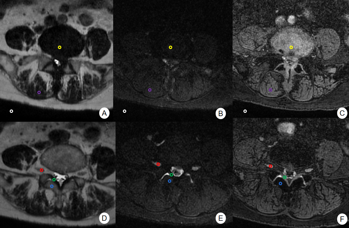
A total of 51 patients who underwent lumbar disc herniation surgery in West China Hospital of Sichuan University from June 2021 to December 2021 were prospectively enrolled. All MRI scan were performed on a 3.0T MRI scanner (Signa Premier, GE Healthcare, Milwaukee, USA). 3D MR sequences (Cube, Cube stir, and MENSA) were used for the lumbar MR imaging. The signal to noise ratio (SNR) and the contrast noise ratio (CNR) of nerve, herniated disc, ligament, and soft tissue were calculated. Objective scores were calculated by one-way ANOVA. The image quality was scored by a 5-point method. Fridman test was used to compare the subjective scores of image quality, and Kappa test was used to evaluate the consistency of two readers' scores.Results
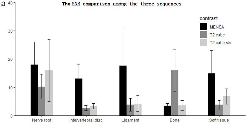
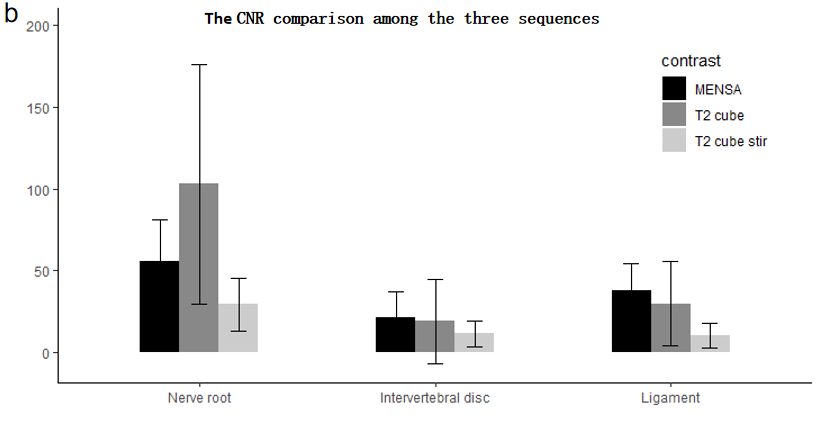
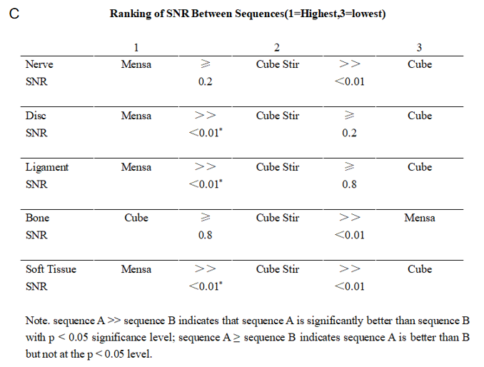
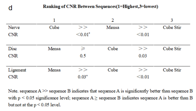
The nerve root SNR in the MENSA and Cube stir groups were significantly higher than in the Cube group (P<0.05), but there was no substantial statistical difference between the two groups. The herniated disc and ligamentum flavum SNRs of the MENSA group were much greater than those of the Cube stir and Cube groups (P<0.05). Soft tissue SNR was significantly greater in the MENSA group compared with the Cube stir group (P<0.05), which was significantly greater than it in the Cube group (P<0.05). The nerve root CNR of the Cube group was significantly greater than that of the MENSA group, which was significantly higher than it in the Cube Stir group. The herniated disc CNR was significantly higher in the MENSA and Cube groups than in the Cube Stir group. The CNR of LF was significantly greater in the MENSA group compared to the Cube group, which was significantly greater than the Cube Stir group. Comprehensive comparison of SNR, CNR and subjective score showed that MENSA sequence was better than cube and cube stir. Among the subjective ratings of the two reviewers, the MENSA sequence scored highest in the qualitative measures of image quality. In the consistency test of the scores of the three groups of images by the two readers, the Kappa values were all greater than 0.73, and the P values of the test were all less than 0.05. Above all indicating that the consistency test results of the two readers were statistically significant and consistent.Discussion and Conclusions
In conclusion, the preoperative 3D MRI MENSA sequence is able to clearly depict the nerve roots and offer desirable contrast between the nerve roots, ligamentum flavum, bone, and intervertebral discs. Patients with lumbar degeneration can effectively benefit from the MENSA sequence since it provides informative imaging information to help understand disc herniation and compression of adjacent tissues when developing preoperative surgical strategies.Acknowledgements
No acknowledgement found.References
[1] Thoomes EJ, Scholten-Peeters GG, de Boer AJ, et al. Lack of uniform diagnostic criteria for cervical radiculopathy in conservative intervention studies: a systematic review. Eur Spine J 2012;21:1459-70.[2] Jin C, Li H, Li X, et al. Temporary Hearing Threshold Shift in Healthy Volunteers with Hearing Protection Caused by Acoustic Noise Exposure during 3-T Multisequence MR Neuroimaging. Radiology. 2018 Feb;286(2):602-608. doi: 10.1148/radiol.2017161622. Epub 2017 Aug 16. PMID: 28813235.
[3] Bell GR, Ross JS. Diagnosis of nerve root compression: myelography, computed tomography, and MRI.Orthop Clin North Am,1992;23:405-19.
[4] Brown BM, Schwartz RH, Frank E, Blank NK. Preoperative evaluation of cervical radiculopathy and myelopathy by surface-coil MR imaging. AJR Am J Roentgenol, 1988;151:1205-12.
[5] Kunogi J, Hasue M. Diagnosis and operative treatment of intraforaminal and extraforaminal nerve root compression. Spine 1991;16:1312-20.
[6] Aota Y, Niwa T, Yoshikawa K, Fujiwara A, Asada T, Saito T. Magnetic resonance imaging and magnetic resonance myelography in the presurgical diagnosis of lumbar foraminal stenosis. Spine 2007;32:896-903.
[7] Friedenberg ZB, Miller WT. Degenerative disc disease of the cervical spine. J Bone Joint Surg Am,1963;45:1171-8.
[8] Christina A. Chen, Richard Kijowski, Lauren M. Shapiro,Michael J. Tuite,et al. Cartilage Morphology at 3.0T: Assessment of Three-Dimensional Magnetic Resonance Imaging Techniques. JOURNAL OF MAGNETIC RESONANCE IMAGING,2010,32:173–183.
[9] Rongli W,Masatoshi H,Hiromitsu O,et al.Effects of reconstruction technique on the quality of abdominal CT angiography:A comparison between forward projected model-based iterative reconstruction solution(FIRST) and conventional reconstruction methods[J]. European Journal of Radiology,2018,106:100-105.
[10] Pillastrini P, Gardengh I, Bonetti F,et al. An updated overview of clinical guidelines for chronic low back pain management in primary care. Jt Bone Spine2011;79:176-185.[11] European Society of Skeletal Radiology. Guidelines for MRI maging of Sports Injuries. https://essr.org/content-essr/uploads/2016/10/ESSR-MRI-Protocols-Spine.pdf. AccessedMay31,2018.
[12] Fast spin echo sequences with very long echo trains: design of variable refocusing flip angle schedules and generation of clinical T2 contrast.
[13] The brachial plexus: normal anatomy, pathology, and MR imaging.
[14] Bruder H, Fischer H, Graumann R, Deimling M. A new steady-state imaging sequence for simultaneous acquisition of two MR images with clearly different contrasts. Magn Res Med 1988;7:35-42.
[15] Thakkar RS, Fammang AJ, Chhabra A et al. 3T MR imaging of cartilage using 3D dual echo steady state (DESS). MAGNETOM Flash,2011:3:33-36.
[16] Kijowski R,Gold GE. Routine 3D magnetic resonance imaging of joints. J Magn Reson Imaging 2011;33:758–771.14.
[17] Tins B, Cassar-Pullicino V, Haddaway M, Nachtrab U. Three-dimensional sampling perfection with application-optimised contrasts using a different flip angle evolutions sequence for routine imaging of the spine: preliminary experience. Br J Radiol 2012;85:480-489.
Figures