2229
Design of a Two-dimensional Ultrashort Echo Time Simultaneous Multi-slice Pulse Sequence1Computer Science, Mathematics, Physics and Statistics, University of British Columbia, Kelowna, BC, Canada, 2UBC MRI Research Centre, Department of Radiology, Faculty of Medicine, University of British Columbia, Vancouver, BC, Canada, 3SFU ImageTech Lab, Simon Fraser University, Surrey, BC, Canada, 4Philips Canada, Mississauga, ON, Canada, 5Biomedical Engineering and Imaging Institute, Icahn School of Medicine at Mount Sinai, New York, NY, United States
Synopsis
Keywords: Pulse Sequence Design, New Signal Preparation Schemes
Simultaneous multi-slice (SMS) pulse sequences have allowed for reductions in scan time with minimal signal-to-noise ratio loss. However, when ultrashort echo times (UTEs) are desired, SMS pulse sequences have been challenging to implement. In this work, we explore a UTE SMS pulse sequence that makes use of a power independent of number of slices prepulse to shape the transverse magnetization profile and a whole volume hard excitation to excite the remaining longitudinal magnetization. The novel pulse sequence is estimated to reduce scan times by a factor of approximately 7.6 when compared to UTE pulse sequences with three-dimensional acquisitions.Introduction
Ultrashort echo time (UTE) pulse sequences are essential for the magnetic resonance imaging (MRI) of species with short T2 such as sodium (23NA) or cortical bone1,2. UTE pulse sequences have used center-out three-dimensional (3D) acquisitions to achieve echo times (TEs) of 0.2 ms; however, 10-18 min are needed to fully sample 3D k-space3-6. Two-dimensional (2D) acquisitions are difficult to implement with UTE pulse sequences due to slice selectivity requirements6.Simultaneous multi-slice (SMS) pulse sequences with 2D acquisitions have reduced scan times with minimal signal-to-noise ratio (SNR) loss7. SMS pulse sequences have frequently involved multi-band (MB) pulses that exhibit a higher specific absorption rate (SAR) and longer minimum TE than single-band pulses6,8. The power independent of number of slices (PINS) pulse has allowed for the excitation of multiple slices with SAR comparable to single-band pulses8. Recently, half PINS pulses have been used to demonstrate the feasibility of UTE SMS pulse sequences for proton MRI9. However, scan times may be further reduced with the use of a whole volume hard excitation. We explore the design of a UTE SMS pulse sequence to achieve frequency selective suppression of all magnetization outside of specific bands prior to broadband excitation, allowing for the measurement of multiple slices simultaneously with minimal TE.
Methods
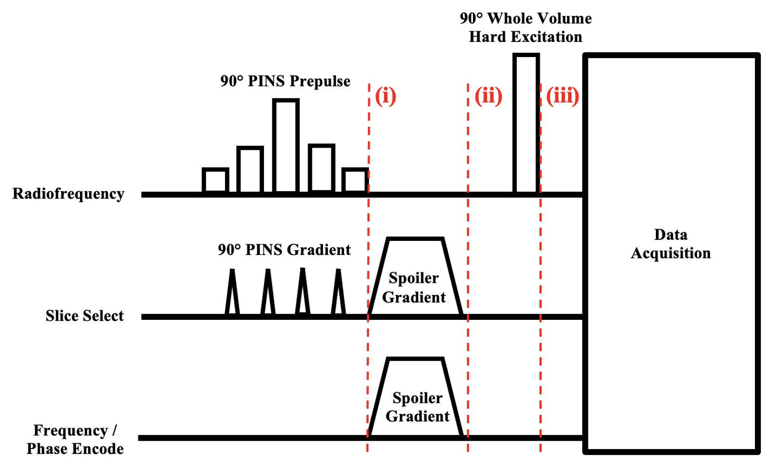
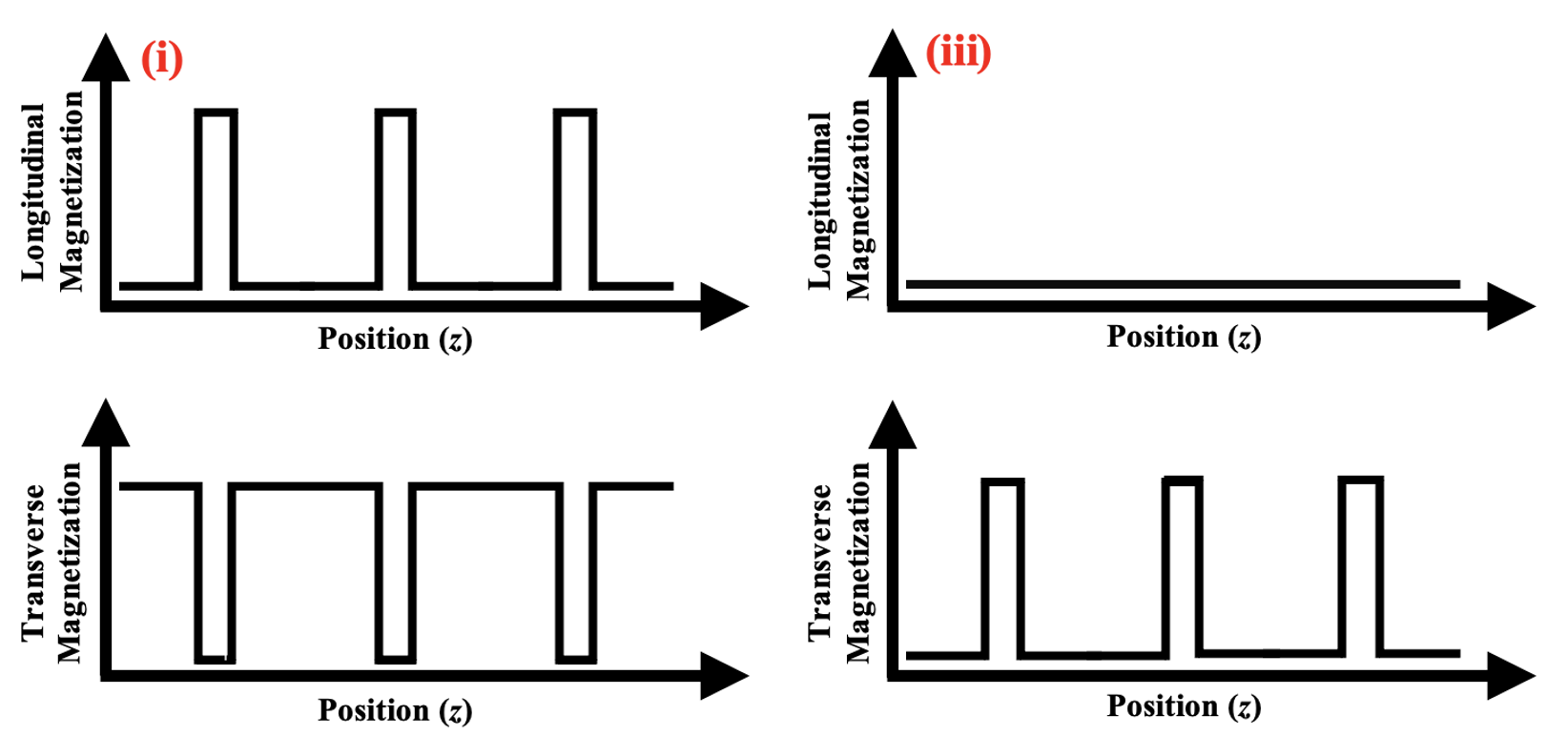
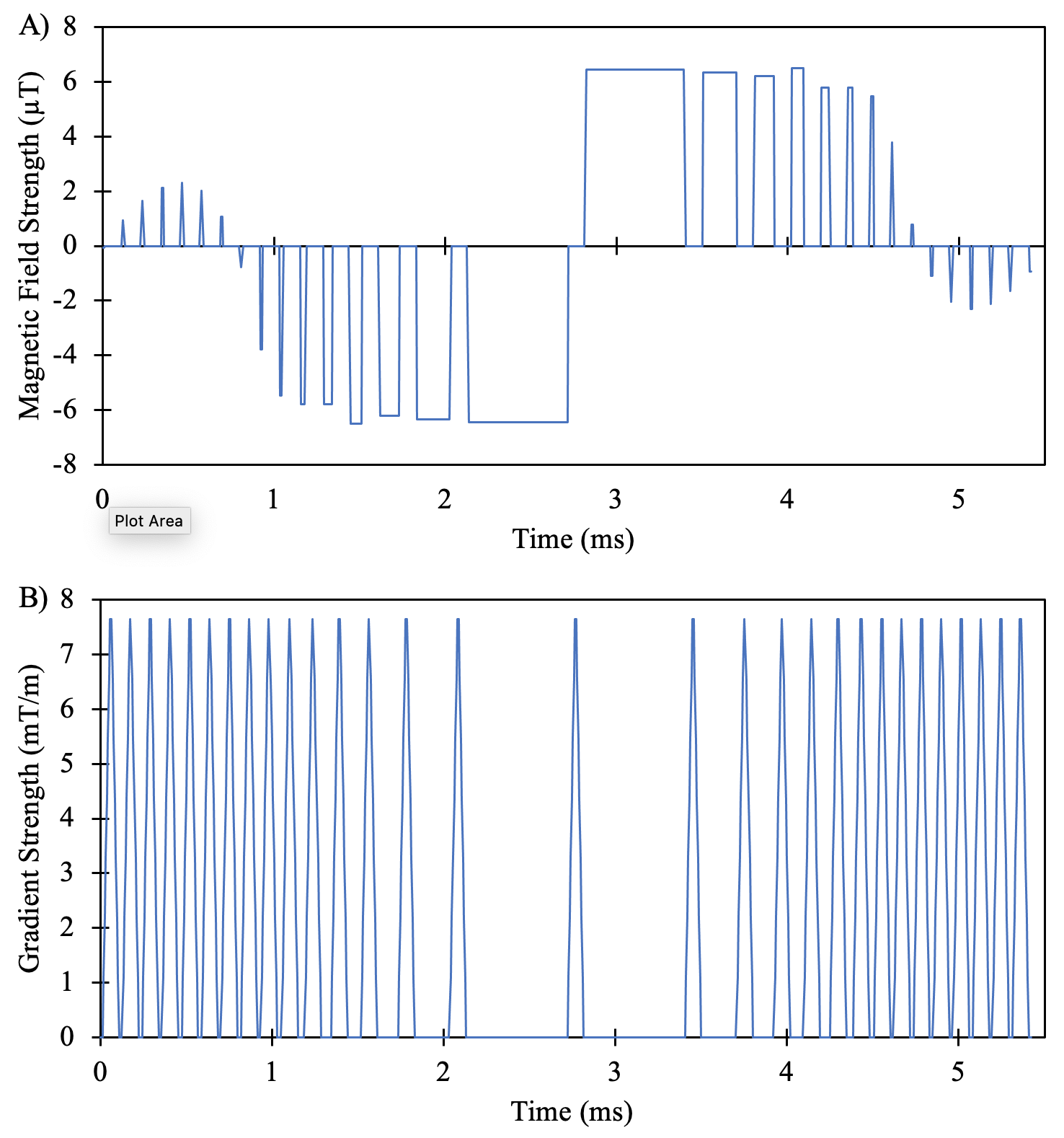
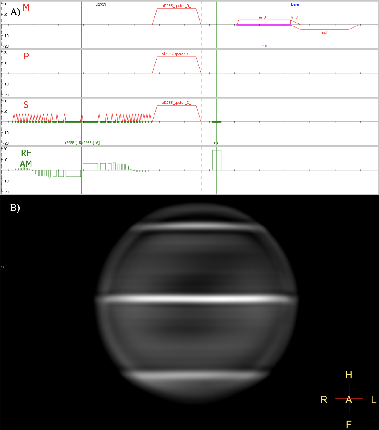
The UTE SMS pulse sequence begins with a power independent of number of slices (PINS) prepulse to excite most of the magnetization, while leaving specific bands of magnetization along the longitudinal axis. Next, spoiler gradients dephase the unwanted transverse magnetization. Finally, a hard excitation is used to excite the specific bands of longitudinal magnetization into the transverse plane to be measured. Data acquisition from the resulting excited slices can begin immediately after the transmit/receive switching time2. The pulse sequence and magnetization profile at various time points are illustrated in Figure 1 and 2.Pulse sequence design was carried out to meet the hardware specifications of a Philips Ingenia Eletion X which allows for a minimum dwell time of 6.4 µs, maximum gradient strength of 45 mT/m, and maximum slew rate of 220 T/m/s. The maximum magnetic field strength of the PINS prepulse was limited to 6.5 µT to prevent head SAR limits from being exceeded during the pulse sequence. The PINS prepulse was designed in MATLAB using a modified Multiband RF Toolbox with linear phase, a flip angle of 90°, a time bandwidth product of 29.55, and a slice gap of 4.60 mm to excite 3 slices over 18 cm8,10. The spoiler gradients were designed using the maximum slew and maximum gradient strength such that complete dephasing was achieved over 1 mm. The whole volume hard excitation pulse was designed with a flip angle of 90° while minimizing its duration. Simulations were conducted in MATLAB to investigate the transverse magnetization profile after the PINS prepulse and at the end of the pulse sequence10.
Results
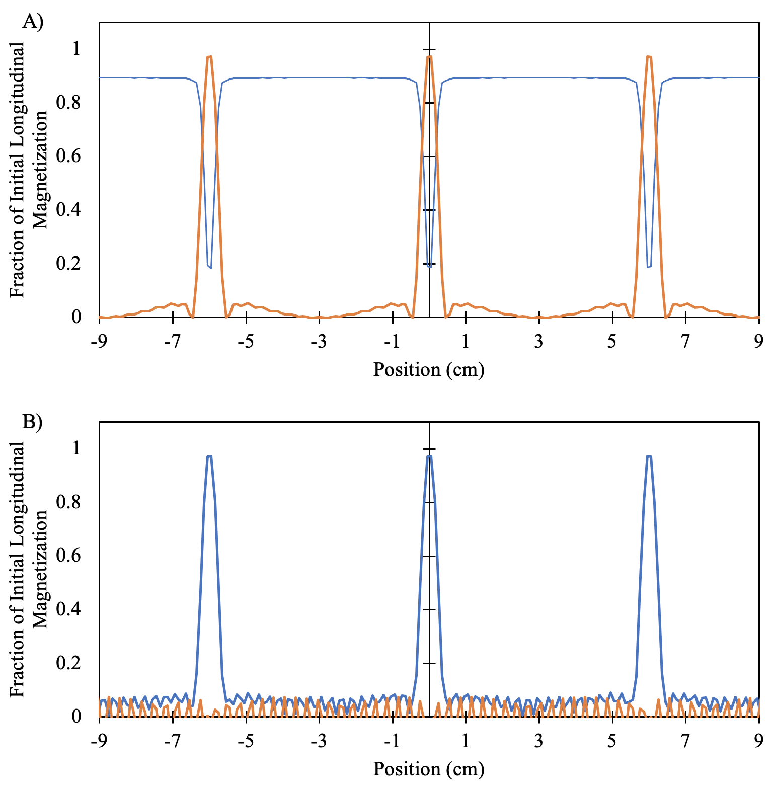
The PINS prepulse with time bandwidth product of 29.55 and slice separation of 4.60 mm along with its gradient and corresponding transverse magnetization profile are shown in Figure 2. The simulated transverse and longitudinal magnetization profiles after the PINS prepulse and at the end of the pulse sequence are shown in Figure 3. The pulse sequence with a repetition time of 23 ms as displayed in Philips scanner software is shown in Figure 4.Discussion
UTE MRI techniques have achieved TEs of 0.2 ms with a 300 µs hard excitation, a 50 µs transmit/receive switching time, and a centre-out 3D acquisition3,4. With the same hard excitation, this novel pulse sequence will allow for the same TE. For example, the use of this novel pulse sequence to image 23NA, with a short T2 component of 2 ms accounting for 60 % and a long T2 component of 23 ms accounting for 40 % of the relaxation, will allow acquisition to begin when the transverse magnetization has decayed by an 6.1 %11. If the PINS prepulse had been used in a typical SMS pulse sequence, 50.0 % of the transverse magnetization would have decayed by the time acquisition could begin. Thus, the novel pulse sequence is estimated to increase signal strength by a factor of 1.8 for 23NA SMS MRI.Techniques such as functional and diffusion MRI, which make use of 2D acquisitions, would benefit from reduced scan times for proton MRI and may even be enabled for 23NA MRI7. 2D acquisitions also allow for more flexibility in resolution, manual selection of the number of slices acquired, and acquisition of fewer lines in k-space to fulfill the Nyquist criterion, allowing for scan times to be reduced beyond that of 3D acquisitions12. With the same field of view and resolution as the centre-out 3D acquisitions discussed above, the pulse sequence may reduce scan times from 6.60 to 0.84 min and from 4.68 to 0.63 min, resulting in the reduction of scan times by a factor of approximately 7.6 3,4. However, the pulse sequence will not have the SNR benefit of a whole volume acquisition. Further, for current UTE SMS techniques, which make use of half pulses, scan times may be reduced by a factor of 2 with the use of a hard excitation10.
Acknowledgements
We acknowledge the support of a Natural Sciences and Engineering Research Council of Canada (NSERC) Discovery Grant and a Canada Foundation for Innovation (CFI) John R Evans Leaders Fund.
References
1. Madelin G and Regatte RR. Biomedical Applications of Sodium MRI in Vivo. J Magn Reson Imaging. 2013;38(3):511-529.
2. Larson PEZ, Han M, Krug R et al. Ultrashort echo time and zero echo time MRI at 7T. Magn Reson Mater Phys. 2016;29(3):359–370.
3. Riemer F, Solanky BS, Stehning C, et al. Sodium (23Na) ultra-short echo time imaging in the human brain using a 3D-Cones trajectory. Magn Reson Mater Phys. 2014;27(1):35-46.
4. Milani B, Delacoste J, burnier M, and Pruijm M. Exploring a new method for quantitative sodium MRI in the human upper leg with a surface coil and symmetrically arranged reference phantoms. Quant Imaging Med Surg. 2019;9(6):985-999.
5. Nielles-Vallespin S, Weber MA, Bock M, et al. 3D Radial Projection Technique with Ultrashort Echo Times for Sodium MRI: Clinical Applications in Human Brain and Skeletal Muscle. Magn Reson Med. 2007;57(1):74-81.
6. Bangerter NK, Tarbox GJ, Taylor MD, and Kaggie JD. Quantitative sodium magnetic resonance imaging of cartilage, muscle, and tendon. Quant Imaging Med Surg. 2016;6(6):699-714.
7. Barth M, Breuer F, Koopmans PJ, Norris DG, and Poser BA. Simultaneous multislice (SMS) imaging techniques. Magn Reson Med. 2016;75(1):63-81.
8. Norris DG, Koopmans PJ, Boyacioglu R, and Barth M. Power Independent of Number of Slices (PINS) Radiofrequency Pulses for Low-Power Simultaneous Multislice Excitation. Magn Reson Med. 2011;66(5):11234-1240.
9. Rettenmeier C and Stenger VA. RF Phase Encoded Half Pulses in Simultaneous Multislice UTE Imaging. Mafn Reson Med. 2019;81(6):3720-3733.
10. Seada SA, Price AN, Schneider T, Hajnal JV, and Malik SJ. Multiband RF pulse design for realistic gradient performance. Magn Reson Med. 2019;81(1):362-376.
11. Qian Y, Panigrahy A, Laymon CM, et al. Short-T2 imaging for quantifying concentration of sodium (23 Na) of bi-exponential T2 relaxation. Magn Reson Med. 2015;74(1):162-174.
12. Konstandin S, Nagel AM, Heiler PM, and Schad LR. Two-dimensional radial acquisition technique with density adaption in sodium MRI. Magn Reson Med. 2011;65(4):1090-1096.
Figures