2214
The effect of MultiBand acquisition on cerebral inversion recovery intravoxel incoherent motion imaging1School for Mental Health & Neuroscience, Maastricht University, Maastricht, Netherlands, 2Department of Intensive Care, Maastricht University Medical Center, Maastricht, Netherlands, 3Department of Radiology and Nuclear Medicine, Maastricht University Medical Center, Maastricht, Netherlands, 4Department of Electrical Engineering, Eindhoven University of Technology, Eindhoven, Netherlands
Synopsis
Keywords: Parallel Imaging, Parallel Imaging
The MultiBand (MB) imaging technique can reduce scan time considerably, which can be especially relevant for clinical application of techniques with extensive scan protocols, such as intravoxel incoherent motion (IVIM) imaging. However, quantitative IVIM parameter estimates may be affected by the use of MB. This study is a first step to assess the comparability between IVIM acquisitions with and without MB, which has considerable implications for interpretation of IVIM results across different datasets.
Introduction
Intravoxel incoherent motion (IVIM) imaging is an application of diffusion weighted imaging (DWI) used to assess diffusion in the brain parenchyma and microvasculature simultaneously (1). With IVIM, three parameters are usually estimated: the parenchymal diffusivity (D), the microvascular diffusivity (pseudo-diffusion; D*), and the perfusion volume fraction (f) (1) These parameters have previously been shown to be relevant markers in cerebral pathology (2).IVIM images are acquired with an extensive b-value sampling scheme, which can drastically increase scan time. To increase the clinical feasibility of IVIM, shorter scan times are desired. One way to reduce acquisition time, whilst retaining the number of b-values, is to use MultiBand (MB). With MB, multiple slices are excited simultaneously, which allows for a considerable shortening of scan time without reduction in signal-to-noise (SNR) (3).
Although MB does not come with a penalty in SNR, it remains undetermined how MB affects IVIM measures. Moreover, cerebrospinal fluid (CSF) is often suppressed to increase accuracy of the perfusion volume fraction f (2). Yet, it is unknown whether CSF suppression is as successful using MB compared to acquiring IVIM images without MB.
This study aims to explore the effect of MB acquisition on IVIM measures by comparing the quantitative IVIM parameter values from MB and non-MB MB acquisitions in two subjects.
Methods
Subjects: Two healthy subjects were included in this study (subject 1 = 55y/female; subject 2 = 22y/female).MRI acquisition: Imaging for both subjects was performed on a 3.0 Tesla MR system (Philips, Ingenia CX; Philips Healthcare, Best, The Netherlands), using a 32-channel head coil (subject 1) or 16-channel head coil (subject 2). Diffusion MR images – with and without MB – were acquired in the anterior-posterior direction, including a reversed phase encoding direction b=0 image, in the same session for both subjects. Anatomical T1-weighted images were also acquired (Table 1).
Diffusion MR image processing: Diffusion MR images were corrected for susceptibility induced distortions (topup, FSL version 6.0.4) (4), head displacements, and eddy currents (ExploreDTI version 4.8.6) (5). A mean image was computed from the b=0 volume of the MB and non-MB dataset. MB and non-MB images were coregistered to this mean image (FLIRT, FSL (6); mri_vol2vol, FreeSurfer version 7.1.0).
Anatomical image processing: T1-weighted images were automatically segmented using FreeSurfer SAMSEG tool (7). Regions of interest (ROIs) included white matter (WM), cortical grey matter (cGM) and deep grey matter (dGM) and were coregistered to the mean diffusion MR image (FLIRT, FSL) (6).
IVIM analysis: To obtain D and f images, a voxel-wise model fitting procedure was employed using the two-step biexponential IVIM model fitting approach (8). D* was also included in the model fitting procedure but is, due to its limited robustness (9), not further discussed. A modified version of the conventional two-compartment IVIM model was used to account for CSF suppression and different T1-relaxation and T2-relaxation times of blood and tissue, as previously described (2).
Statistics: Due to the exploratory nature of this study, only descriptive statistics were extracted from the data in MATLAB R2020a (MathWorks, Natick, Massachusetts).
Results
Estimated values of D and f from the IVIM data acquired with and without MB are reported in Table 2 for all ROIs in both subjects. Overall, D and f values are of similar order when comparing the MB and non-MB datasets, but higher variability in the MB dataset is apparent (Figure 1). Similar distribution patterns of parameter values were observed in all ROIs. Spatial distributions of D and f reveal differences between the MB and non-MB, particularly in the f map (Figure 2).Discussion
This study explored the effect of employing MB on IVIM measures. Obtained parameter values were of similar order as previously reported (2), reflecting the validity of the current values. At first glance, values obtained using the MB acquisition seem similar to the non-MB acquisition, although discrepancies exist particularly for the perfusion volume fraction f. Whether these differences are substantial cannot be concluded from the current results due to a limited sample size.Since both imaging strategies were acquired within the same subjects in the same scanning session, no physiological changes were expected. Moreover, good reproducibility of IVIM imaging with inversion recovery has been previously reported (10). Therefore, the observed differences in D and f values can mainly be attributed to the acquisition strategy, although intrasubject variability remains a contaminating factor.
Of note, less variance was observed for D and f values obtained from the non-MB acquisition compared to the MB dataset. This study used an inversion recovery pulse to achieve suppression of CSF, but its effectiveness may have been affected by the employment of MB. When CSF is not adequately suppressed more partial voluming can occur, possibly resulting in more spatial variance within the IVIM maps.
Conclusion
The results of this explorative study suggest that comparison of IVIM measures from MB and non-MB acquisitions is not straightforward. Obtained IVIM measures generally seem similar between MB and non-MB acquisitions but employing MB results in more spatial variance within the IVIM measures. Acquisition strategy should be taken into careful consideration when interpreting IVIM results across datasets. Future studies with larger sample sizes should verify these results.Acknowledgements
No acknowledgement found.References
1. Le Bihan D, Breton E, Lallemand D, Aubin ML, Vignaud J, Laval-Jeantet M. Separation of diffusion and perfusion in intravoxel incoherent motion MR imaging. Radiology 1988;168(2):497-505.
2. Wong SM, Zhang CE, van Bussel FC, et al. Simultaneous investigation of microvasculature and parenchyma in cerebral small vessel disease using intravoxel incoherent motion imaging. Neuroimage Clin 2017;14:216-221.
3. Barth M, Breuer F, Koopmans PJ, Norris DG, Poser BA. Simultaneous multislice (SMS) imaging techniques. Magn Reson Med 2016;75(1):63-81.
4. Andersson JL, Skare S, Ashburner J. How to correct susceptibility distortions in spin-echo echo-planar images: application to diffusion tensor imaging. Neuroimage 2003;20(2):870-888.
5. Leemans A, Jeurissen B, Sijbers J, Jones DK. ExploreDTI: a graphical toolbox for processing, analyzing, and visualizing diffusion MR data. 2009.
6. Jenkinson M, Bannister P, Brady M, Smith S. Improved optimization for the robust and accurate linear registration and motion correction of brain images. Neuroimage 2002;17(2):825-841.
7. Puonti O, Iglesias JE, Van Leemput K. Fast and sequence-adaptive whole-brain segmentation using parametric Bayesian modeling. Neuroimage 2016;143:235-249.
8. Federau C, O'Brien K, Meuli R, Hagmann P, Maeder P. Measuring brain perfusion with intravoxel incoherent motion (IVIM): initial clinical experience. J Magn Reson Imaging 2014;39(3):624-632.
9. Wu WC, Chen YF, Tseng HM, Yang SC, My PC. Caveat of measuring perfusion indexes using intravoxel incoherent motion magnetic resonance imaging in the human brain. Eur Radiol 2015;25(8):2485-2492.
10. Wong SM, Backes WH, Zhang CE, et al. On the Reproducibility of Inversion Recovery Intravoxel Incoherent Motion Imaging in Cerebrovascular Disease. AJNR Am J Neuroradiol 2018;39(2):226-231.
Figures
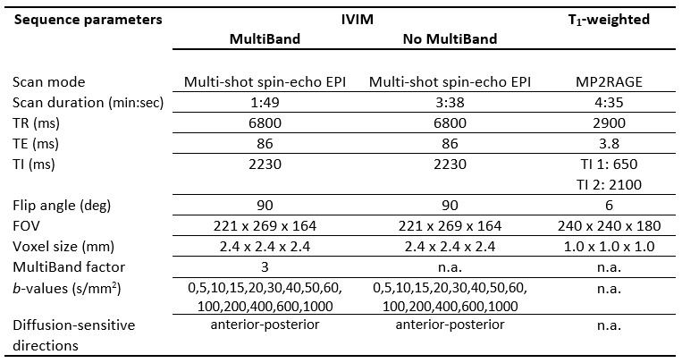
Table 1. Summarized sequence parameters.
Abbreviations: IVIM = intravoxel incoherent motion, EPI = echo planar imaging, MP2RAGE = magnetization prepared 2 rapid gradient echo, TR = repetition time, TE = echo time, TI = inversion time, FOV = field of view.
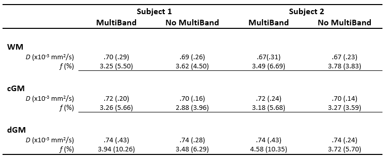
Table 2. Estimated values of parenchymal diffusivity D and perfusion volume fraction f in IVIM with and without MultiBand. Median (IQR) is reported.
Abbreviations: IVIM = intravoxel incoherent motion, MB = MultiBand, WM = white matter, cGM = cortical grey matter, dGM = deep grey matter, D = parenchymal diffusivity, f = perfusion volume fraction.
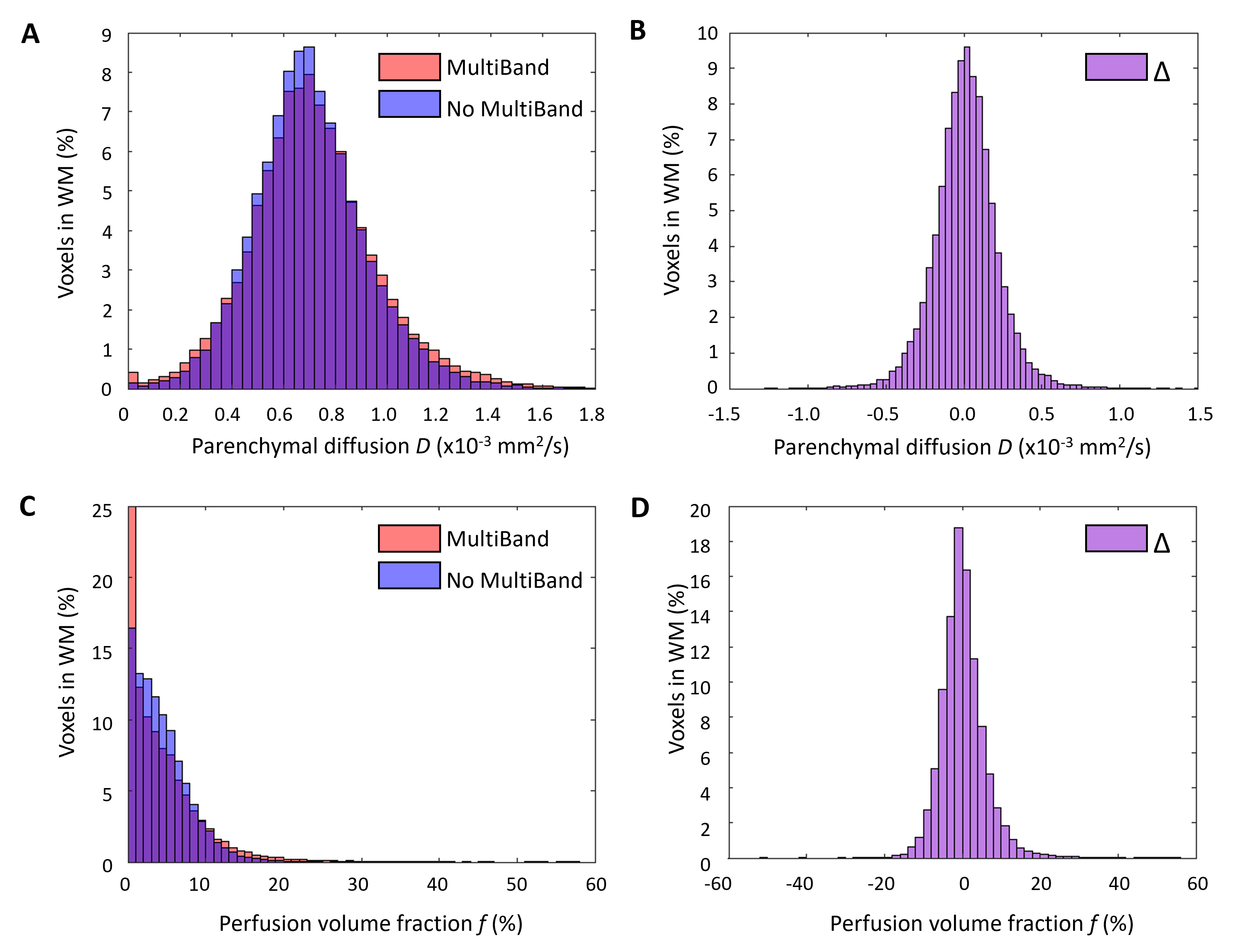
Figure 1. Effect of MultiBand on IVIM parameter values in the white matter. Distributions of estimated IVIM parameters in the white matter (WM) for the MultiBand dataset (red) and non-MultiBand dataset (blue) for the parenchymal diffusion D the perfusion volume fraction f (A) – and its corresponding difference (delta Δ) histogram (MultiBand – non-MultiBand) (B) – and for the perfusion volume fraction f (C) – and its corresponding difference histogram (D). Distributions are shown for subject 1.
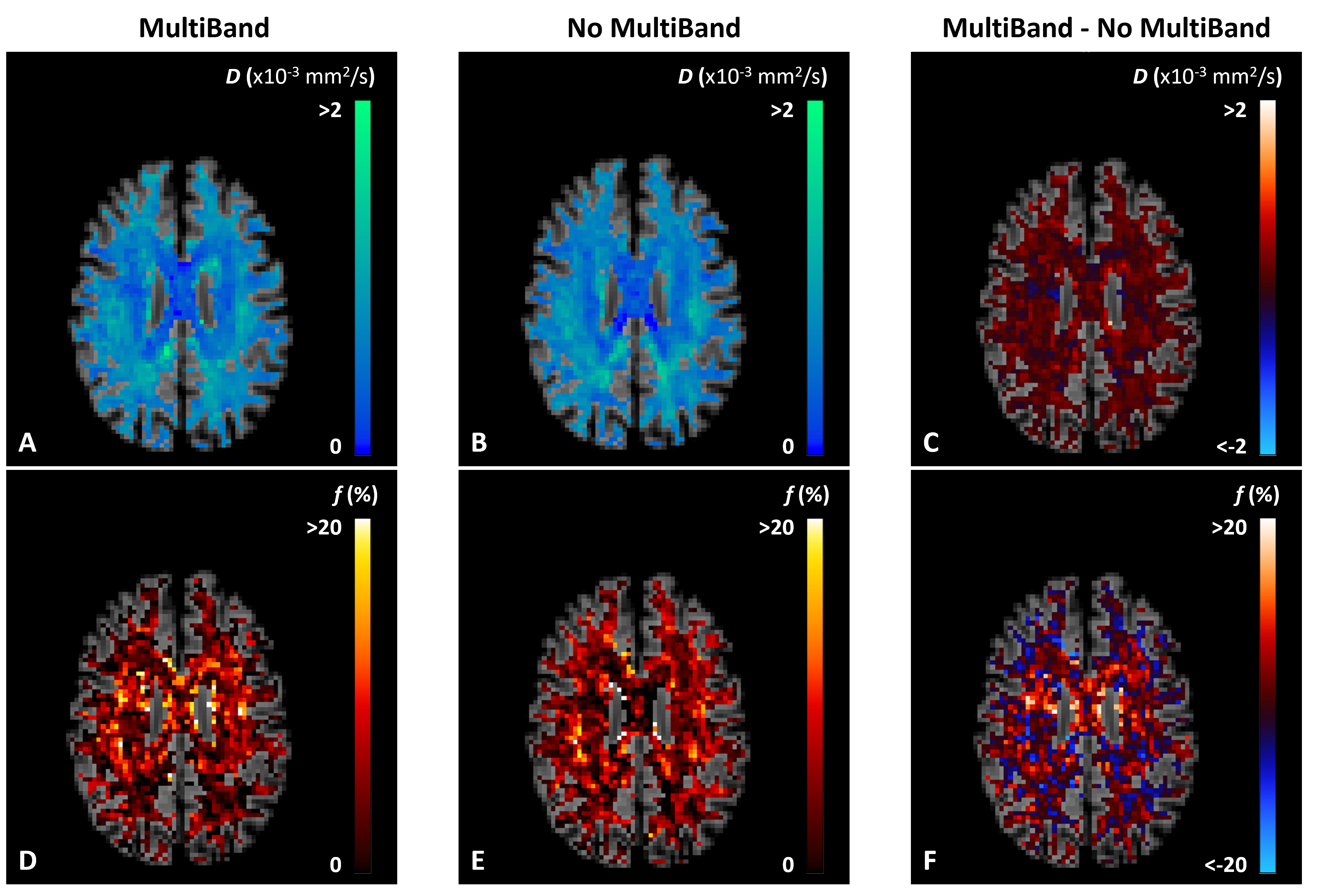
Figure 2. Effect of MultiBand on IVIM parameter maps in the white matter. Parenchymal diffusion (D) maps for the white matter with MultiBand (A), without MultiBand (B), and the corresponding difference map (C). Perfusion volume fraction (f) maps are also shown for the white matter with MultiBand (D), without MultiBand (E), and the corresponding difference map (F). IVIM maps of the white matter are shown for subject 1.