2208
Sub-second T2 mapping of the whole brain via multiband SENSE multiple overlapping-echo detachment imaging and deep learning1Department of Electronic Science, Xiamen University, Xiamen, China, 2Department of Radiology, Zhongshan Hospital of Xiamen University, School of Medicine, Xiamen University, Xiamen, China, 3MSC Clinical & Technical Solutions, Philips Healthcare, Beijing, China
Synopsis
Keywords: Image Reconstruction, Quantitative Imaging
Most quantitative magnetic resonance imaging (qMRI) methods are time-consuming. Multiple overlapping-echo detachment (MOLED) imaging can achieve quantitative parametric mapping for a single slice within hundred milliseconds. To further accelerate MOLED, we combine MOLED with multiband SENSE (MB-SENSE) technology to achieve simultaneous multi-slice T2 mapping. To solve the problem of reconstructed image quality degraded caused by a high multiband factor MB, a plug-and-play (PnP) approach with prior denoisers was applied for image restoration to realize denoising at a high MB. The proposed multiband multiple overlapping-echo detachment (MB-MOLED) imaging can achieve sub-second T2 mapping of the whole brain with a high MB.
Introduction
Quantitative magnetic resonance imaging (qMRI) can provide imaging biomarkers and blot out influences of pulse sequences and experimental parameters, which makes different scans comparable. However, most qMRI methods have the disadvantage of low temporal resolution. Multiple overlapping-echo detachment (MOLED) imaging is a qMRI method which can map a single slice within hundred milliseconds1, so it has great potential in real-time dynamic mapping or motion scenarios. It is meaningful to further accelerate the MOLED to satisfy the requirement of higher temporal resolution (such as for fMRI). Multiband SENSE (MB-SENSE)2,3 is an acceleration approach that is not limited by the evolution of the single-slice signal. This approach obtains images of multiple slices by simultaneously exciting and acquiring multi-slice signals. It can shorten the scan time. However, if the multiband factor MB is higher, the geometric factor (g-factor) will become higher, thus the quality of the reconstructed image will be degraded. In this work, we combine MOLED with MB-SENSE, and apply a plug-and-play (PnP) algorithm4 and deep learning to improve the reconstruction quality of T2 maps from multiband multiple overlapping-echo detachment (MB-MOLED) imaging at a high MB.Methods
Experiments: A BrainWeb numerical model was used for simulation. Different signal-to-noise ratio (SNR) levels (varied from 15 dB to 40 dB with an increment of 5 dB) were simulated. In vivo experiments were carried on a 3.0T MR scanner (Ingenia CX, Philips Healthcare) equipped with a 32-channel head coil. This study was approved by the IRB at Zhongshan Hospital of Xiamen University. The MB-MOLED pulse sequence is shown in Figure 1. Three healthy volunteers (male, age = 28; male, age = 31; female, age = 24) were imaged. The blipped-controlled aliasing in parallel imaging (b-CAIPI)3 was enabled. Twenty-four slices were acquired to cover the whole brain with 4 mm slice thickness. The acquisition matrix was 128 × 128. The slice-to-slice gap of simultaneous excitation was 96/MB mm. The sequence parameters were: TE1 = 23.25 ms, TE2 = 43.75 ms, TE3= 64.25, TE4=84.75 ms. In-plane SENSE factor R = 2. FOV = 220 mm ×220 mm. MB = 1, 2, and 4 respectively. MB = 1 means MOLED without MB-SENSE.Image reconstruction: The flowchart of image reconstruction is shown in Figure 2. The raw data was pre-processed by a Philips Recon2.0 Server. The reconstruction matrix was 256 × 256 (with k-space zero-padding). DRUNet4 was used as CNN1 for denoising, and U-Net was used as CNN2 for reconstruction of T2 maps. Synthetic virtual imaging objects were used for generating the training samples. The readers are encouraged to refer to literature5-7 for more details.
Results
The results of numerical simulation are shown in Figure 3. The peak signal to noise ratios (PSNRs) were calculated and marked on T2 maps. The results of in vivo experiments for MB = 1, MB = 2 without PnP algorithm, MB = 4 without PnP algorithm, and MB= 4 with PnP algorithm are shown in Figure 4(a). The reconstructed T2 maps are very noisy for MB = 4 w/o PnP. The PnP algorithm can significantly improve the quality of the reconstructed T2 maps. Several representative slices are shown in Figure 4(b), and some regions of interest (ROIs) are enlarged to illustrate the impact of noise on reconstruction. The reconstructed results for MB = 1 were taken as the benchmarks, and the PSNRs were calculated and marked on T2 maps. From the enlarged ROIs and PSNRs, we can see that MB-SENSE can be well combined with MOLED to accelerate T2 mapping, and the PnP algorithm can effectively improve the quality of the reconstructed T2 maps under a high MB. When MB = 4, T2 mapping of whole brain can be done within 600 ms.Conclusion
MOLED and MB-SENSE can be combined effectively. The method can achieve sub-second T2 mapping of the whole brain. The PnP algorithm can improve the quality of reconstructed T2 maps.Acknowledgements
This work was supported by the National Natural Science Foundation of China under grant numbers 11775184, 82071913 and 22161142024.
References
1. Zhang J, Wu J, Chen SJ, et al. Robust single-shot T2 mapping via multiple overlapping-echo acquisition and Deep Neural Network. IEEE Trans Med Imaging. 2019;38(8):1801-1811.
2. Larkman DJ, Hajnal JV, Herlihy AH, Coutts GA, Young IR, Ehnholm G. Use of multicoil arrays for separation of signal from multiple slices simultaneously excited. J Magn Reson Imaging. 2001;13(2):313-317.
3. Setsompop K, Gagoski BA, Polimeni JR, Witzel T, Wedeen VJ, Wald LL. Blipped-controlled aliasing in parallel imaging for simultaneous multislice echo planar imaging with reduced g-factor penalty. Magn Reson Med. 2012;67(5):1210-1224.
4. Zhang K, Li Y, Zuo W, Zhang L, Van Gool L, Timofte R. Plug-and-play image restoration with deep denoiser prior. IEEE Trans Pattern Anal Mach Intell.2022;44(10):6360-6376.
5. Ma LC, Wu J, Yang QQ, et al. Single-shot multi-parametric mapping based on multiple overlapping-echo detachment (MOLED) imaging. Neuroimage. 2022;263:119645-119645.
6. Ouyang BY, Yang QZ, Wang XY, et al. Single-shot T2 mapping via multi-echo-train multiple overlapping-echo detachment planar imaging and multitask deep learning. Med Phys. 2022; doi:10.1002/mp.15820.
7. Yang QQ, Lin YH, Wang JC, et al. MOdel-based SyntheTic Data-driven Learning (MOST-DL): Application in single-shot T2 mapping with severe head motion using overlapping-echo acquisition. IEEE Trans Med Imaging. 2022; 41:3167-3181.
Figures
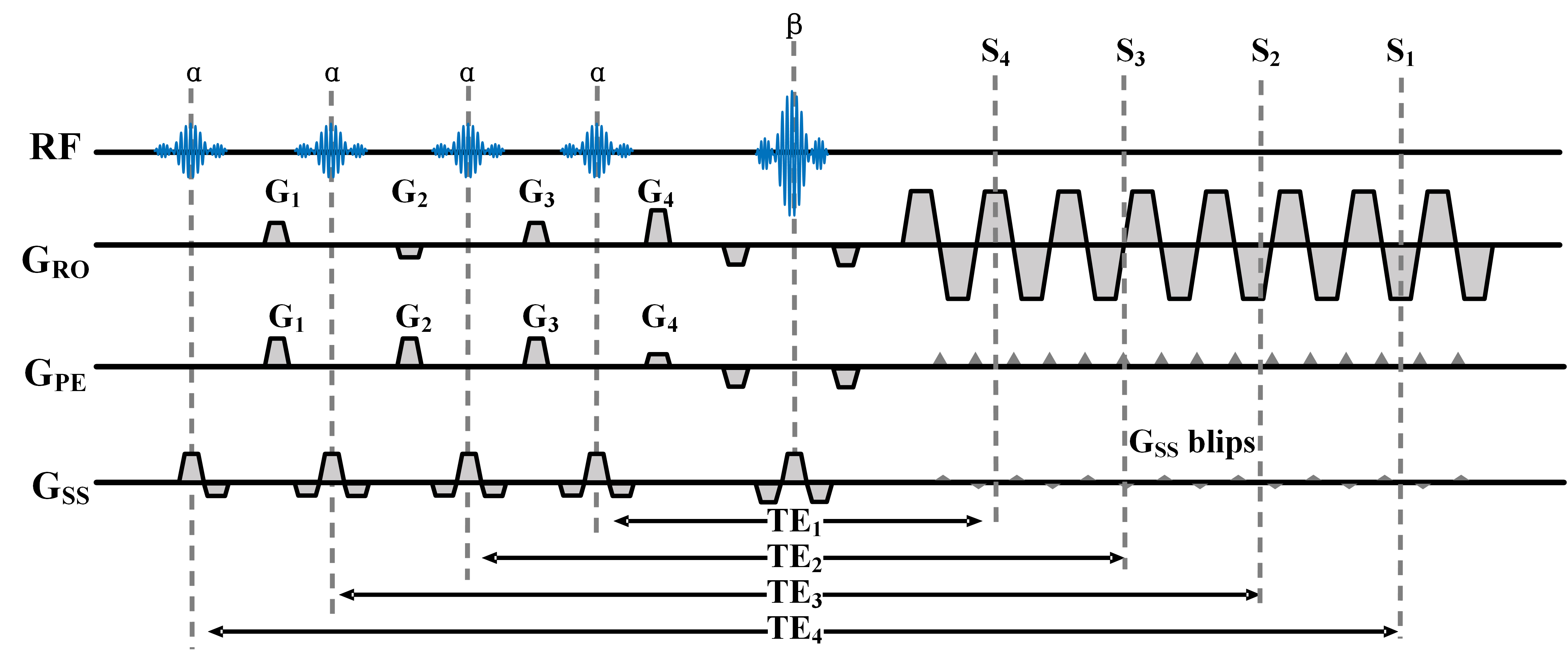
Figure 1 MB-MOLED T2 mapping sequence. The excitation pulse α and the refocusing pulse β are both multiband pulses. The pulse flip angles are α = 30° and β = 180°. Four echo-shifting gradients G1, G2, G3, and G4 are used to shift the four main echoes S1, S2, S3, S4 from the center of k-space. Gss blips are the blip gradients of the b-CAIPI.
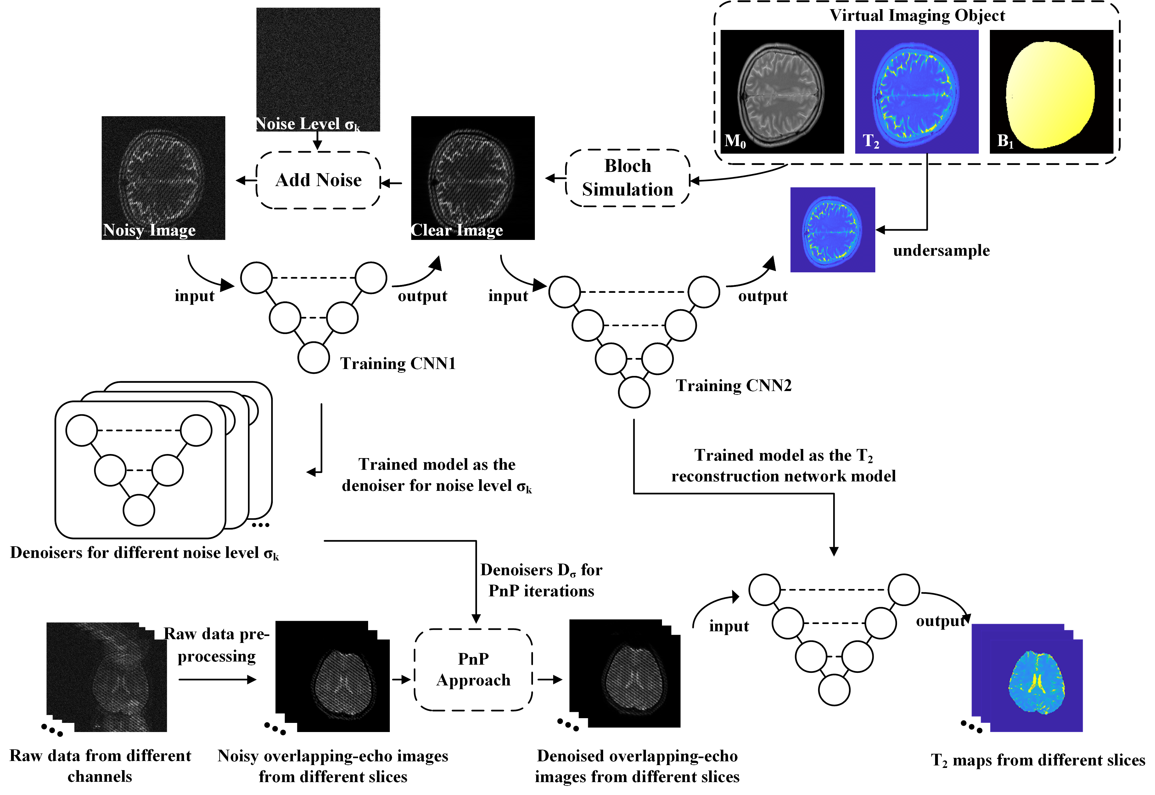
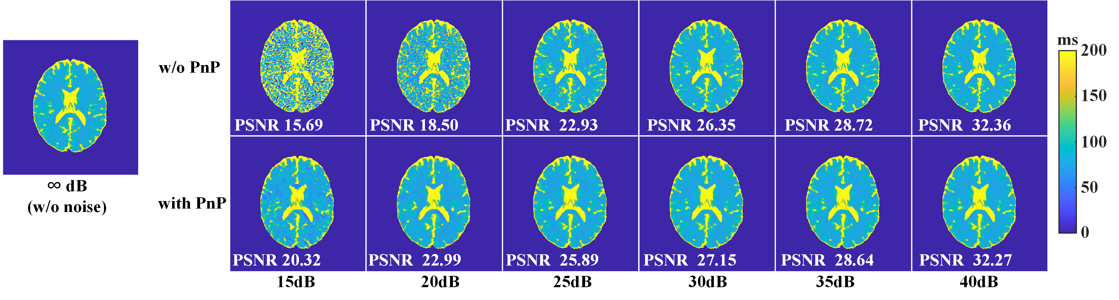
Figure 3 The reconstructed results of the numerical human brain.
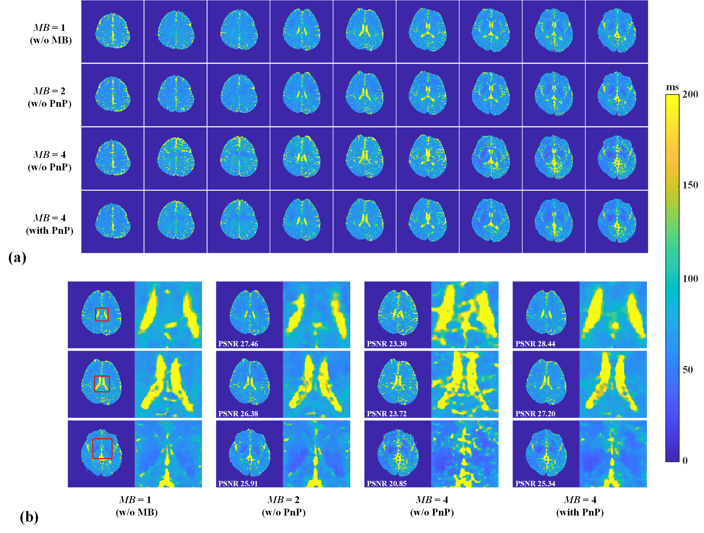
Figure 4 Reconstructed images of in vivo experiments. (a) MB = 1, MB = 2 without PnP algorithm, MB = 4 without PnP algorithm, and MB= 4 with PnP algorithm, respectively. (b) Several representative slices and enlarged ROIs.