2198
3D Extension of the multi parametric acquisition Multi-Phase balanced non-Steady State Free Precession
Riwaj Byanju1, Gyula Kotek1, Mika W. Vogel2, Juan A Hernandez-Tamames1, and Dirk H. J. Poot1
1Erasmus MC, Rotterdam, Netherlands, 2GE Healthcare, Hoevelaken, Netherlands
1Erasmus MC, Rotterdam, Netherlands, 2GE Healthcare, Hoevelaken, Netherlands
Synopsis
Keywords: Pulse Sequence Design, Quantitative Imaging
We propose a 3D extension of the novel MP-b-nSSFP sequence, which interleaves RF pulse types for Multiparametric mapping from the transient response. Comparing selective versus non-selective refocusing pulses we observe lower bias with non-selective pulses, despite modelling the spatially varying effect of the pulses in the fitting process. Phantom and in-vivo comparison to QRAPTEST (MAGIC) are performed.INTRODUCTION
Multi-parameter QMRI, such as MRF or MAGIC, can enhance the utility of MR imaging by providing better diagnostic capability through measuring tissue parameters. These acquisitions map tissue parameters and potentially system-specific parameters such as B0 and B1 inhomogeneity. Recently, Kotek1 proposed a novel multi-parameter mapping technique MP-b-nSSFP and demonstrated it with 2D imaging experiments. In this work a block of RF and gradient pulses is repeated and the transient magnetization response is expressed as an algebraic description that allowed in-silico optimization of acquisition settings for multi-parameter mapping experiments. In this work we aim to extend that novel MP-b-nSSFP method to 3D in a clinically acceptable scan time. We address the challenges for performing a 3D scan and compare its performance to MAGIC2.METHODS
A special feature of the MP-b-nSSFP acquisition is that it interleaves two types of RF pulses, a slice selective 30 degree pulse with high quality slice profile ($$$\alpha$$$) and a hard refocusing pulse, surrounded by crusher gradients, with substantially larger spatial coverage ($$$\gamma$$$).For the 3D version we used a similar selective pulse $$$\alpha$$$ (3.2 ms) for slab excitation and evaluated two version of the hard refocussing pulse $$$\gamma$$$, one with slab size similar to the slab size of the $$$\alpha$$$ pulse and the second without slab select gradient during the RF. Spatial encoding was performed by 3D stack of Spirals, with a readout duration of 20 ms and 32 ms between echoes. Image reconstruction was performed in two stages with a subspace constrained reconstruction to allow an acceleration factor of 5.33: First (effectively) a B0 field of 0 was assumed during the reconstruction and the parameters were quantified using dictionary interpolation3. Subsequently the resulting B0 map was unwrapped and used for a second pass of image reconstruction while compensating B0 induced phase evolution during the readout4 followed by the final fitting.
A separate dictionary was generated for each slice by full Bloch simulation of the sequence, using the RF and gradient waveforms as played out on the scanner and hence including the spatial variation in effect of the RF pulses across the slab.
Experiments: Scans were performed on a 1.5T GE MRI system with 8 channel head coil and IRB approval and informed consent were obtained for the volunteer study. Phantom acquisitions with the 3D MP-b-nSSFP method with both types of refocussing pulse and the commercial T1 and T2 mapping sequence MAGIC were performed with settings given in table 1. Additionally these settings were used for an in-vivo acquisition with the selective $$$\gamma$$$ pulse and MAGIC scan. The resulting maps are shown and for the phantom compared quantitatively through ROI analysis of the tubes with specified reference values.
RESULTS
Figure 1 shows the maps obtained by the different sequences. A substantial variation in the slice direction of the estimated T1 is observed in 1A. This is strongly reduced in 1B. T2 is not affected by the choice of $$$\gamma$$$ pulse.Figure 2 shows the ROI mean and standard deviation of each of the tubes in different slices for all acquisitions. For all acquisitions the values are somewhat below the reference values. For the selective $$$\gamma$$$ pulse the deviation is larger away from the central slice for T1.
Figure 3 shows the in-vivo T1, T2, ad B0 maps. Theres is no apparent trent in T1 values along the slice direction (S/I). The values of T1 and T2 are in the range reported for 1.5 T scanners.
DISCUSSION
In the fitting a separate dictionary is used for each slice. These dictionaries include the variation of the RF pulses in the slab encoding direction and hence would be expected to avoid bias in T1 and T2. However, the results clearly show that the T1 is biased with the selective $$$\gamma$$$ pulse and this can be avoided by making the $$$\gamma$$$ pulse non-selective. In 3D imaging the imaged volume is a much larger fraction of the total tissue volume inside the coils. Hence, out of slab signals are proportionally less of an issue in 3D than in 2D experiments. With the non-selective $$$\gamma$$$ pulse accuracy in T1 and T2 similar to the reference acquisition was obtained. The in-vivo scan is visually not affected as much by the T1 bias.CONCLUSION
We successfully extended the novel MP-b-nSSFP sequence to a 3D acquisition and are able to obtain in-vivo T1, T2, and B0 maps simultaneously.Acknowledgements
No acknowledgement found.References
- Kotek et al, From signal‑based to comprehensive magnetic resonance imaging, Sci Rep 2021, 11:17216, doi: 10.1038/s41598-021-96791-w
- Hagiwara et al. SyMRI of the brain: rapid quantification of relaxation rates and proton density, with synthetic MRI, automatic brain segmentation, and myelin measurement. Invest Radiol 2017;52:647–57. doi: 10.1097/RLI.0000000000000365
- Valenberg et al, An Efficient Method for Multi-Parameter Mapping in Quantitative MRI Using B-Spline Interpolation, TMI 2020, 38:1681, doi: 10.1109/TMI.2019.2954751
- Fessler et al, Toeplitz-Based Iterative Image Reconstruction for MRI With Correction for Magnetic Field Inhomogeneity, TSP 2005, 53:3393, doi: 10.1109/TSP.2005.853152
Figures
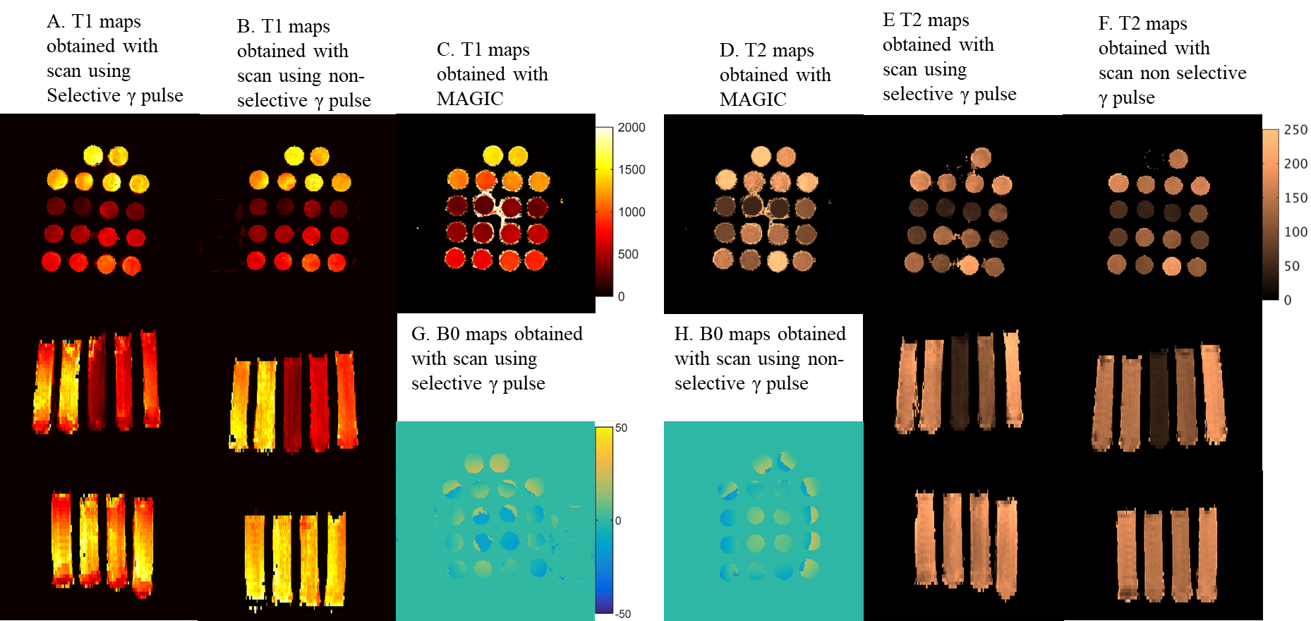
Figure 1: Maps of the T1, T2 in ms and B0 in Hz of the two versions of 3DMP-b-nSSFP and T1 and T2 maps of MAGIC
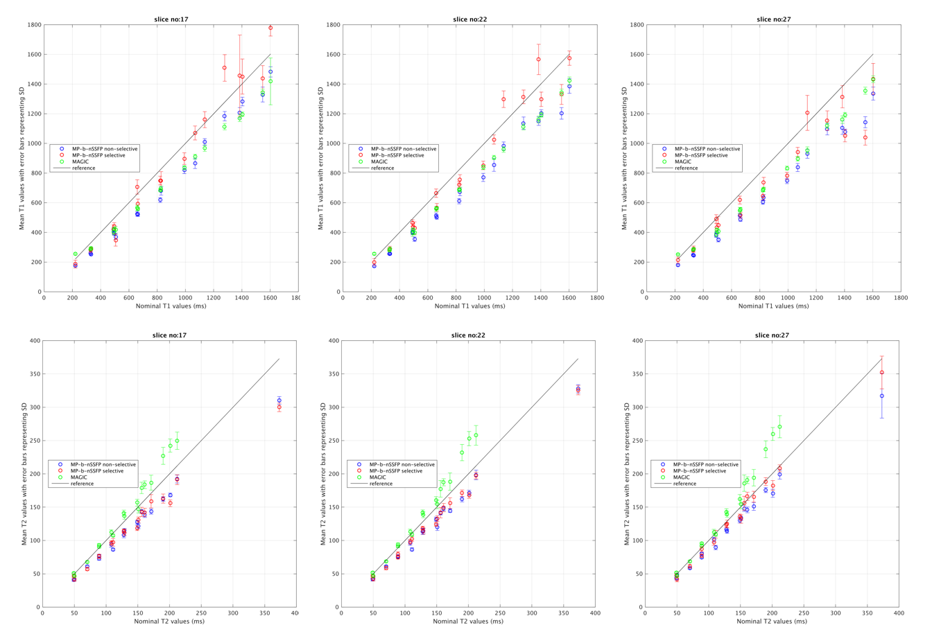
Figure2: ROI comparison of the different acquisitions. Comparison of T1 and T2 maps obtained with scan using Selective and non-Selective $$$\gamma$$$ pulse MP –b-nSSFP with MAGIC for three slices: two from opposite ends (slice17 and 27) and center (slice22)
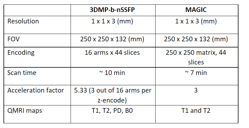
Table 1: Acquisition settings used for the phantom and in-vivo experiments. The 3DMP-b-nSSFP is acquired both with slab-selective as well as non-selective \gamma pulse in the phantom.
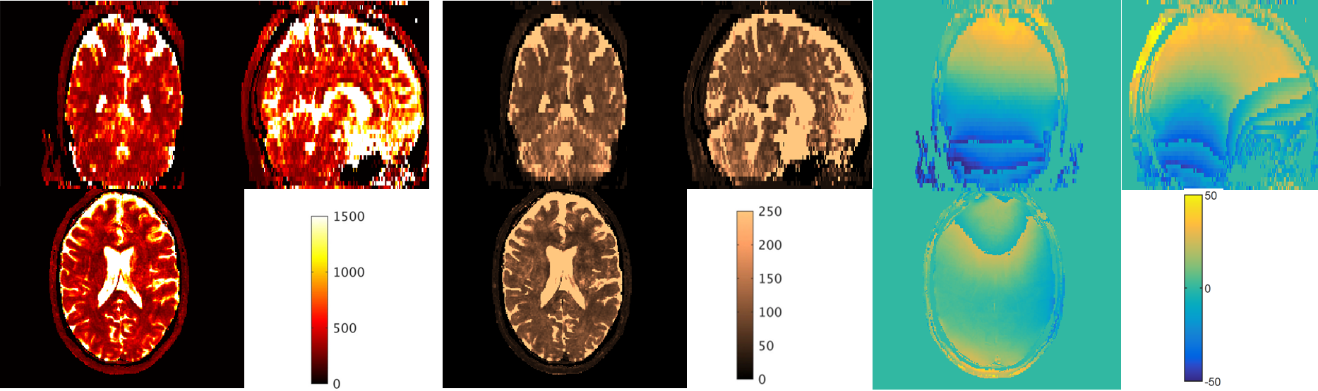
Figure 3: In-vivo maps of the 3D MP-b-nSSFP acquisition with slab selective $$$\gamma$$$ pulse.
DOI: https://doi.org/10.58530/2023/2198