2169
Three-dimensional black-blood T1 mapping of arterial plaque and venous thrombosis: a concept-proof study1School of Biomedical Engineering, Guangzhou Medical University, Guangzhou, China, 2The First People’s Hospital of Qinzhou, Qinzhou, China, 3Siemens Shenzhen Magnetic Resonance Ltd, Shenzhen, China
Synopsis
Keywords: Quantitative Imaging, Cardiovascular, T1 mapping
Quantitative T1 mapping have showed the potential to characterize arterial plaque and venous thrombotic components. However, owing to the interference of blood signals, it is a challenge for T1 mapping technique to combine with black-blood imaging, which may otherwise be a confounder for plaque and thrombus component. In this study, we present a black-blood T1 mapping technique based on DANTE black-blood preparation and MP2RAGE sequence. Experiment results demonstrated that BB-MP2RAGE provided accurate measurement of T1 relaxation time with effective blood suppression. The technique has the potential to be a quantitative tool for thrombosis and plaque characterization.Purpose
Quantitative T1 maps have shown the potential to identify arterial plaque and venous thrombotic components. The stage of intraplaque hemorrhage can be classified based on T1 relaxation time, which provided a basis for improved risk stratification of patients1,2. Effective treatment of deep vein thrombosis requires accurate assessment of thrombus distribution, and T1 relaxation time is a good predictor of successful thrombolysis3. In view of the potential impact of quantitative T1 mapping at high fields, Marques et al. proposed the Magnetization Prepared 2 Rapid Acquisition Gradient Echoes (MP2RAGE) sequence to achieve perfect sufficient insensitivity of transmit B1 field4. However, owing to the interference of blood signals, especially for slow or complex flow patterns at carotid bifurcation and cerebral venous sinus, it is a challenge for MP2RAGE to combine with blood suppression, which may otherwise be a confounder for plaque and thrombus component characterization. To address this issue, we developed a black-blood T1 mapping technique based on DANTE black-blood prepared MP2RAGE (BB-MP2RAGE) for quantitative evaluation of arterial plaque and venous thrombosis. The feasibility of the technique in vivo was then demonstrated in healthy volunteers as well as in patients with arterial plaque or deep vein thrombosis.Methods
MR Sequence Design and OptimizationThe BB-MP2RAGE is equivalent to an inversion recovery sequence, where two gradient echo images (INV1 and INV2 images) acquired at different inversion times are combined to obtain a synthetic image (UNI image). T1 map was estimated by interpolation using a signal model. The BB-MP2RAGE was optimized based on Bloch's equation, which should meet the following objectives: (i) the T1 map resulted from the BB-MP2RAGE should be as accurate as the respective T1 values gained with MP2RAGE; and (ii) ensuring sufficient flow signal suppression.
Phantom and In Vivo Study
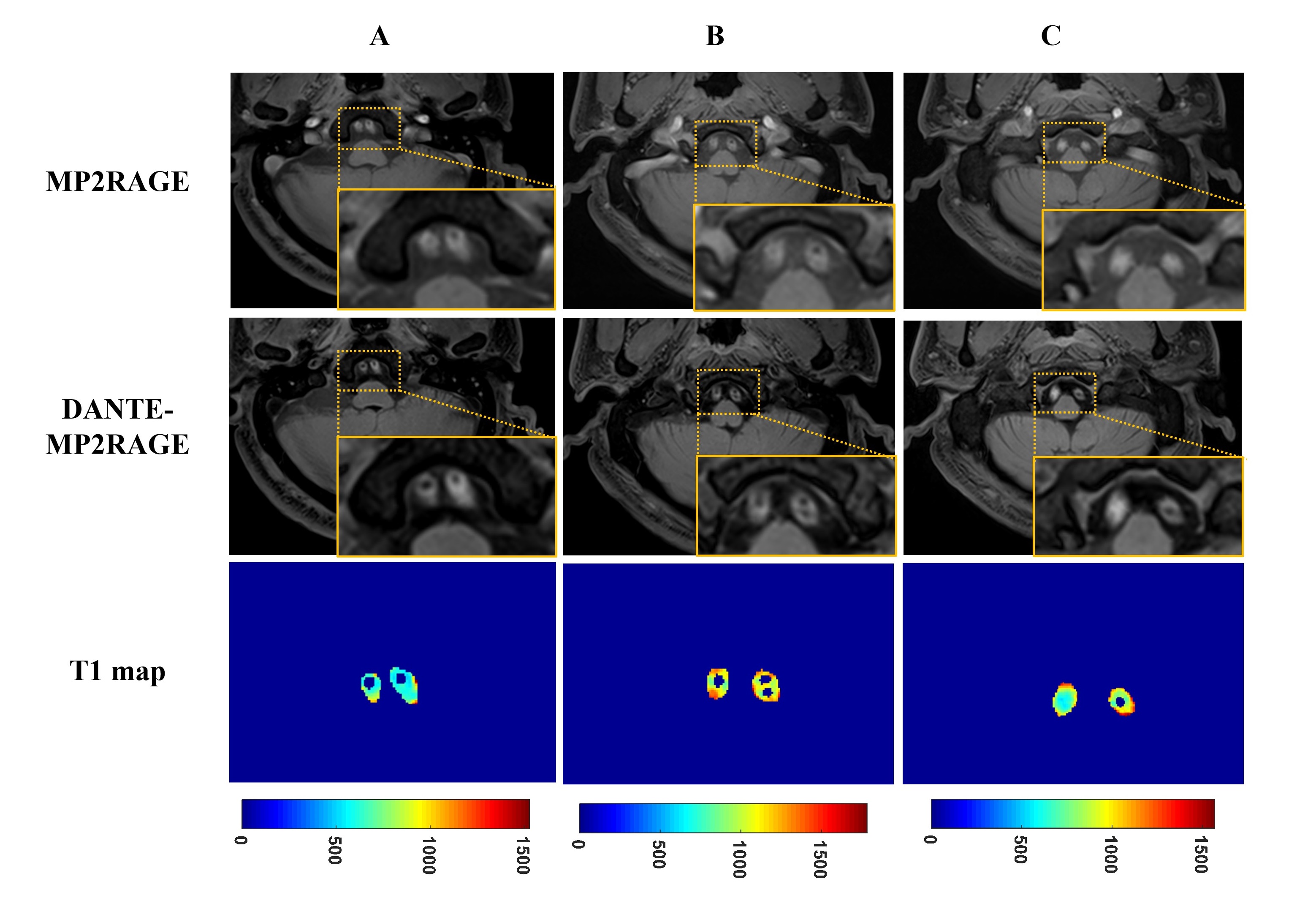
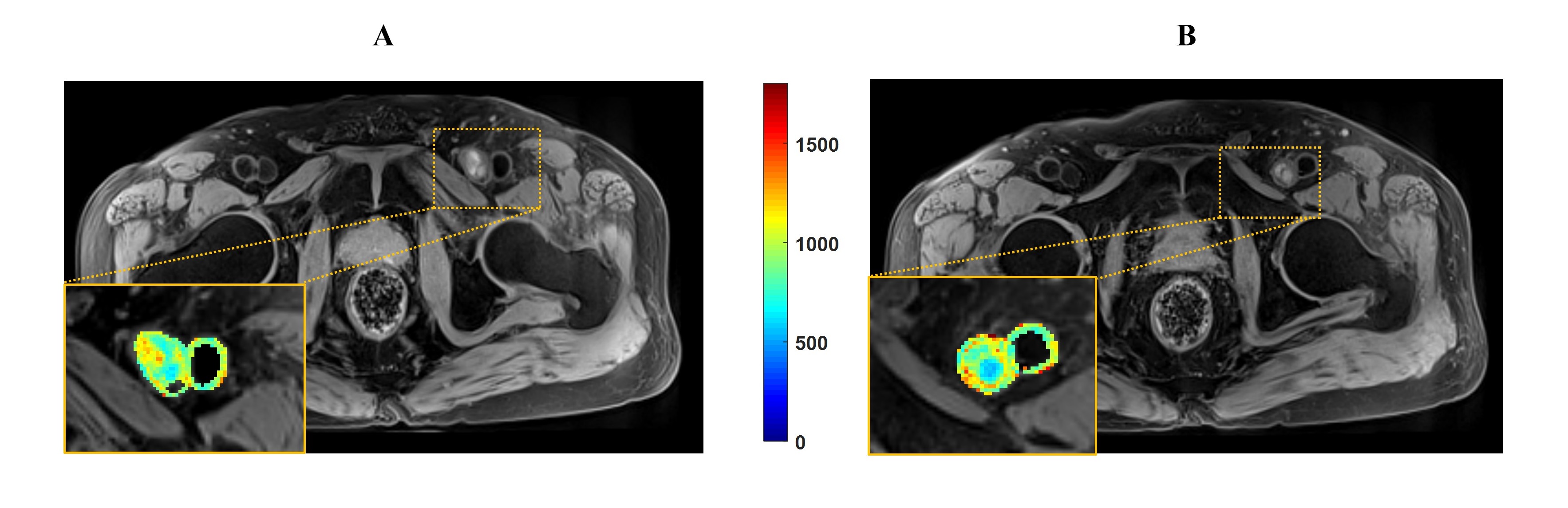
Quantitative T1 mapping was performed in a phantom to verify the accuracy of the proposed BB-MP2RAGE. In vivo head imaging experiments by BB-MP2RAGE and conventional MP2RAGE were performed at 3T Siemens scanner (Siemens AG, Germany) in 8 healthy volunteers. And BB-MP2RAGE were conducted for plaque and thrombus imaging on 3 patients (i.e., one patient with bilateral vertebral artery stenosis and two patients with subacute deep vein thrombosis).
Image Processing and Analysis
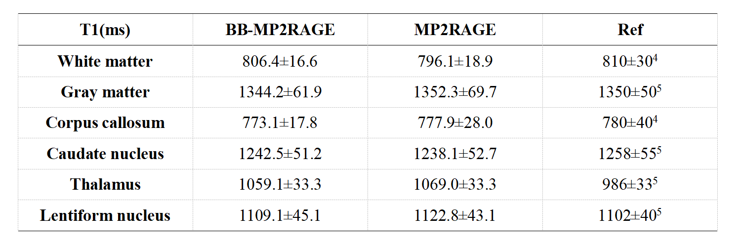
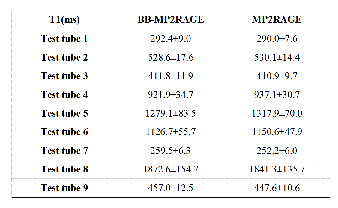
For phantom images by BB-MP2RAGE and conventional MP2RAGE, the regions of interest (ROIs) were drawn in different tubes to compare the T1 values. For human subjects, identical ROIs were drawn on two T1 maps using ITK-SNAP to obtain the mean T1 values for different brain tissues. The black-blood effect of BB-MP2RAGE was quantitatively assessed by signal-to-noise ratio (SNR) and contrast-to-noise ratio (CNR). For phantom results, the mean T1 values by BB-MP2RAGE were compared with MP2RAGE using Pearson correlation and Bland-Altman analysis. Student's t-tests were performed for two sets of in vivo data obtained by conventional and black-blood T1 mapping.
Results
Phantom and in vivo results showed excellent correlation of T1 maps between BB-MP2RAGE and conventional MP2RAGE (in vivo: white matter: 806.4±16.6ms vs. 796.1±18.9ms; gray matter: 1344.2±61.9ms vs. 1352.3±69.7ms; p=0.38, r2= 0.99) (Table 1 and Table 2). BB-MP2RAGE effectively suppressed the arterial and venous blood signals, and showed 172.6% improvement in the static tissue-to-blood CNR (250.5±66.6 vs. 91.9±35.9, p=0.0054). Patient data revealed the feasibility of BB-MP2RAGE for quantitative measurement of plaque components and venous thrombosis (Fig. 1 and Fig. 2).Discussion
In this study, a BB-MP2RAGE technique was developed for black-blood T1 mapping. The T1 values of phantom using BB-MP2RAGE agreed well with conventional MP2RAGE. In vivo studies showed sufficient blood suppression and robust T1 measurements in the brain ROIs. The preliminary patient study revealed that BB-MP2RAGE may contribute to the characterization of plaque or thrombus components. The presented BB-MP2RAGE protocol has several advantages. First, it provided robust slow flow suppression, nearly pure T1 weighting and high SNR. Also, the kept information of phase change facilitated more precise model simulations. Second, the proposed BB-MP2RAGE provided low SAR and minimizes the effect of B1 field, which suggests a potential for assessing the plaque components by high-field T1 mapping. Third, the proposed protocol avoided the effect of image co-registration while providing large anatomical coverage and isotropic resolution. And the use of the central view order and DANTE preparation did not increase the inversion time additionally.Conclusion
BB-MP2RAGE technique provided accurate measurement of T1 relaxation time with effective blood suppression. The sequence has the potential to be a quantitative tool for venous thrombosis and arterial plaque characterization.Acknowledgements
No acknowledgement found.References
1. Qiao H, Li D, Cao J, et al. Quantitative evaluation of carotid atherosclerotic vulnerable plaques using in vivo T1 mapping cardiovascular magnetic resonaonce: validation by histology. J Cardiovasc Magn Reson. 2020;22(1):38.
2. Qi H, Sun J, Qiao H, et al. Carotid Intraplaque Hemorrhage Imaging with Quantitative Vessel Wall T1 Mapping: Technical Development and Initial Experience. Radiology. 2018;287(1):276-284.
3. Saha P, Andia ME, Modarai B, et al. Magnetic Resonance T1 Relaxation Time of Venous Thrombus Is Determined by Iron Processing and Predicts Susceptibility to Lysis. Circulation. 2013, 128:729-736.
4. Marques JP, Kober T, Krueger G, van der Zwaag W, Van de Moortele PF, Gruetter R. MP2RAGE, a self bias-field corrected sequence for improved segmentation and T1-mapping at high field. NeuroImage. 2010;49(2):1271-1281.
5. Lu H, Nagae-Poetscher LM, Golay X, Lin D, Pomper M, van Zijl PCM. Routine clinical brain MRI sequences for use at 3.0 Tesla. J Magn Reson Imaging. 2005;22(1):13-22.
Figures