2147
The value of MRI 3D-DESS technique in preoperative evaluation of polydactyly in children1Xijing Hospital, Air Force Military Medical University, Xi 'an, China
Synopsis
Keywords: Joints, Pediatric
Polydactyly is very common disease in pediatric orthopeadic. In this study, 18 patients with polydactyly were included. All the 3D-DESS image were evaluated by two senior diagnostic radiologists using a double-blind 3-point scale, the intraoperative results were used as the “gold standard” to analyze the consistency of image results and intraoperative results. The purpose of this study is to assess whether the 3D-DESS sequence can provide the growth and development information of cartilage epiphysis, tendon and bone tissue of polydactyly in children before surgery, which has value for preoperative evaluation and preparation of surgical treatment.Introduction
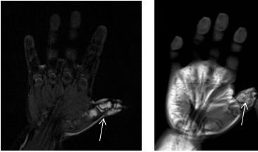
Polydactyly is very common disease.1 If the epiphysis is not properly handled during the operation for these children, it may lead to malformed growth (Figure 1).2 More importantly, children are too young at the time of surgery, and routine preoperative X-ray examination is radioactive and difficult to distinguish soft tissues such as epiphysis and tendons (Figure 2).3 In this study, the MRI 3D-DESS ( 3 Dimensions - Double Echo steady state) sequence was used here to evaluate the clinical value of this technique on preoperative evaluation and treatment plan formulation of polydactylyis. The consistency of image diagnosis and intraoperative results were also compared.Method
This on-going study was approved by the Ethics Committee, and informed consents were obtained from all participants. 18 children (mean age 1.5 years ; age ranging 8 month-2.5 years) were prospectively enrolled in this study, who were clinically diagnosed as polydactyly in the Department of Orthopaedics of our hospital from May 2021 to April 2022. MR scans were performed on a 1.5T MR system (AERA, Siemens, Germany). Commercially available scanning sequence 3D-DESS was used. Detailed scan parameters are as follow: field of view 120mm×120mm , slice thickness 0.8mm , matrix 256×190, TR 19.98ms, TE 7.42mm, receiving bandwidth 195KHz, number of averaging 1. Image quality was analyzed by two senior radiologists using a double-blinded 3-point scale, and the intraoperative results were used as the “gold standard” to analyze the consistency of image results and intraoperative results.Results
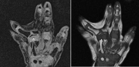
All the 18 children performed MRI scanning successfully. The epiphysis, tendon and bone tissue were clearly illustrated (Figure 3). All the image quality met the diagnostic requirements, of which the score of 16 cases (88.9%)was 3 point, with an average score of 2.6±0.3. ICC value of two doctors' assessments of image quality was 0.8579 ( 95% CI:0.7253-0.9658). The image results of all the cases were consistent with intraoperative findings.Conclusion
Non-invasive and radiation-free MR 3D-DESS technique can provide the growth and development information of cartilage epiphysis, tendon and bone tissue of polydactyly in children before surgery, which has high value for preoperative evaluation and preparation of surgical treatment.Discussion
With the development and popularization of magnetic resonance technology, the epiphysis can be clearly displayed, so as to provide an objective basis for the clinician's accurate diagnosis and the choice of surgical plan. The 3D-DESS technique has better SNR and resolution. In addition, this sequence is a three-dimensional thin-layer scan, which can be reconstructed in any orientation. The comprehensive effect of this technique is better than PD and T2 technique for epiphysis .4 Our study showed that the image diagnosis results using 3D-DESS technique are completely consistent with the intraoperative results. In addition,3D-DESS technique can provide doctors with soft tissue information such as cartilage epiphysis and muscle hyperplasia in preoperative evaluation of children with polydactyly.Our study also had several limitations. Due to the small number of enrolled cases in this study, the quantitative analysis of epiphysis was not performed,and further studies are needed to provide more detailed information for clinicians.
Summary of Main Findings
The 3D-DESS technique can provide the growth and development information of cartilage epiphysis, tendon and bone tissue of polydactyly in children before surgery, which has high value for preoperative evaluation and preparation of surgical treatment.Acknowledgements
No acknowledgement found.References
1. Braun T, Trost J,Pederson W.Syndactyly Release[J]. Seminars in Plastic Surgery, 2016, 30(04):162-170.DOI:10.1055/s-0036-15934782.
2. Liu Q, Lei Z,Hong L,et al. Preoperative percutaneous arthrography provides detailed information for treatment of Wassel type IV thumb duplication[J]. Journal of Plastic Reconstructive & Aesthetic Surgery, 2018, 71(12):1717-1722.DOI:10.1016/j.bjps.2018.08.0033.
3. Tian X,Chan P,Li A,et al. Analysis of Causes for Congenital Ulnar Deviated Thumbs at the Distal Phalanx Level in 157 Thumbs[J]. The Journal of Hand Surgery, 2019, 44(10):860-867.DOI:10.1016/j.jhsa.2019.04.0134.
4.Moriya S,Miki Y,Matsuno Y,et al. Three-dimensional double-echo steady-state (3D-DESS) magnetic resonance imaging of the knee: establishment of flip angles for evaluation of cartilage at 1.5 T and 3.0 T[J]. Acta Radiologica, 2012, 53(7):790-794.DOI:10.1258/ar.2012.110532
Figures