2138
Evaluation of Pediatric Musculoskeletal Disease using CT-like MR of Pointwise Encoding Time Reduction with radial Acquisition sequence1Hunan Children's Hospital, Changsha, China, 2MR Scientific Marketing, Siemens Healthineers, Wuhan, China
Synopsis
Keywords: Body, Bone
Cortical bone can be clearly displayed by CT-like MRI using ultrashort TE sequence. This study aimed to investigate the value of CT-like MRI using PETRA (ct-PETRA) sequence in the pediatric musculoskeletal diseases. Our results showed that ct-PETRA had no significant difference in measuring the C-E index and acetabulum index of children's hip joint compared with X-ray, and had no significant difference in visualization of lesions compared with X-ray and CT. Moreover, ct-PETRA has more advantages than X-ray in displaying trabecular fracture of fine bone. CT-like PETRA can be used as conventional complementary sequence for some assessments of pediatric musculoskeletal diseases.Abstract
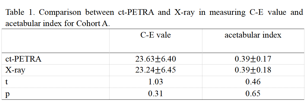
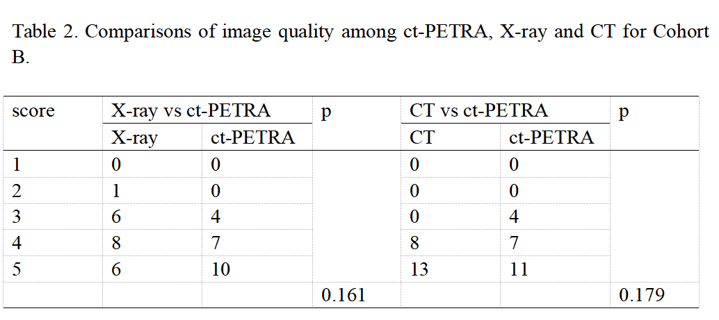
Introduction The diagnosis of musculoskeletal system diseases often require comprehensive imaging that contains soft tissues and bone structures. Frequently, multiple cross-sectional examinations, including X-ray plain film, computed tomography (CT) and magnetic resonance (MR) imaging, are needed to assess soft-tissue stabilizers and osseous support of musculoskeletal system diseases for diagnosis or surgical planning. Pediatric patients often require evaluation of joints development, as well as multiple reexaminations after treatment of trauma, tumor and tumor-like lesions. Pediatric patients are more sensitive to ionizing radiation, which exists in X-ray and CT imaging. Traditional MR imaging pulse sequences provide superior soft tissue contrast, but depict cortical bone as a signal void because of bone’s inherent short T2 relaxation time [1-4]. As we know, tissues and tissue components with ultrashort-T2 relaxation time of less than approximately 1 ms can’t be reliably detected by conventional MRI pulse sequences due to limitations on the minimum achievable echo time (TE) [5-7]. Pointwise encoding time reduction with radial acquisition (PETRA) MR sequence is a kind of ultrashort echo time (UTE) pulse sequences, which used specialized acquisition and reconstruction techniques to enable detection of ultrashort-T2 components in vivo. Moreover, CT-like MRI using UTE technique has also been investigate in the bone [8-10]. In this study, we aimed to evaluate the feasibility of CT-like PETRA in the pediatric musculoskeletal disease trough comparisons with X-ray and CT.Method 63 pediatric patients with musculoskeletal system diseases were recruited from November 2021 through November 2022. All patients underwent conventional MR and PETRA examination on a 3T MRI scanner (MAGNETOM Prisma; Siemens Healthcare, Erlangen, German). The imaging parameters of PETRA were as follows: TR 3.32ms,TE0.07ms,FOV300mm,thickness0.94mm,FOV phase 100%,basae resolution 320, radial views 60000, edge enhancement 3, smoothing 3. After PETRA acquisition, the CT-like PETRA (ct-PETRA) image was calculated according to the method in the study of Breighner et. al [11]. All pediatric patients were divided into two cohorts. Cohort A was patients with hip joint dysplasia, such as developmental dysplasia of the hip, acetabular dysplasia, and so on. Cohort B was patients with pathological lesion, which was divided two subgroups: Cohort B1, ct -PETRA to conventional X-ray; Cohort B2, ct-PETRA to conventional CT. For cohort A, C-E angle and acetabular index was used to assess the development of children's hip joint between ct-PETRA and X-ray. For the qualitative evaluation of image quality, the overall pathological diagnostic acceptability of ct-PETRA, CT and X-ray was scored by using a five-point scale (from 1 nondiagnostic to 5 outstanding). Statistical analyses were performed using SPSS 25.0 version . Image quality score between X-ray, CT and ct-PETRA were compared using the wilcoxon test, and p<0.05 was considered statistically significant.
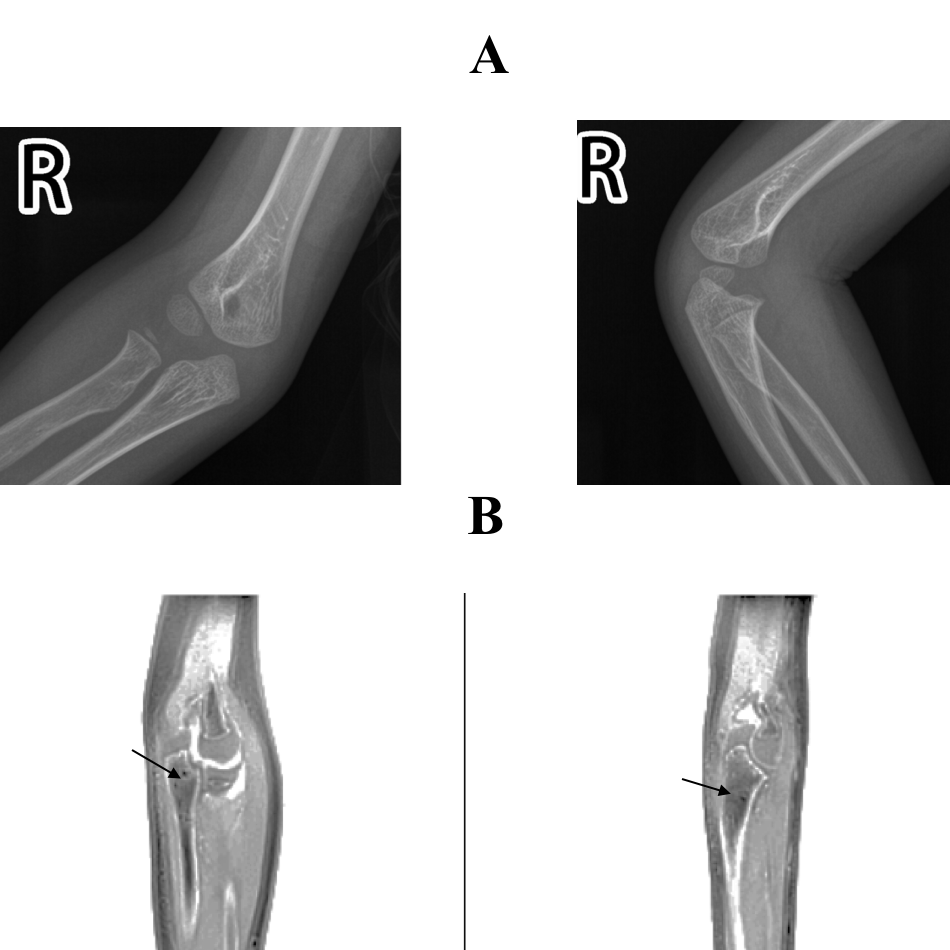
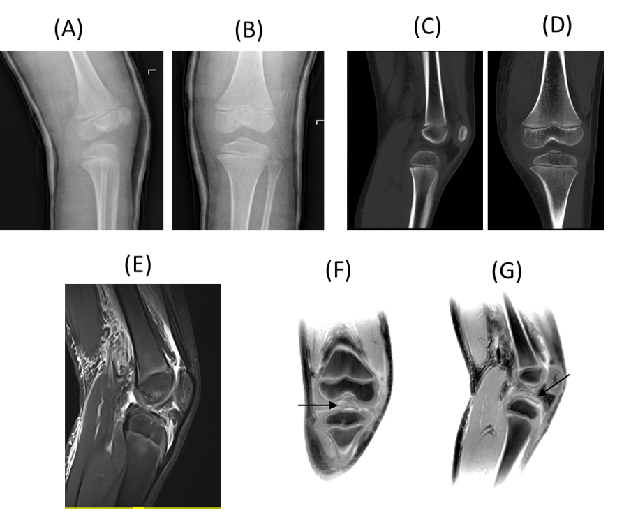
Result In total, we identified 42 cases for cohort A, 21 cases for cohort B. For the cohort A, there were no significant differences in C-E angle and acetabular index between ct-PETRA and X-ray (Table 1.) . For cohort 2, there was no significant difference in the structural display and diagnosis acceptability of lesions between X-ray and ct-PETRA, and between ct-PETRA and CT (Table 2). However, ct-PETRA was superior to X-ray in displaying bone trabecular fracture (Figure 1), and superior to CT in detecting subtle cortical erosions (Figure 2). Lesions delineations were rated diagnostic or better (score of 3,4 or 5) for ct-PETRA, as shown in Figure 1.
Discussion Our results confirmed that CT-like PETRA images provided accurate imaging of bone morphology. Ct-PETRA images could clearly display children's bone and bone cortex, which was similar to traditional X-ray and CT images. The display of bone mass, fracture of bone cortex, callus formation and periosteal reaction had no difference from that in X-ray and CT images, and basically met the diagnosis requirements. For patients with special fractures (e.g. trabecular fracture), traditional X-ray images often couldn’t detect the lesions, but ct-PETRA could clearly show. In X-ray, avulsion fracture may be missed due to anatomical position, and free bone may be covered by normal anatomical structure, while they can be clearly and accurately detected by ct-PETRA. It is difficult to discern subtle cortical erosion with CT, but ct-PETRA can provide greater conspicuity. Furthermore, positive contrast for bone provided by ct-PETRA facilitates more intuitive interpretation of images by surgeons and clinicians. ct-PETRA also has some limitations. First, ct-PETRA MR imaging inherently has lower spatial resolution compared with conventional CT and X-ray, resulting poor display in anatomical structures with thinner cortices. Besides, some artifacts were present for patients with motion due to long scan time.
Conclusion CT-like PETRA imaging can provide additional morphologic information comparable to CT and X-ray, and may replace CT imaging for some assessments of the pediatric musculoskeletal diseases.
Acknowledgements
The authors thanks ZHT of the MR Scientific Marketing, Siemens Healthineers for data analysis.References
1. Reichert, IL, Robson, MD, Gatehouse, PD, et al. Magnetic resonance imaging of cortical bone with ultrashort TE pulse sequences. MAGN RESON IMAGING. 2005; 23 (5): 611-8. doi: 10.1016/j.mri.2005.02.017.
2. Du, J, Hermida, JC, Diaz, E, et al. Assessment of cortical bone with clinical and ultrashort echo time sequences. MAGNET RESON MED. 2012; 70 (3): 697-704. doi: 10.1002/mrm.24497
3. Larson, PE, Han, M, Krug, R, et al. Ultrashort echo time and zero echo time MRI at 7T. MAGN RESON MATER PHY. 2015; 29 (3): 359-70. doi: 10.1007/s10334-015-0509-0
4. Chang, E, Du, J, Statum, S, et al. Quantitative bi-component T2* analysis of histologically normal Achilles tendons Muscles Ligaments Tendons J. 2019; 05 (02): 58. doi: 10.32098/mltj.02.2015.02.
5. Sneag, DB, Shah, P, Koff, MF, et al. Quantitative Ultrashort Echo Time Magnetic Resonance Imaging Evaluation of Postoperative Menisci: a Pilot Study. HSS J. 2014; 11 (2): 123-9. doi: 10.1007/s11420-014-9420-x.
6. Jang, H, Ma, Y, Carl, M, et al. Feasibility of an Inversion Recovery-Prepared Fat-Saturated Zero Echo Time Sequence for High Contrast Imaging of the Osteochondral Junction. Front Endocrinol (Lausanne). 2021; 12 777080. doi: 10.3389/fendo.2021.777080.
7. Chong, LR, Lee, K, Sim, FY. 3D MRI with CT-like bone contrast - An overview of current approaches and practical clinical implementation. EUR J RADIOL. 2021; 143 109915. doi: 10.1016/j.ejrad.2021.109915.
8. Sgard, B, Khalifé, M, Bouchut, A, et al. ZTE MR-based attenuation correction in brain FDG-PET/MR: performance in patients with cognitive impairment. EUR RADIOL. 2019; 30 (3): 1770-1779. doi: 10.1007/s00330-019-06514-z.
9. Xu, Y, Shi, L, Li, N, et al. Value of zero echo time MR imaging and CT in diagnosis of bone destructions of bone tumors and tumor-like lesions Chin J Acad Radiol. 2020; 3 (2): 108-114. doi: 10.1007/s42058-020-00035-1.
10. Leynes, AP, Yang, J, Wiesinger, F, et al. Zero-Echo-Time and Dixon Deep Pseudo-CT (ZeDD CT): Direct Generation of Pseudo-CT Images for Pelvic PET/MRI Attenuation Correction Using Deep Convolutional Neural Networks with Multiparametric MRI. J NUCL MED. 2017; 59 (5): 852-858. doi: 10.2967/jnumed.117.198051.
11. Breighner RE, Endo Y, Konin GP, et al. Zero Echo Time Imaging of the Shoulder: Enhanced Osseous Detail by Using MR Imaging. Radiology. 2018 Mar;286(3):960-966.
Figures