2112
Following evolving pH changes in the penumbra using a novel MRI method1Advanced Imaging Research Center, University of Texas Southwestern Medical Center, Dallas, TX, United States, 2Brain repair and Rehabilitation, Institute of Neurology, Dallas, TX, United States, 3University College of London, Center for Advanced Biomedical Imaging, London, United Kingdom, 4Wellcome Centre for Integrative Neuroimaging, FMRIB, Nuffield Department of Clinical Neurosciences, University of Oxford, London, United Kingdom, 5A. I. Virtanen Institute for Molecular Sciences, University of Eastern Finland, Kuopio, Finland, Kuopio, Finland, 6Institute of Neurology, Brain repair and rehabilitation, London, United Kingdom, 7Department of Medical Physics & Bioengineering, University College London, UK, London, United Kingdom, 8Brain repair and rehabilitation, Institute of Neurology, London, United Kingdom
Synopsis
Keywords: Stroke, Ischemia, ischemic penumbra
Here, our goal was to visualise dynamic pH changes in a rat model of MCAO and their relation to other imaging techniques for identifying the penumbra zone which is used to guide therapeutic intervention.Introduction
Ischemic tissue acidosis which is represented by the accumulation of lactic acid in hypoperfused brain tissue activates specific ion channels causing cellular neurotoxic calcium accumulation and cytotoxic edema which leads to the loss of the ischemic penumbra. Measurements of the exchange rates of amide protons of mobile proteins and peptides was shown to detect changes in pH non-invasively (1,2). Here, our goal was to visualise dynamic pH changes in a rat model of MCAO and their relation to other imaging techniques for identifying the penumbra zone which is used to guide therapeutic intervention.Methods
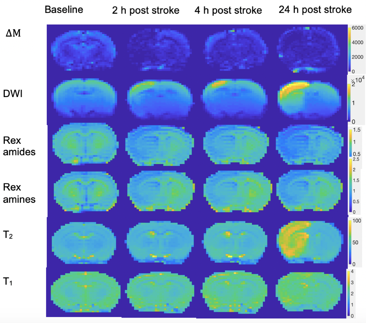
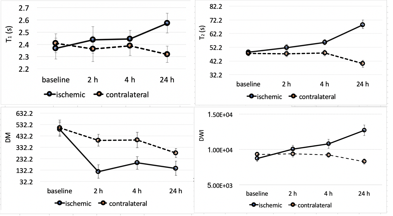
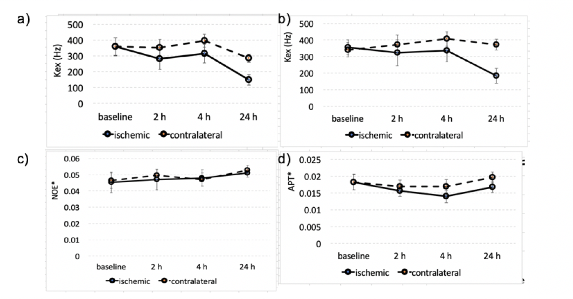
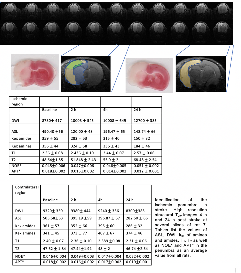
We measured the exchange rates of amide and amine protons using PRO-QUEST (3) which are directly related with tissue pH as well as perfusion and diffusion for delineating the ischemic penumbra based on the established PWI/DWI mismatch. Additional CEST metrics such as APT*, NOE*, MTRasymmetry and the area under the curve of the Z-spectrum were used as complementary indices of metabolic status in stroke. We included 19 rats which underwent stroke and scanned at several timepoints (i.e. baseline, 2h, 4h, 24h) at the level of the penumbra. Dataset from 8 rats which completed the 24 h timepoint are included in the study. Additional 3 rats were scanned in the core region for assessing the validity of our measurements with literature. We then compared multiparametric maps using statistical analyses to assess significant differences in various parameters in the ischemic tissue vs the contralateral and the healthy brain.Results
In stroke, significant changes in tissue pH were measured and found to precede DWI lesion as compared with healthy brain tissue. pH signal was found to drop within the first 2 hours and then recovered following reperfusion. DWI/PWI mismatch overestimates the final infarct region which is identified more precisely in pH maps due to impaired metabolism and energy loss during stroke.Conclusion
Identification of ischemic penumbra using PWI/DWI mismatch is challenging because of the inclusion of regions with benign oligemia. In turn, pH deficits predict the final infarct and describe active compensatory mechanisms after cerebral ischemia more accurately.Acknowledgements
No acknowledgement found.References
1. Sun, P.Z., Zhou, J., Sun, W., Huang, J., and van Zijl, P.C.M. (2007). Detection of the ischemic penumbra using pH-weighted MRI. Journal of Cerebral Blood Flow and Metabolism: Official Journal of the International Society of Cerebral Blood Flow and Metabolism 27, 1129–1136.2.
2.Leigh, R., Knutsson, L., Zhou, J., and van Zijl, P.C. (2018). Imaging the physiological evolution of the ischemic penumbra in acute ischemic stroke. J. Cereb. Blood Flow Metab. 38, 1500–15163.
3.Demetriou, E., Tachrount, M., Zaiss, M., Shmueli, K., and Golay, X. (2018). PRO-QUEST: a rapid assessment method based on progressive saturation for quantifying exchange rates using saturation times in CEST. Magn Reson Med.
Figures