2110
Comparison of 7T MRA and DSA in assessment of the Suzuki staging system for Moyamoya disease
Qi Duan1, Jinhao Lyu1, Caohui Duan1, Xiangbing Bian1, Jianxun Qu2, and Xin Lou1
1Chinese PLA General Hospital, Beijing, China, 2MR Collaboration, Siemens Healthineers Ltd., Beijing, China
1Chinese PLA General Hospital, Beijing, China, 2MR Collaboration, Siemens Healthineers Ltd., Beijing, China
Synopsis
Keywords: Stroke, High-Field MRI, Suzuki staging system
Moyamoya disease (MMD) is an uncommon chronic cerebrovascular disease and a leading cause of stroke in pediatric and young patients. Suzuki staging system based on conventional digital subtraction angiography (DSA) is a useful and important index in clinical. However, DSA is invasive and unnecessary in some situations. 7T TOF-MRA enables the visualization of the "puff of smoke" collateral network in MMD compared to 1.5T and 3T. We tried to grade the Suzuki staging system by using 7T TOF-MRA in MMD patients. Our results indicated that 7T TOF-MRA showed excellent performance as DSA on grading the Suzuki staging system in MMD.BACKGROUND AND PURPOSE
Moyamoya disease (MMD) is an uncommon chronic cerebrovascular disease with progressive steno-occlusion of the internal carotid arteries and "puff of smoke" collateral network compensatory1. MMD is a leading cause of stroke in pediatric and young patients2. Suzuki staging system based on conventional digital subtraction angiography (DSA) is considered the gold standard for diagnosing and assessing MMD. However, DSA is an invasive procedure and is always not feasible for pediatric patients. And DSA is unnecessary when internal carotid arteries occlusion and Moyamoya vessels are founded by Magnetic resonance angiography (MRA). Time-of-flight MRA (TOF-MRA) is widely accepted as a noninvasive diagnostic modality for evaluating moyamoya disease (MMD). Compared to 1.5T and 3T MRA, ultra-high-field strengths 7T TOF-MRA enables the visualization of the smaller distal intracranial vessels, even the "puff of smoke" collateral network in MMD3. To our knowledge, no other study has focused on comparing 7T TOF-MRA and DSA in diagnosing MMD. Therefore, the aim of our study was to systematically compare 7T TOF-MRA images and DSA for Suzuki staging system grading.METHODS
7T TOF-MRA data and Digital subtraction angiography (DSA) from each patient were collected. 7T TOF-MRA acquisition by 7T whole-body MR scanner (MAGNETOM Terra, Siemens Healthcare, Erlangen, Germany) equipped with an 8-channel transmitting and 32-channel receiving head coil. 7T TOF-MRA acquisition time of approximately 4 minutes 52 seconds, repetition time = 23.0 ms, echo time = 3.42 ms, field of view = 180 × 200 mm2, slice thickness = 0.5 mm, voxel size = 0.3 × 0.3 × 0.5 mm3, flip angle = 24°, bandwidth = 187 Hz/Px, number of slices = 136. Grading the Suzuki staging system of each symptomatic hemisphere by using 7T TOF-MRA and DSA. (Figure 1). The Suzuki staging system of each symptomatic hemisphere was assessed by two experienced radiologists with blinded clinical information independently. The diagnostic consistency and interobserver agreement for the Suzuki staging on the 7T TOF-MRA and DSA were assessed by using the Weighted Kappa value respectively. The correlation of Suzuki staging system grades on 7T TOF-MRA and DSA was tested by Spearman's correlation.RESULTS
Fifteen MMD patients with 28 symptomatic hemispheres were enrolled (mean age, 45.27 years ±11.42, 9 men). (Figure 2). The Weighted Kappa was 0.796 (95%CI, 0.669-0.923) for the results of the assessment Suzuki score between 7T TOF-MRA and DSA. The value 0.806 (95%CI, 0.6309-0.982) of Weighted Kappa was excellent for the Suzuki assessment using 7T TOF-MRA images between two readers. The result of Spearman’s correlation showed that the correlation of Suzuki staging system grading on 7T TOF-MRA and DSA was strong (rs=0.894, p<0.001).DISCUSION AND CONCLUSION
7T TOF-MRA enables the visualization of the "puff of smoke" collateral network for MMD patients in a noninvasive way. Our findings indicated that 7T MRA was as good as DSA for grading the Suzuki staging system of MMD patients.Acknowledgements
This work was supported by the National Natural Science Foundation of China (Nos. 81825012, 81730048 and 82151309 to Xin Lou; Nos. 81901708 to Jinhao Lyu).References
1. Kim JS. Moyamoya Disease: Epidemiology, Clinical Features, and Diagnosis. J Stroke. 2016 Jan;18(1):2-11.
2. Park EK, Lee YH, Shim KW, Choi JU, Kim DS. Natural history and progression factors of unilateral moyamoya disease in pediatric patients. Childs Nerv Syst. 2011 Aug;27(8):1281-7.
3. Su J, Ni W, Yang B, Xiao W, Gao X, Yang H, Li Y, Lei Y, Jiang H, Wang H, Gu Y, Mao Y. Preliminary Study on the Application of Ultrahigh Field Magnetic Resonance in Moyamoya Disease. Oxid Med Cell Longev. 2021 Jan 13;2021:5653948.
Figures
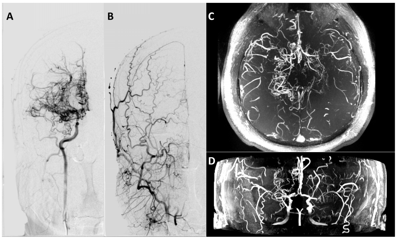
(A) and (B) shows the Right digital subtraction angiography (DSA) of a 40 years old man, the Suzuki staging system was 4. (C) and (D) shows the 7T TOF-MRA of the same patient, "puff of smoke" collateral network start to reducing and extracranial collateral network start to building, the the Suzuki staging system was 4.
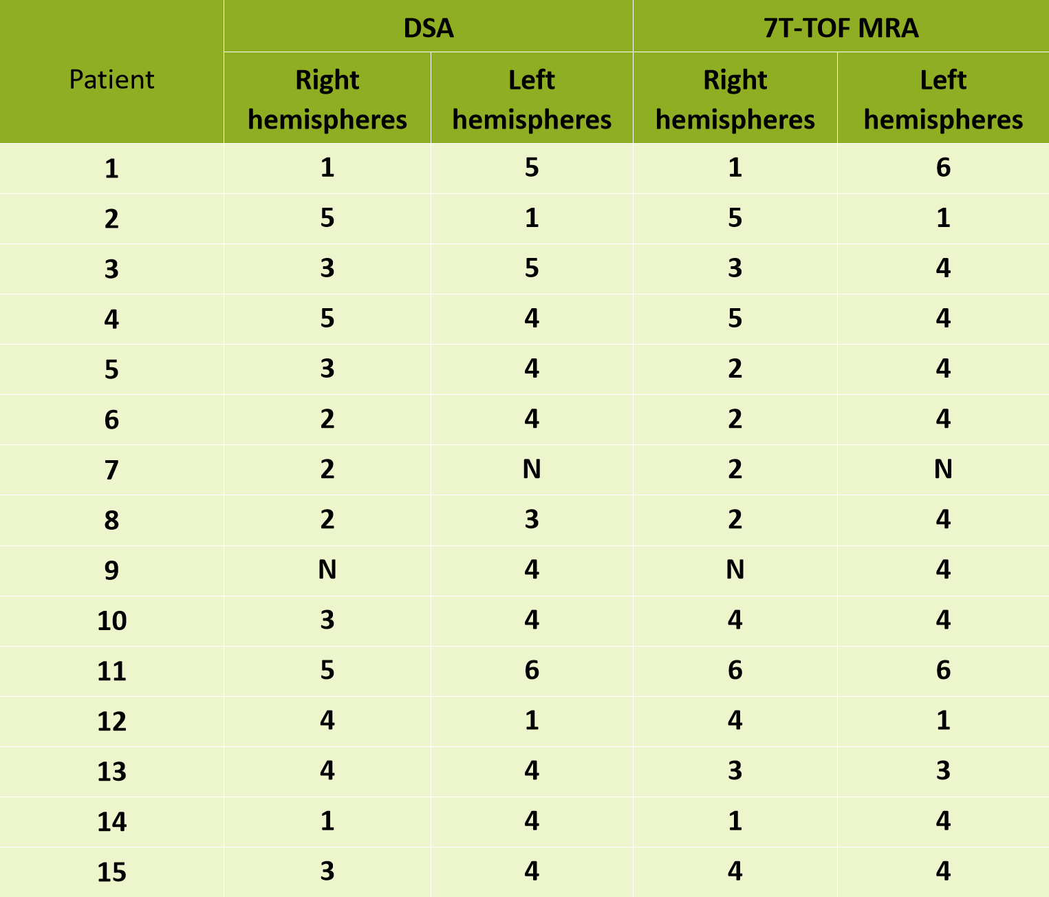
Grades of Suzuki staging system for each symptomatic hemispheres.
DOI: https://doi.org/10.58530/2023/2110