2107
Identification of intracranial atherosclerotic plaque features associated with recurrent stroke: a multi-modality imaging study
Lingling Wang1, Beibei Sun1, Maysam Orouskhani2, Mahmud Mossa-Basha2, Jianrong Xu1, Yan Zhou1, Chengcheng Zhu2, and Huilin Zhao1
1Radiology, Renji Hospital, Shanghai Jiao Tong University School of Medicine, Shanghai, China, 2University of Washington, Seattle, United States, Seattle, WA, United States
1Radiology, Renji Hospital, Shanghai Jiao Tong University School of Medicine, Shanghai, China, 2University of Washington, Seattle, United States, Seattle, WA, United States
Synopsis
Keywords: Stroke, Atherosclerosis, Calcification
Intracranial atherosclerotic plaque is a major cause of stroke. Both CTA and vessel wall magnetic resonance imaging (VW-MRI) can identify high risk plaque features. We aim to investigate the intracranial atherosclerotic plaque features associated with recurrent stroke using a multi-modality imaging approach by combining CTA and whole brain VW-MR. We found that higher calcium burden quantified by volume and Agatston score of intracranial arteries, as well as whole-brain plaque number and culprit plaque burden were independently associated with recurrent acute stroke.Background and Purpose
Background and Purpose: Intracranial atherosclerotic plaque is a major cause of stroke. Both computed tomography angiography (CTA) and vessel wall magnetic resonance imaging (VW-MRI) can identify high risk plaque features. However, the two imaging modalities have rarely been used together. We aim to investigate the intracranial atherosclerotic plaque features associated with recurrent stroke using a multi-modality imaging approach by combining CTA and whole brain VW-MRI.Methods
Methods: Eighty-nine patients with acute ischemic stroke caused by intracranial plaque were included, and they had CTA and VW-MRI within 1 month of stroke. Participants were divided into two group: first-time acute stroke (n=50) and recurrent acute stroke (n=39). Calcium burden was quantified semiautomatically by calculating total spotty calcium number, whole-brain Agatston scores and calcium volumes on thin-section unenhanced CT images (resolution 0.5x0.4x0.4mm3). Meanwhile, whole-brain plaque number, degree of culprit plaque luminal stenosis, culprit plaque burden, intraplaque hemorrhage were assessed on VW-MRI sequences (0.6mm isotropic resolution). Those characteristics were compared between two groups. Multivariate regression analysis was performed to assess the correlation between plaque features and recurrent acute stroke.Results
Results: A total of 89 participants (age 61.3±8.7 years, 54[60.7%] male) were included in this study. More spotty calcium and whole-brain plaques were found in recurrent stroke patients than first-time stroke patients (p=0.041 and P=0.017). Recurrent stroke patients had larger calcium volume (p=0.031), higher calcium score (p=0.008) and greater culprit plaque burden (p=0.002). After adjustment of clinical demographic factors, in multivariate analysis, calcium volume (odds ratio, OR=7.787; p=0.044), calcium score (OR=1.421; p=0.035), whole-brain plaque number (OR=1.378; p=0.032) and culprit plaque burden (OR=3.087; P=0.002) were all independently associated with recurrent acute stroke compared to the first-time acute stroke. The AUC of MRI was 0.745 and CT was 0.723, and the combination of both CT and MRI achieved AUC of 0.803.Conclusions
Conclusions: Higher calcium burden quantified by volume and Agatston score of intracranial arteries, as well as whole-brain plaque number and culprit plaque burden were independently associated with recurrent acute stroke. The use of both CTA and VW-MRI achieved better performance of differentiating high risk intracranial plaque than a single imaging modality.Acknowledgements
Not applicable.References
[1] Fan Zhang et al. Stroke. 2019 Apr;50(4):859-866.
[2] Sung Soo Ahn et al. Radiology. 2013 Sep;268(3):842-9
[3] Philip J Homburg et al. Stroke. 2011; 42:1244-1250
Figures
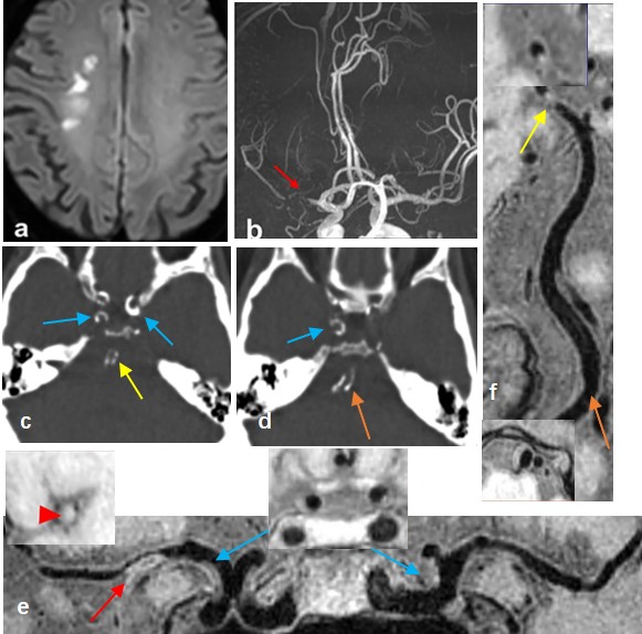
A case presented with recurrent acute stroke.
(a) DWI detects high-signal-intensity lesions in the right corona radiata. (b) TOF-MRA
shows severe stenosis on the M1 segment of right middle cerebral artery (MCA) (red
arrow). (c-d) CT images show diffuse calcification and spotty calcium in the bilateral
C4-C7 segments of ICA, right vertebral artery (VA). (e–f) Curved planar
reconstruction images from T1-VISTA vessel wall imaging show multiple plaques
in the right MCA (red arrow) and bilateral C7 segments of ICA (blue arrow), right
PCA (yellow arrow), and RVA (orange arrow)
DOI: https://doi.org/10.58530/2023/2107