2100
Temporal dynamic change patterns of intrinsic brain activity in multidomain brain networks in subcortical stroke patients1The First Affiliated Hospital of Zhengzhou University, Zhengzhou, China, 2Tianjin Medical University General Hospital, Tianjin, China, 3Tianjin Huanhu Hospital, Tianjin, China, 4GE Healthcare MR Research China, Beijing, China
Synopsis
Keywords: Stroke, fMRI, ischemic, cognitive, dynamic
This study aimed to explore the temporal dynamic change patterns and the mechanisms of verbal memory deficit in chronic subcortical stroke patients with motor pathway based on the static and dynamic amplitude of low-frequency fluctuations. A total of 136 patients and 88 normal controls were included in the study. We found verbal memory deficits in the patients of chronic subcortical stroke involving the motor pathway, especially in patients with partial recovery. Moreover, subcortical stroke-induced functional deficits may not only occur in the motor system but also in the cognitive functional system.Purpose
In this study, we sought to characterize the abnormality of spatio-temporal alteration patterns and to explore mechanisms of verbal memory deficit in chronic subcortical stroke patients involving motor pathway.Methods
A total of 136 right-handed patients with ischemic stroke involving chronic subcortical motor pathway (Figure 1) and 88 healthy controls were recruited to undergo imaging and behavioral tasks. All patients were first-onset and showed motor deficits in both the upper and lower extremities. MR images were acquired on a 3.0 Tesla GE Discovery 750 MR scanner. Functional MRI data were obtained using a single-shot GRE-EPI sequence with the following parameters: TR/TE = 2000/41 ms; field of view = 220 mm × 220 mm; matrix = 64 × 64; flip angle = 90°; slice thickness = 4 mm; 0.5 mm gap; 32 slices; 190 time points. Anatomical images were acquired using a 3D T1-weighted sequence with the following parameters: TR/TE = 8.2/3.2 ms; FOV = 256 mm × 256 mm; matrix = 256 × 256; slice thickness = 1.0 mm, no gap; 188 slices. The Rey Auditory Verbal Learning Test (RAVLT)1 was used to evaluate the verbal memory function including the verbal short-term memory (VSTM) and the verbal long-term memory (VLTM). The National Institutes of Health Stroke Scale was used to assess global neurological deficits and the Fugl-Meyer Assessment of the whole extremity (WE_FM) was used to evaluate motor deficits in the chronic subcortical stroke patients. Then the patients were divided into partial recovery (PR, WE_FM <100) and complete recovery (CR, WE_FM = 100) subgroups according to the WE_FM scores3.The indexes of static and dynamic amplitude of low-frequency fluctuations (ALFF) were extracted by using the toolbox of Data Processing & Analysis of Brain Imaging (DPABI) and the Dynamic Brain Connectome (DynamicBC)2. The Shapiro-Wilk test and one-sample t-test were conducted to assess intra-group patterns of static ALFF (sALFF) and dynamic ALFF (dALFF) maps. Then general linear mode (GLM), Kruskal-Wallis test, one-way ANOVA, Spearman correlation were performed for further statistical analyses. The height level family-wise error (FWE) method was used for multiple comparisons correction ( p < 0.05).Results
Both the PR and CR patients performed worse than controls in the VSMT and VLMT, particularly the PR patients (Figure 2). The differences of sALFF and dALFF were displayed in Figure 3 and 4. In the left stroke group, there were not significant difference in the static analysis when compared with the normal controls, whereas the patients displayed significant alterations in the dynamic analysis (FWE correction, p < 0.05). Noteworthy, the trend of change in patients was similar to that in the dynamic analysis when multiple comparison correction was not applied in the static analysis (uncorrected, p <0.001). In the right stroke patient group, the static and temporal dynamic analyses showed similar patterns of brain network changes.In the left stroke group, the dALFF of the region with increased value was significantly negatively correlated with the VSMT scores (r=-0.2329, p=0.0031; Figure 5A), while of the region with decreased value dALFF was significantly positively correlated with both VSMT and VLMT scores (r=0.2160, p=0.0063, and r=0.1759, p=0.0275, respectively; Figure 5B, C). In the right stroke group, only dALFF of the region with increased value was significantly negatively correlated with both the VSMT and VLMT scores (r=-0.2164, p=0.0074, and r=-0.2480, p=0.0021, respectively; Figure 5D, E).Discussions
In this study, the patients of chronic subcortical stroke involving the motor pathway showed both short- and long-term verbal memory deficits, especially the PR stroke patients. Compared with the normal controls, there were significant differences of the alteration characteristic of the static and dynamic ALLF in patients, particularly in patients with left-hemisphere stroke. Interestingly, the findings of dynamic analysis were more impactful than those of static analysis, suggesting that the temporal dynamic analysis is more capable to capture uncontrolled but recurring brain networks patterns than the static analysis. Moreover, the altered dALFF was significantly correlated with the VSMT and VLMT scores in both left- and right-stroke patients. The results indicated that there were abnormalities of temporal dynamic variances in whole-brain in chronic subcortical stroke involving motor pathway. The subcortical stroke-induced functional deficits may not only occur in motor system but also in cognitive functional system, and especially in patients with poor motor function recovery. It further suggests that stroke can cause imbalances between segregation and integration of temporo-spatial patterns of neural activity in the whole-brain network, which may be the underlying mechanism of verbal memory impairment in these subcortical stroke patients.Conclusions
In conclusion, there were abnormalities of temporal dynamic variances in brain networks in patients with subcortical stroke involving motor pathway. The subcortical stroke-induced functional deficits may not only occur in the motor system but also in the cognitive functional system, and especially in patients with poor motor function recovery. It further suggests that the stroke can cause imbalances between segregation and integration of temporo-spatial patterns of neural activity in the whole brain network, which may be the underlying mechanism of verbal memory impairment in these subcortical stroke patients.Acknowledgements
The authors thank patients and their caregivers for generously supporting this study.References
1. Ferreira CA, Campagna OI. (2014). The Rey Auditory Verbal Learning Test: Normative data developed for the Venezuelan population. Archives of Clinical Neuropsychology, 29, 206–215.
2. Liao W, Wu GR, Xu Q, et al. (2014). DynamicBC: a MATLAB toolbox for dynamic brain connectome analysis. Brain Connect. 4, 780–790.
3. Liu J, Wang C, Qin W, et al. (2022) Cortical structural changes after subcortical stroke: Patterns and correlates. Hum Brain Mapp. doi: 10.1002/hbm.26095. Epub ahead of print.
Figures
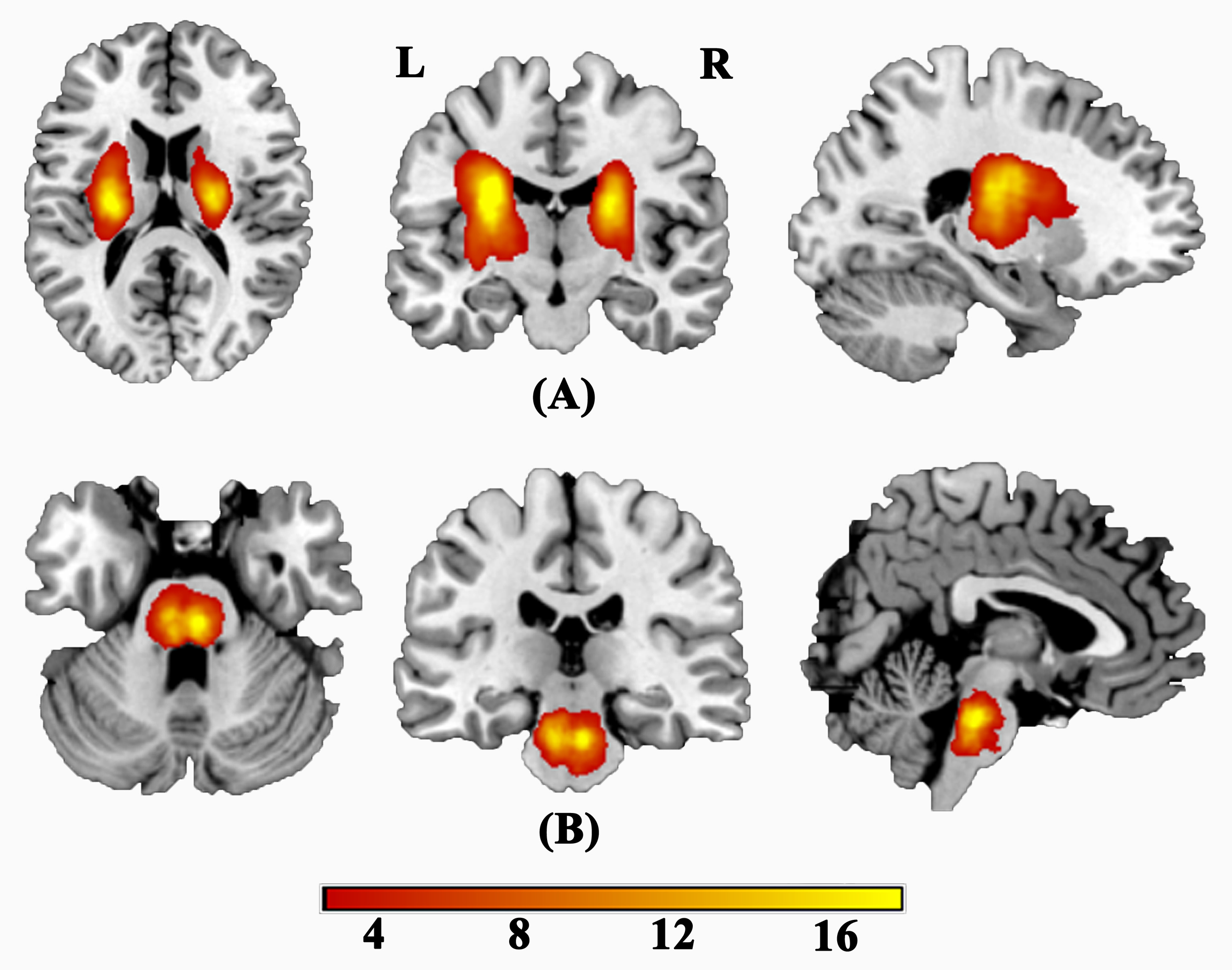
Figure 1: Lesion incidence map for patients with stroke. (a) For patients with stroke at the level of internal capsule. (b) For patients with stroke at the level of pontine.
Color indicates the lesion incidence frequency.
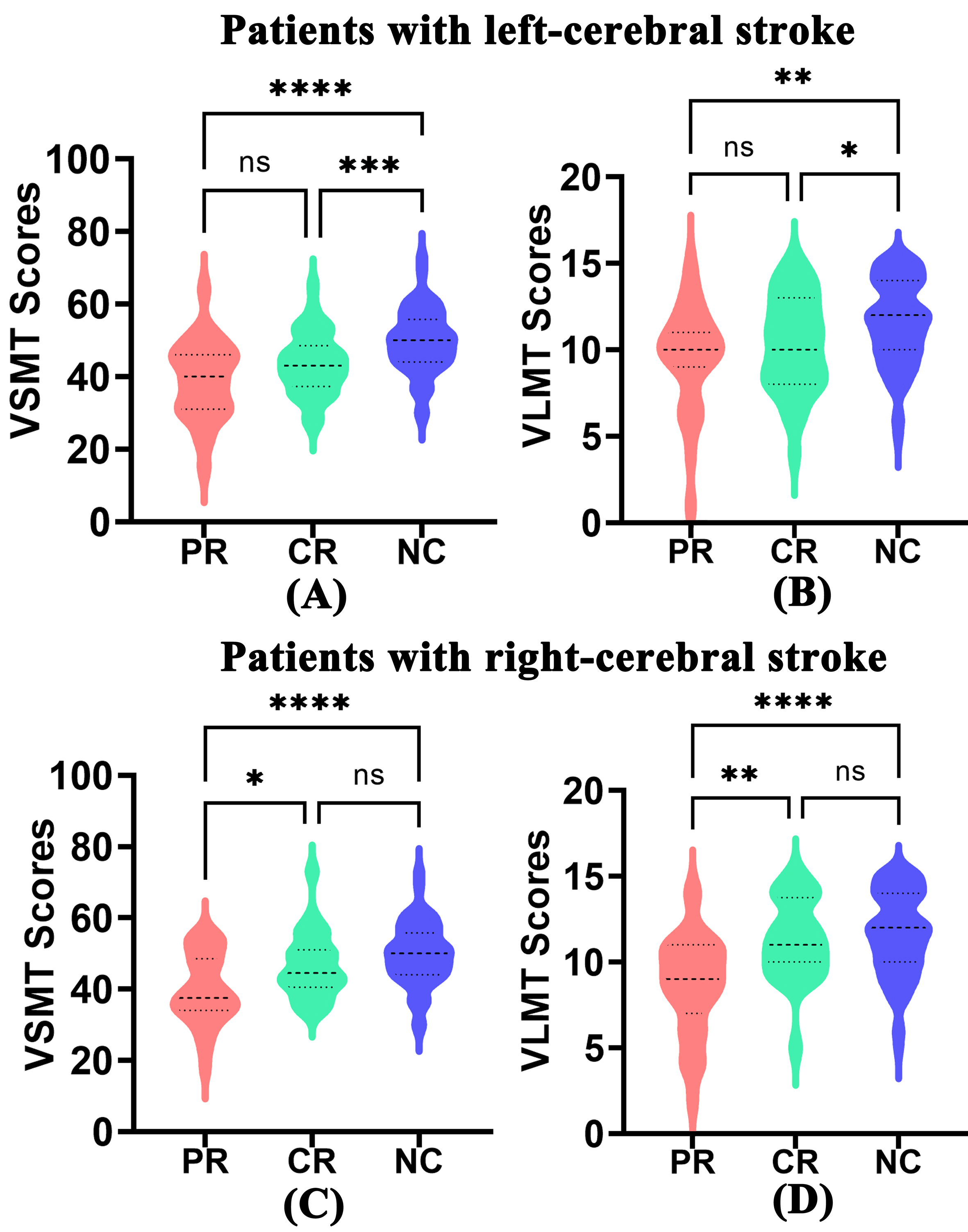
Figure 2: The significant group differences in VLMT and VSMT in patients with left- and right-cerebral stroke.
Abbreviations: CR, complete recovery; PR, partial recovery; NC, normal control; VLMT, verbal long-term memory; VSMT, verbal short-term memory
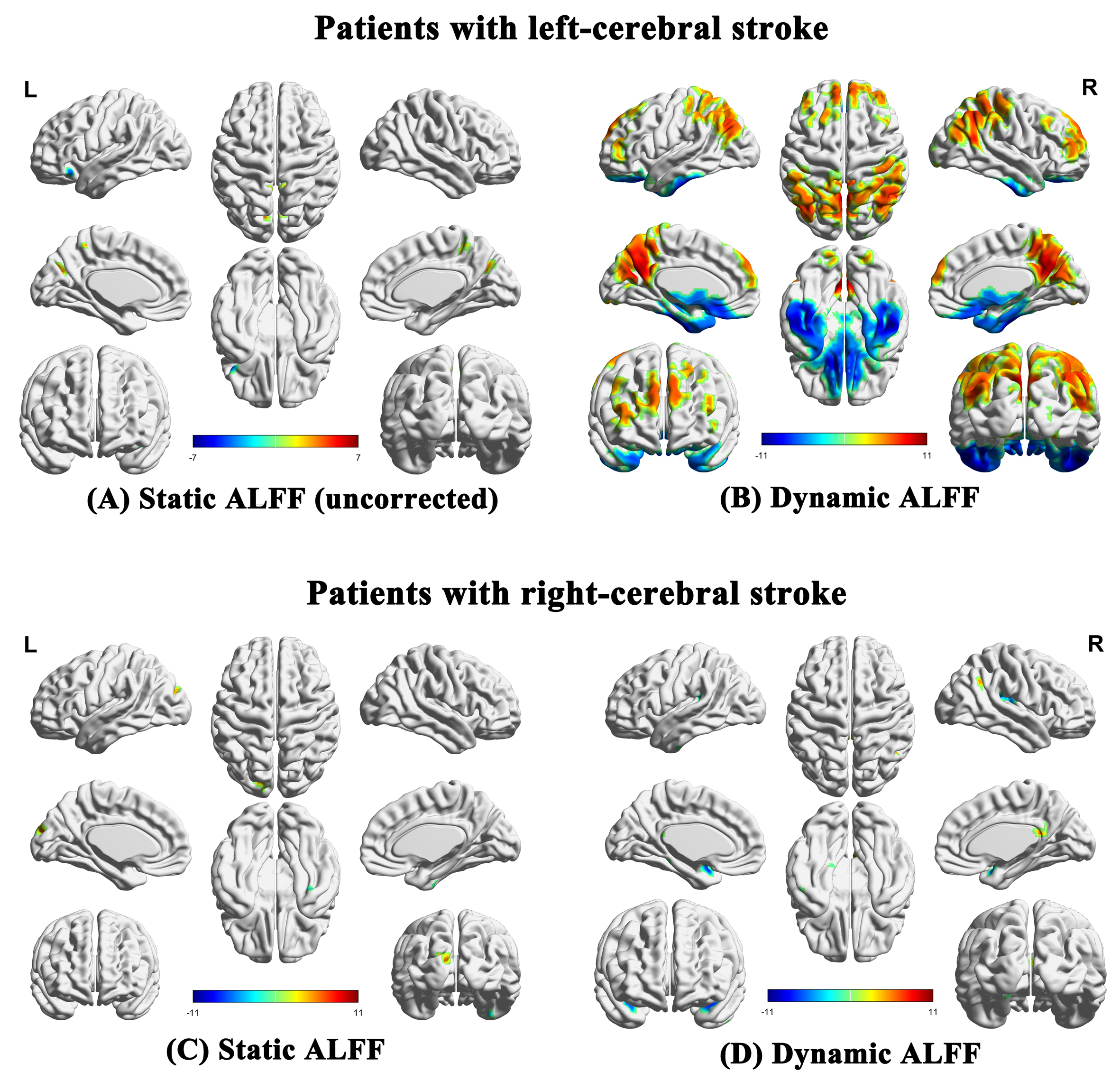
Figure 3: Brain regions showing significant differences in the sALFF and dALFF analyses (FWE correction, P < 0.05 ).
Abbreviations: ALFF, amplitude of low-frequency fluctuations; L, left; R, right
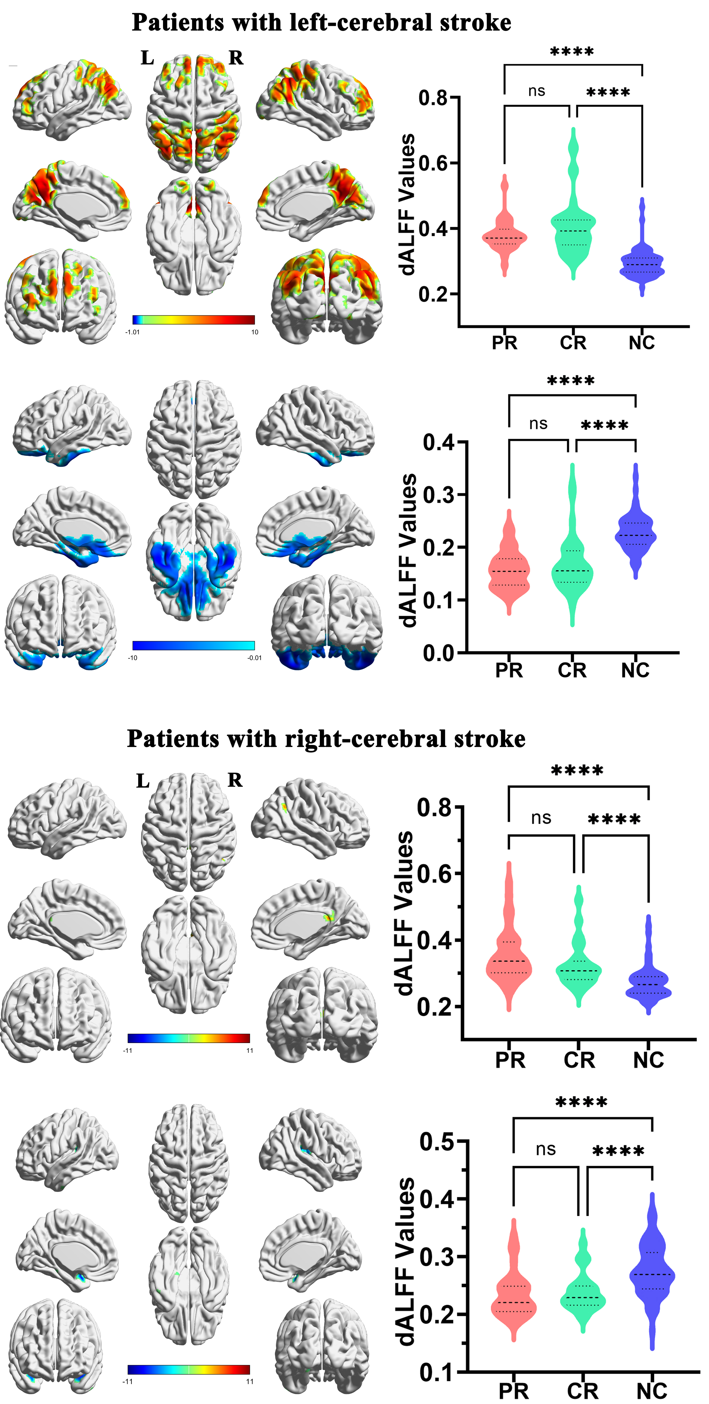
Figure 4: The comparison of dALFF in increased (A, C) and decreased (B, D) regions among the PR, CR and NC groups.
Abbreviations: CR, complete recovery; dALFF, dynamic amplitude of low-frequency fluctuations; L, left; NC, normal control; PR, partial recovery; R, right
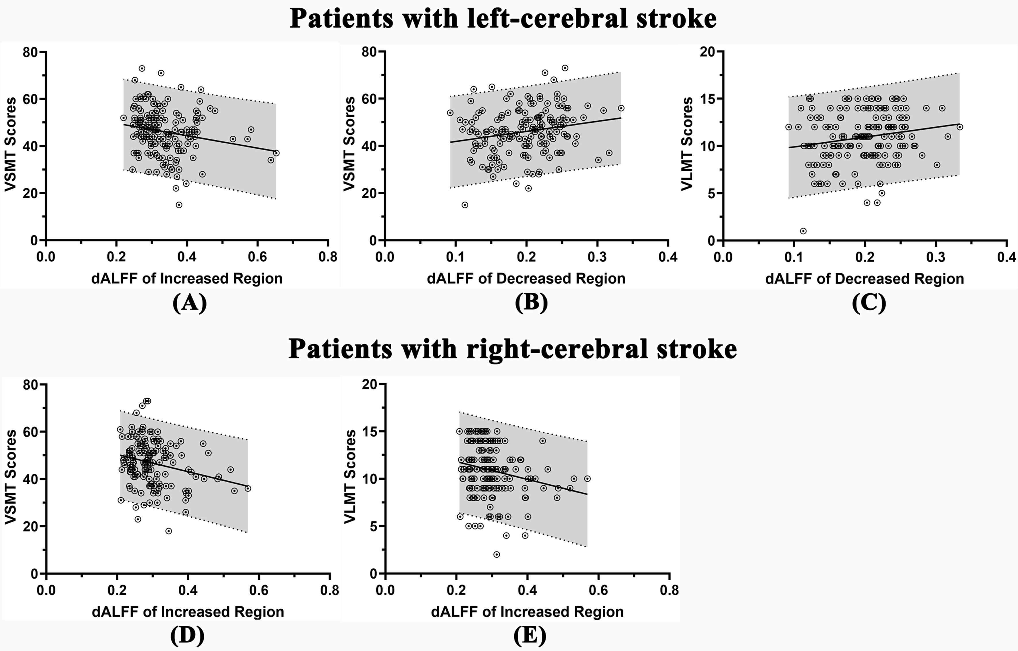
Figure 5: In patients with left-cerebral stroke, the correlation between dALFF of increased region and VSMT score (A), of decreased region and VSMT score (B), and of decreased region and VLMT score (C). In patients with right-cerebral stroke, the correlation between dALFF of increased region and VSMT score (D) and of increased region and VLMT score (D).
Abbreviations: dALFF, dynamic amplitude of low-frequency fluctuations; VLMT, verbal long-term memory; VSMT, verbal short-term memory