2096
Flow augmentation after occlusion maintains functional connectivity and mean diffusivity1Radiology, University of Chicago, Chicago, IL, United States, 2Mount Carmel, Columbus, OH, United States
Synopsis
Keywords: Stroke, fMRI (resting state)
Through the use of resting state functional MRI, diffusion tensor imaging (DTI), and a unique canine stroke model, our study explored the functional and structural effects of maintaining perfusion pressure as a means of extending the window of opportunity for thrombectomy. In our preliminary results, the employment of flow augmentation indicated a maintenance of functional connectivity, mean T2* signal intensity, and mean diffusivity in the ischemic region. Through this we have shown a potential method of extending the "door-to-needle time" in stroke treatment and possible therapeutic effects of flow augmentation therapy in acute stroke.Introduction, Methods, Results, and Discussion
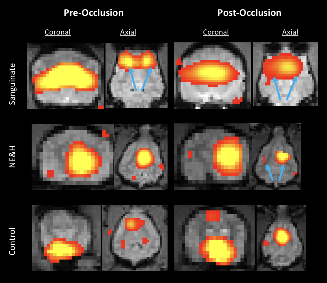
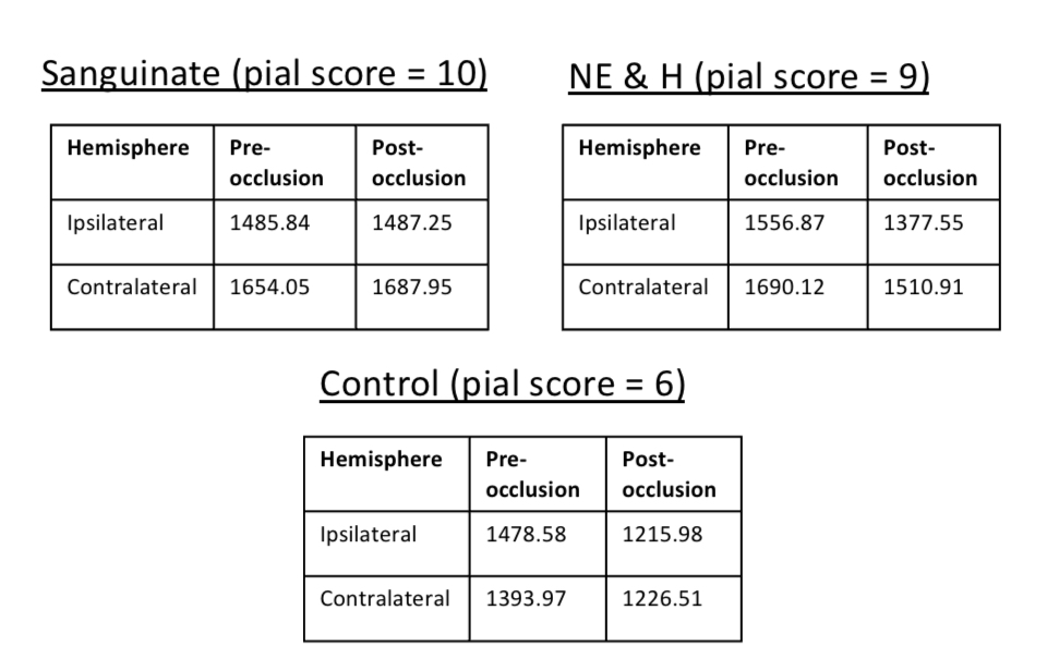
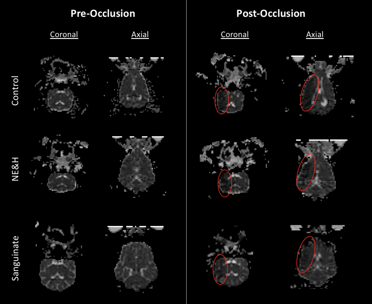
Introduction: Methods for extending the “door-to-needle time” in stroke treatment is a clinically relevant problem that has garnered significant attention from the research community. Beyond the removal of the arterial occlusion, is the critical loss of functional behavior that is a hallmark of stroke survivors. We compare the use of two potential treatments that extend the “door-to-needle time” by maintaining perfusion pressure and functional and structural connectivity in the ischemic region, thereby extending the window of opportunity for thrombectomy. We compare 1) a combination of norepinephrine and hydralazine (NE&H) and 2) Sanguinate. Sanguinate is purified bovine hemoglobin (Hgb) that has been pegylated and combined with carbon monoxide to suppress vasoconstriction and provide anti-inflammatory effects1. The Hgb then carries oxygen for targeted release to hypoxic tissue (i.e., with low partial pressure of oxygen) in the presence of robust blood supply to the affected hemisphere through dilated pial arteries and associated anastomoses (pial collateral supply)1,2. We seek to evaluate the therapeutic effects of flow augmentation therapy in acute stroke. The functional and structural effects of these treatments are assessed through resting-state functional magnetic resonance imaging (fMRI) and diffusion tensor imaging (DTI) analysis in a canine stroke model and compared to a pre- and post-occlusion canine control group.Methods: This preliminary study assesses the effect of a middle cerebral artery (MCA) occlusion in three experimental groups: control, NE&H, and Sanguinate all imaged on a 3T Philips Achieva dStream scanner. A BOLD-sensitive EPI sequence (TR = 1400 ms , TE = 20 ms, voxel-size = 2.5mm, number of temporal positions = 300) was used to acquire resting-state functional images of each canine in each group prior to occlusion (“Day 1”) and on a separate day, four hours post-occlusion (“Day2”). On Day 2 the MCA was permanently occluded, and images were re-acquired. Pre-processing of resting-state fMRI data was performed using FMRIB Software Library (FSL v6.01) tools. Each dataset was co-registered to the first timepoint volume using FLIRT. Probabilistic independent component analysis (PICA) was performed with MELODIC (Multivariate Exploratory Linear Optimized Decomposition into Independent Components), distributed with FSL v6.01. MELODIC estimates group-wise spatial maps that correspond mainly to Resting State Networks (RSNS). The anterior Default Mode Network (DMN) in each dataset is identified by visual inspection in comparison to results of the study conducted by Beckmann et al3. Functional connectivity analysis was then conducted through a general Z-score comparison of the anterior DMN. In addition to a functional connectivity comparison, a comparison of the mean T2* signal intensity averaged over all timepoints is conducted through ROI analysis. ROIs were selected in the fMRI images ipsilateral and contralateral to the region of occlusion. The functional analysis was paired with structural analysis through DTI. The diffusion weighted images were acquired with a SE-EPI sequence (TR = 2993 ms, TE = 83 ms, slice thickness = 2 mm, number of diffusion directions = 33). Pre-processing of the diffusion data was done in FSL and DTIFIT was used to fit the diffusion tensor model at each voxel. Scalar maps of the principle direction of diffusion, fractional anisotropy (FA), and mean diffusivity (MD) were generated and compared for Day 1 and Day 2 of a representative subject in each group. Lastly, the results of the above analyses were viewed in the context of the pial collateral score of each subject2. A representative subject is shown for each experimental group (Figure 1). The pial collateral scores of the subject in each group were as follows: control = 6, NE&H = 9, Sanguinate =10.
Results: Functional connectivity maps with Z-scores ranging from 1.75 to 10 show an asymmetry and shifting of the DMN post-occlusion (Figure 1). Attenuation of the DMN as well as the mean T2* signal intensity is seen comparing pre- to post-occlusion data in all experimental groups except for Sanguinate. The Sanguinate group maintained functional connectivity as well as signal intensity and had minimal loss in MD in the occluded region.
Discussion: We have found that functional connectivity is maintained using flow augmentation (Figure 1). Preliminary results also indicate a retention of mean T2* signal intensity using flow augmentation (Figure 2). Lastly, MD maps support functional results by the drop in values seen in the ischemic region with alteration being minimal in the treatment group post-occlusion (Figure 3).
Conclusion: The functional and structural effects of flow augmentation therapy have been assessed and found to maintain functional connectivity, mean T2* signal intensity, and MD.
Acknowledgements
Research reported in this publication is supported by NIH Grant 5R25GM109439-09, NIH Grant R01NS093908, and NSF DGE-1746045.
References
[1] G. A. Christoforidis, N. Saadat, M. Liu, Y. I. Jeong, S. Roth, M. Niekrasz, and T. Carroll, Effect of Early Sanguinate (PEGylated Carboxyhemoglobin Bovine) Infusion on Cerebral Blood Flow to the Ischemic Core in Experimental Middle Cerebral Artery Occlusion, J NeuroIntervent Surg neurintsurg (2021).
[2] M. Liu, N. Saadat, Y. I. Jeong, S. Roth, M. Niekrasz, T. Carroll, and G. A. Christoforidis, Augmentation of Perfusion with Simultaneous Vasodilator and Inotropic Agents in Experimental Acute Middle Cerebral Artery Occlusion: A Pilot Study, J NeuroIntervent Surg neurintsurg (2022).
[3] K. M. Beckmann, A. Wang-Leandro, H. Richter, R. N. Bektas, F. Steffen, M. Dennler, I. Carrera, and S. Haller, Increased Resting State Connectivity in the Anterior Default Mode Network of Idiopathic Epileptic Dogs, Sci Rep 11, 23854 (2021).
Figures