2095
Evaluation of the Glymphatic System Using the DTI-ALPS Index in Patients with Spontaneous Intracerebral Haemorrhage1Department of Radiology, Affiliated Hospital of Xuzhou Medical University, Xuzhou, China, 2MR Research China, GE Healthcare, Beijing, China
Synopsis
Keywords: Stroke, Diffusion Tensor Imaging, glymphatic system
We evaluated the function of the human glymphatic system (GS) in patients with spontaneous intracerebral haemorrhage (sICH) using diffusion tensor imaging analysis along with the perivascular space (DTI-ALPS). Twenty patients with sICH and 31 healthy controls (HCs) were recruited for analysis. The results showed that DTI-ALPS index on the lesion side was significantly decreased, but not in the contralateral side in sICH or in HCs. And, the decreased DTI-ALPS index was significantly correlated with disease duration. This study confirmed the presence of GS dysfunction only ipsilateral to the lesion, indicating the GS may be a separate system in bilateral hemispheres.Introduction
Spontaneous intracerebral haemorrhage (sICH) refers to a nontraumatic intraparenchymal haemorrhage caused by spontaneous rupture of cerebral blood vessels [1]. Despite some advances in early prevention and acute interventions over the past decade, the high mortality rate of ICH has not been reduced [2]. Therefore, understanding the pathophysiology of sICH may contribute to the exploration of therapeutic approaches and assessments of treatment effects.The glymphatic system (GS) was first discovered by Iliff et al. [3] through two-photon imaging of small fluorescent tracers in 2012. GS is involved in the pathological process of stroke, including brain oedema, blood–brain barrier (BBB) disruption, immune cell infiltration, neuroinflammation, and neuronal apoptosis [4]. As suggested by the above studies, diffusion tensor imaging analysis along with the PVS (DTI-ALPS) can be used as a quantitative imaging biomarker to monitor the ability of the GS [5]. Thus, the aim of this study was to investigate the pathological features of GS in patients with sICH using this new method of DTI analysis.Materials and Methods
SubjectsWe recruited a convenience sample of 23 hospitalized patients with sICH. All patients received conservative treatment, and magnetic resonance imaging (MRI) was performed on each patient one month later.
MRI Data Acquisition
All subjects underwent 3.0 Tesla MRI scanning (GE Medical Systems, Signa HD, Waukesha, WI, USA) with an eight-channel head coil. DTI data were obtained using the following parameters: TR/TE = 9000/90 ms, matrix = 128 × 128 mm2, FOV = 256 × 256 mm2, number of diffusion gradient directions = 64, b value =1000 and providing voxels of 3 mm × 2 mm × 2 mm. The SWI with the following parameters: TR/TE = 44.5 ms/5.5 ms, bandwidth = ± 41.67 kHz, slice thickness/slice spacing was 2 mm/0 mm, and the reversal angle was 15°.
MRI Data Processing
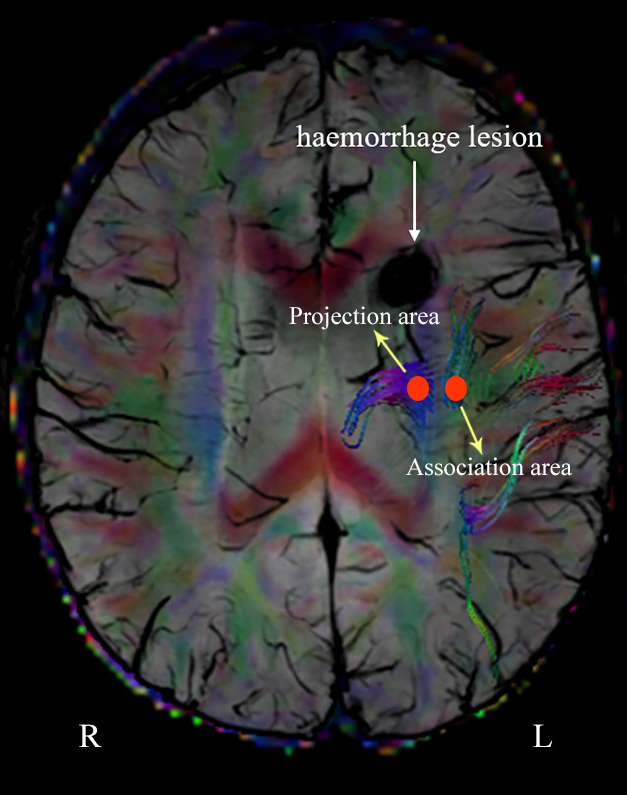
DTI Studio software was used in this study to measure DTI metrics (https://www.mristudio.org/). Briefly, the steps were as follows: (i) raw DTI data of a single individual were imported into the software; (ii) automatic image registration was performed; and (iii) the diffusion tensor was calculated, including a colour-coded fractional anisotropy (FA) map and diffusivity in the directions of the x-axis, y-axis, and z-axis (Figure 1). We evaluated the diffusivity along the direction of the PVS to further calculate the DTI-ALPS index [5-7]. SWI scans were performed to accurately visualize these fine veins (Figure 1). A 4-mm-diameter ROI was placed in the area of the projection fibres and the area of the association fibres in the bilateral hemisphere. Then, each DTI parameter, measured by two radiologists, was averaged as the final result of each individual. The DTI-ALPS index was calculated by using the DTI parameters measured above, according to the equation: DTI-ALPS index = mean (Dxxproj, Dxxassoc)/mean (Dyyproj, Dzzassoc).
Statistical Analysis
A chi-square test was used to observe sex differences between participants in the sICH and HC groups. Intraclass correlation coefficient (ICC) statistics were used to assess interobserver agreement for DTI parameter measurements. A two-sample t test was employed to find intergroup differences in age and DTI-ALPS index. Differences in the DTI-ALPS index between the left and right cerebral hemispheres of all the subjects were tested using paired t tests. Pearson correlation analyses were carried out to test whether DTI-ALPS data of patients with sICH can indicate disease duration. The threshold for the significance level was set at 0.05.
Results
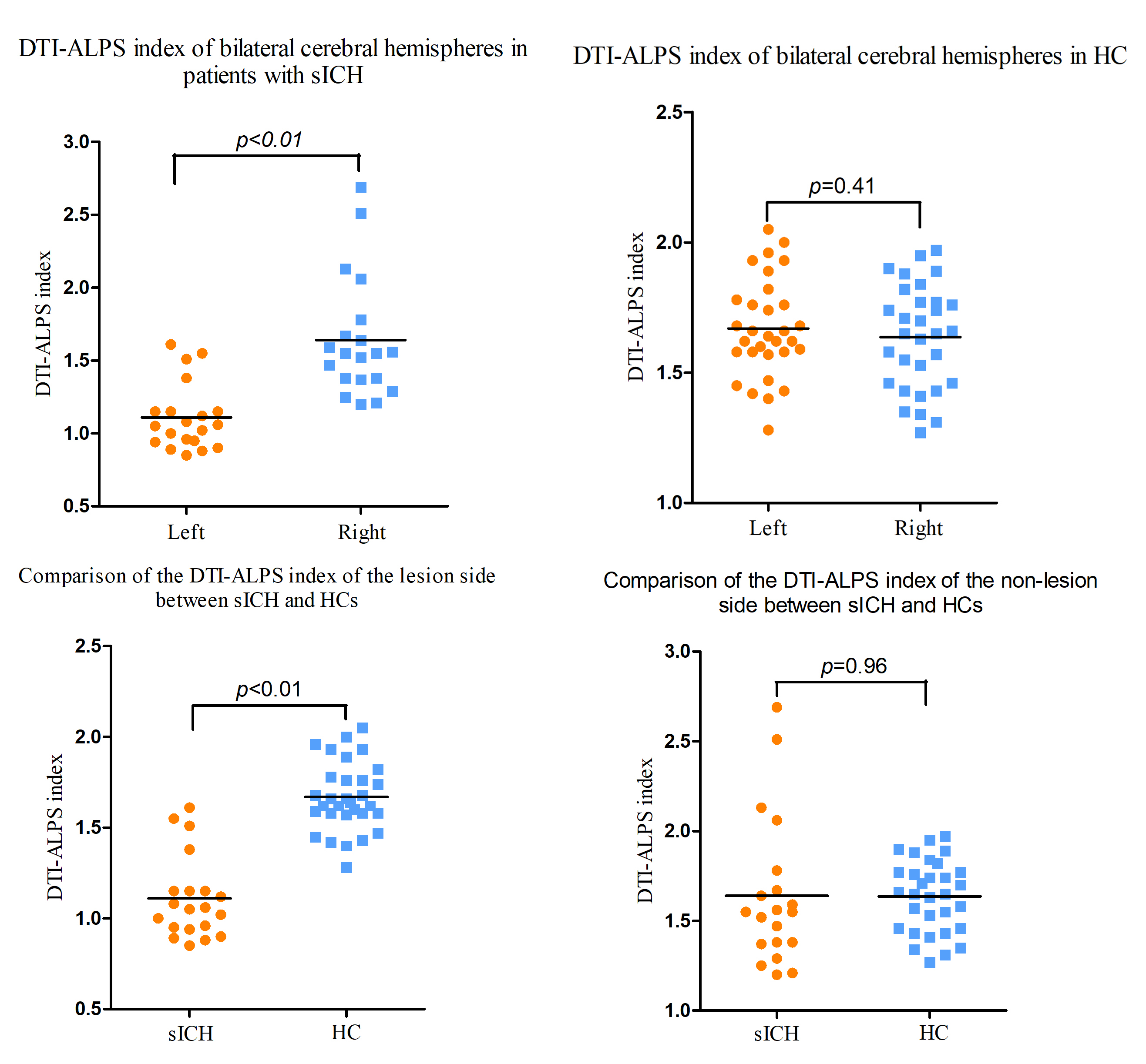
The DTI-ALPS index of the patient's lesion side was 1.11 ± 0.23, and the DTI-ALPS index of the contralateral side was 1.64 ± 0.41. The DTI-ALPS index of the left and right cerebral hemispheres of the HCs were 1.67 ± 0.19 and 1.63 ± 0.19, respectively. The DTI-ALPS index on the lesion side was significantly lower than that of the contralateral side (p < 0.01, t = -5.77); in addition, DTI-ALPS index on the lesion side was significantly lower than that of the ipsilateral side of the HCs (p < 0.01, t = -9.50) (Figure 2). No significant differences were found in the DTI-ALPS index on the nonlesion side between patients and HCs (p = 0.96, t = 0.05) or between the left and right cerebral hemispheres of HCs (p = 0.41, t = -0.83) (Figure 2). The DTI-ALPS index of the lesion side was significantly correlated with disease duration (p = 0.018, r = 0.537) (Figure 3).Discussion
The main goal of the current study was to determine the pathological changes in the GS in patients with sICH by means of DTI analysis. The lower DTI-APLS index indicated a weaker ability of the GS after intracerebral haemorrhage. Interestingly, we found no significant difference in DTI-ALPS on the same side between participants in the HC and sICH groups, but it was significantly reduced on the lesion side. The possible reason is that blood vessels are one of the most important parts of the GS, and the bilateral cerebral hemispheres are fed by separate blood vessels. Therefore, we speculate that the separate blood supply system may be the structural basis of GS that is functionally independent. Overall, these findings have significant implications for the understanding of sICH from a new perspective.Acknowledgements
The authors thank the patients who participated in this study and the staff of the Department of Radiology and Department of Rehabilitation.References
[1] B. A. Gross, B. T. Jankowitz and R. M. Friedlander, “Cerebral intraparenchymal hemorrhage: a review,” JAMA, vol. 321, no. 13, pp. 1295–1303, 2019.
[2] C. Weimar and J. Kleine-Borgmann, “Epidemiology, prognosis and prevention of non-traumatic intracerebral hemorrhage,” Current Pharmaceutical Design, vol. 23, no. 15, pp. 2193–2196, 2017.
[3] J. J. Iliff, M. Wang, Y. Liao et al., “A paravascular pathway facilitates CSF flow through the brain parenchyma and the clearance of interstitial solutes, including amyloid β,” Science Translational Medicine, vol. 4, no. 147, pp. 147ra111, 2012.
[4] T. Lv, B. Zhao, Q. Hu and X. Zhang, “The glymphatic system: a novel therapeutic target for stroke treatment,” Frontiers in Aging Neuroscience, vol. 13, pp. 689098, 2021.
[5] T. Taoka, Y. Masutani, H. Kawai et al., “Evaluation of glymphatic system activity with the diffusion MR technique: diffusion tensor image analysis along the perivascular space (DTI-ALPS) in Alzheimer's disease cases,” Japanese Journal of Radiology, vol. 35, no. 4, pp. 172–178, 2017.
[6] G. Yang, N. Deng, Y. Liu, Y. Gu and X. Yao, “Evaluation of glymphatic system using diffusion MR technique in T2DM cases,” Frontiers in Human Neuroscience, vol. 14, pp. 300, 2020.
[7] X. Ma, S. Li, C. Li et al., “Diffusion tensor imaging along the perivascular space index in different stages of Parkinson's disease,” Frontiers in Aging Neuroscience, vol. 13, pp. 773951, 2021.
Figures