2071
3D Distortionless Diffusion Weighted Imaging in the Prostate using a Diffusion Prepared Fast Spin-Echo Sequence
Jeremiah Joseph Hess1, Philip Kenneth Lee2, Xuetong Zhou1, Andreas Markus Loening2, and Brian Andrew Hargreaves1,2,3
1Bioengineering, Stanford University, Stanford, CA, United States, 2Radiology, Stanford University, Stanford, CA, United States, 3Electrical Engineering, Stanford University, Stanford, CA, United States
1Bioengineering, Stanford University, Stanford, CA, United States, 2Radiology, Stanford University, Stanford, CA, United States, 3Electrical Engineering, Stanford University, Stanford, CA, United States
Synopsis
Keywords: Prostate, Diffusion/other diffusion imaging techniques
Diffusion-Weighted Imaging (DWI) of the prostate is commonly used for tumor detection and characterization. However, the echo planar imaging (EPI) based DWI methods commonly used in prostate imaging fail in the setting of field inhomogeneities most related to hip prosthesis and rectal gas. Cartesian Fast Spin-Echo (FSE) is an alternative to EPI that is more robust to off-resonance. In this study, we compare in vivo 3D FSE to multi-shot EPI (MUSE) for DWI prostate imaging, demonstrating that an FSE DWI sequence achieves high image quality while avoiding geometric distortion seen in EPI based DWI.Introduction
Diffusion weighted imaging (DWI) in the prostate is commonly used for tumor detection and assessment1. However, the current sequences used for prostate DWI, single-shot or multi-shot Echo Planar Imaging (EPI), are limited by sensitivity to off-resonance. Especially in cases with patients that have total hip replacements, or air-tissue interfaces adjacent to the structure (such as rectal gas adjacent to the prostate gland), EPI suffers from severe geometric distortion and signal pile-up2. Cartesian Fast Spin-Echo (FSE) sequences are an alternative that are far more robust to off-resonance effects and, as such, suffer less geometric distortion.In this work, we present a 3D Cartesian FSE sequence and reconstruction for distortionless DWI of the prostate. Previous work on 3D FSE for prostate DWI used an M1-nulled sequence without phase navigators3. M1-nulled and M0-nulled 3D FSE sequences with phase navigators were tested in vivo and compared to M1-nulled without phase navigators and multi-shot EPI. Phantom studies with a metal hip replacement were conducted to show a reduction in geometric distortion for 3D FSE compared to EPI.
Methods
The 3D Cartesian FSE sequence (Figure 1) uses a diffusion-prepared stimulated-echo sequence3 to address non-Carr-Purcell-Meiboom-Gill (CPMG) magnetization effects. 2D projection phase navigators, consisting of 4 lines in the central region of k-space with an R=2 acceleration factor, are acquired to correct shot-to-shot phase from motion-sensitizing diffusion gradients. This assumes that the phase variations in the slab direction are small due to the relatively small slab thickness. Similar strategies have been applied in the lumbar spine4. All acquisitions were performed on a 3T MRI (Signa Premier, GE Healthcare, Milwaukee, WI) with b values of 0 s/mm2 and 800 s/mm2 for each scan.The reconstruction is outlined in the flowchart in Figure 2. Sensitivity maps are obtained by performing ESPIRIT calibration on the b = 0 s/mm2 data, and phase navigators are reconstructed through an iterative SENSE reconstruction. Full reconstruction is performed using an L1-wavelet-regularized linear reconstruction (formulation in Figure 2).
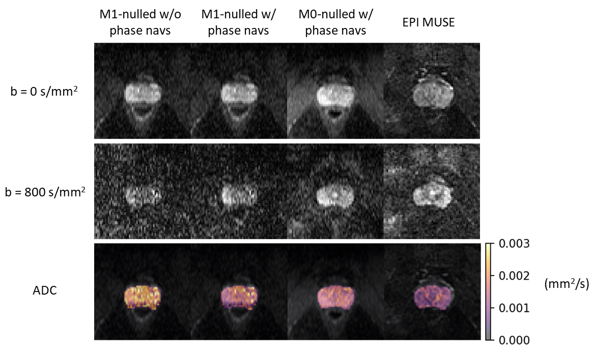
To compare M0-nulled and M1-nulled sequences, in vivo data was acquired in a healthy volunteer following IRB approval and informed consent using both versions of the 3D FSE sequence. A 2D axial commercial multi-shot EPI DWI (MUSE5, GE Healthcare) with 4 shots was acquired as a comparison (Scan time: 2:30, TE: 55 ms, TR: 4000 ms). FOV for all scans was 40cm x 40cm with 3mm slice thickness for 24 slices. Relevant scan parameters for the 3D FSE sequences are: TEprep single-refocused M0-nulled – 35 ms, TEprep twice-refocused M1-nulled – 65 ms, TR 2500 ms, ETL 68, variable flip angle readout, scan time: 9:00 for combined b0/b800.
To demonstrate distortion correction, phantom data was acquired using an HPD diffusion phantom Model 128 (High Precision Devices, Boulder, CO) with the head of a metal hip replacement placed near it. EPI MUSE was acquired alongside the M0-nulled 3D FSE sequence. A MAVRIC SL6 sequence was also acquired as a distortion-free reference image. FOV for the scans was 24cm x 24cm with 3mm slice thickness for 24 slices.
Results
Prostate images for both b values and ADC maps for the M0-nulled and M1-nulled 3D FSE sequences are shown in Figure 3. Compared to the M1-nulled sequences, the M0-nulled sequence produced the best image quality in both the b = 0 s/mm2 and b = 800 s/mm2 images. The prostate ADC estimate for the M0-nulled sequence was 1.55 ± 0.26 (x10-3 mm2/s). Previous literature showed ADC values in the prostate to be around 1.60 (x10-3 mm2/s) for 3D FSE DWI3.Phantom images for the M0-nulled 3D FSE and EPI MUSE sequences are shown in Figure 4. The EPI MUSE data has visible geometric distortion compared to the 3D FSE sequence. The ADC maps for both sequences generally showed good agreement in the tubes. The central tube in the EPI images suffered from ghosting artifacts, potentially leading to the disparity in the ADC estimate.
Discussion
As shown in Figure 3, the image quality of the M1-nulled sequence is improved by the usage of phase navigators in the reconstruction, indicating that the M1-nulling does not eliminate all varying shot-to-shot phase effects in the prostate. Additionally, the M0-nulled sequence with phase navigators performed the best, with noticeable structure seen in the prostate on the DWI image. Comparable ADC values to literature in the M0-nulled scan suggests that the 2D projection phase navigator is sufficient for correcting shot-to-shot phase in the prostate. For the phantom data, the 3D FSE images suffered no geometric distortion compared to the EPI MUSE images, whose geometric distortion was severe to the point of impacting ADC estimates. However, the SNR efficiency of 3D FSE is about half as much as EPI.In the future, we plan to image patients with total hip replacements using both 3D FSE DWI and EPI MUSE DWI and compare the results to see if the geometric distortion significantly impacts the prostate and ADC measurements.
Conclusion
In cases where EPI-MUSE suffers severe geometric distortion due to off-resonance, 3D FSE DWI suffers no distortion, indicating the potential for 3D FSE for distortionless DWI of the prostate for patients with hip prostheses or bowel gas.Acknowledgements
GE Healthcare. NIH R01-EB009055. NIH R01-CA249893.References
- Maurer MH, Heverhagen JT. Diffusion weighted imaging of the prostate-principles, application, and advances. Transl Androl Urol. 2017 Jun;6(3):490-498.
- Czarniecki M, Caglic I, Grist JT, Gill AB, Lorenc K, Slough RA, Priest AN, Barrett T. Role of PROPELLER-DWI of the prostate in reducing distortion and artefact from total hip replacement metalwork. Eur J Radiol. 2018 May;102:213-219.
- Zhang Q, Coolen BF, Versluis MJ, Strijkers GJ, Nederveen AJ. Diffusion-prepared stimulated-echo turbo spin echo (DPsti-TSE): An eddy current-insensitive sequence for three-dimensional high-resolution and undistorted diffusion-weighted imaging. NMR Biomed. 2017 Jul;30(7).
- Cervantes B, Van AT, Weidlich D, Kooijman H, Hock A, Rummeny EJ, Gersing A, Kirschke JS, Karampinos DC. Isotropic resolution diffusion tensor imaging of lumbosacral and sciatic nerves using a phase-corrected diffusion-prepared 3D turbo spin echo. Magn Reson Med. 2018 Aug;80(2):609-618.
- Chen NK, Guidon A, Chang HC, Song AW. A robust multi-shot scan strategy for high-resolution diffusion weighted MRI enabled by multiplexed sensitivity-encoding (MUSE). Neuroimage. 2013 May 15;72:41-7.
- Koch KM, Brau AC, Chen W, Gold GE, Hargreaves BA, Koff M, McKinnon GC, Potter HG, King KF. Imaging near metal with a MAVRIC-SEMAC hybrid. Magn Reson Med. 2011 Jan;65(1):71-82.
Figures
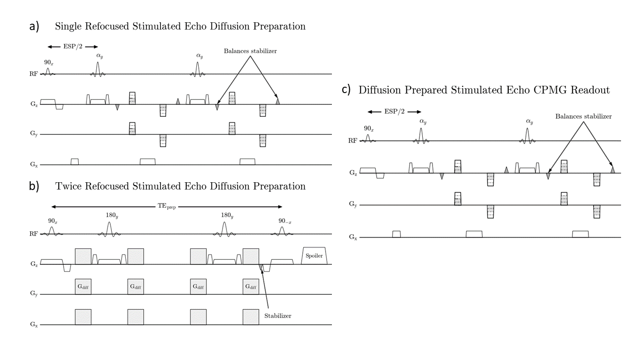
3D
FSE diffusion preparation sequences for a) M0-nulled and b) M1-nulled
sequences. c) the 3D FSE stimulated echo readout used for both sequences.
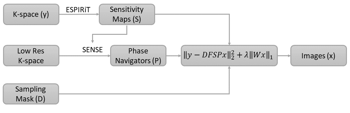
The linear reconstruction pipeline for the 3D FSE diffusion sequences. F represents the 3D Fourier transform linear operator and W represents the Wavelet transform linear operator.
In vivo results for M1-nulled and M0-nulled 3D FSE sequences compared to EPI MUSE.
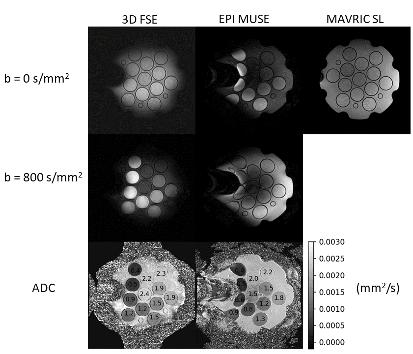
Phantom results for M0-nulled 3D FSE sequence with
phase navigators, compared to EPI MUSE, with a MAVRIC SL image as a
distortion-free reference. The head of a metal hip implant was placed to the
left of the phantom. ADC values (x10-3) were calculated for each
tube.
DOI: https://doi.org/10.58530/2023/2071