2069
Using Multi-parametric MRI Quantitative Metrics as Image Biomarkers to Predict the Tumor Malignant degree of Prostate Cancer1Clinical medicine school of Ningxia Medical University, YinChuan, China, 2Department of Radiology ,the First Hospital Affiliated to Hainan Medical College, Haikou, China, 3Clinical medicine school of Ningxia Medical University, Yinchuan, China, 4GE Healthcare,MR Reseaich, Beijing, Beijing, China, 5Department of Radiology, General Hospital of Ningxia Medical University,, Yinchuan, China
Synopsis
Keywords: Prostate, Data Processing
This study aims to investigate whether the quantitative metrics derived from diffusion weighted imaging (DWI) and dynamic contrast-enhanced (DCE) MRI can be used to predict the tumor malignancy of prostate cancer. The results showed that the apparent diffusion coefficient (ADC) value, Tmax (s), SImax%, Rmax% were correlated with Ki-67 to a certain extent, DWI and DCE-MRI parameters are expected to be a non-invasive examination method to measure the ability of prostatic malignant tumor cells to expand, and provide an important theoretical basis for evaluating the efficacy of prostate cancer and monitoring the prognosis.Summary of Main Findings
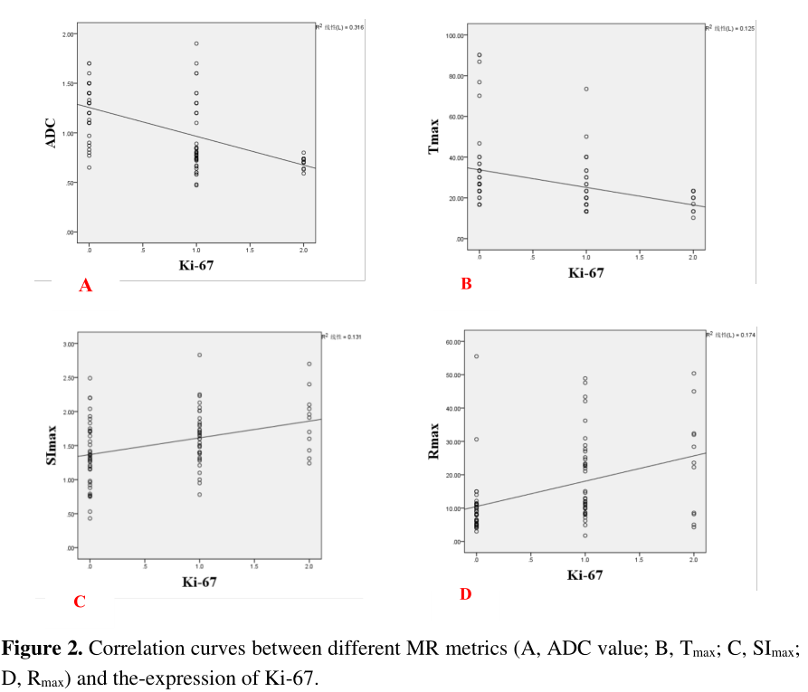
ADC value and Tmax were negatively correlated with Ki-67; SImax and Rmax were positively correlated with Ki-67;there were certain correlations between ADC value, Tmax, SImax, Rmax and Ki-67, indicating that the above imaging parameters are expected to become non-invasive alternative markers of Ki-67 index in the diagnosis and differentiation of prostate cancer, and thus predict the degree of malignancy of tumor cells, providing a certain reference for the selection of reasonable treatment plan in the future and the determination of long-term prognosis.Introduction
Prostate cancer (PCa) has the first incidence rate and the second mortality rate among male malignant tumors in the United States1, and its incidence in China is rising rapidly in recent years. Early diagnosis of PCa may improve the long-term survival of patients2. With the recent development of imaging technology in prostate MRI, multi-parametric MRI, which including conventional anatomical T2-weighted imaging (T2WI), diffusion weighted imaging (DWI) and dynamic contrast-enhanced (DCE) MRI, has been recognized as the best imaging strategy for PCa diagnosis3.Ki-67 is a commonly used biomarker to reflect the level of tumor proliferation. However, the relationship between quantitative metrics derived from multi-parametric MRI and Ki-67 in PCa is seldom reported. This study aims to investigate whether the quantitative metrics of multi-parametric MRI can be used to predict the tumor malignancy of PCa.Methods
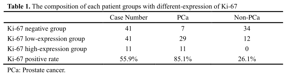
Participants 93 patients (68.9 ± 7.9 years) with pathologically confirmed prostate diseases were recruited in this study. For each patient, the Ki-67 staining index was measured by immunohistochemical method. Then the patients were classified into Ki-67 negative group (41 cases), Ki-67 low-expression group (41 cases), and Ki-67 high-expression group (11 cases). The detailed composition of each group was listed in Table 1.Data acquisition All MR examinations were performed on a 3.0T MR scanner (SIGNA™ Excite HD; GE Healthcare, Milwaukee, WI, USA). Our Institutional Review Board approved the scan protocol and written informed consent was obtained from each patient before the scan. The protocol included T2WI, DWI and DCE-MRI, while the detailed scan parameters were listed in Table.
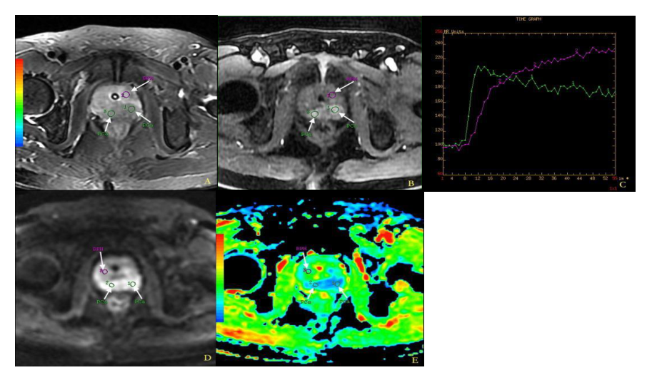
Post-processing The data post-processing was conducted on a GE ADW 4.3 workstation. The regions of interest (ROIs) were drawn at the most prominent location of the lesion. The apparent diffusion coefficient (ADC) maps were calculated from the DWI images. The time-signal intensity (SI-T) curves were plotted based on the DCE-MRI images, and multiple metrics including the peak time (Tmax), the maximum enhancement degree (SImax) and the fastest enhancement rate (Rmax) were measured subsequently. Whereafter, all quantitative metrics were compared between PCa and non-PCa groups, and among groups with different-expression of Ki-67.
Results
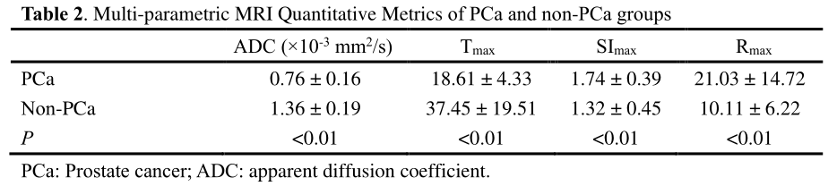
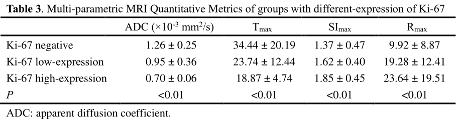
As shown in Table 2, the mean ADC and Tmax values of the PCa group were significantly lower than that of the non-PCa group, while the SImax and Rmax values were significantly higher (all P < 0.01).Ki-67 negative group and Ki-67 positive group (P < 0.01). However, there was no significant difference between low-expression group and high-expression group of Ki-67 (P > 0.05). There were negative correlations between the ADC values, Tmax and Ki-67 (r = -0.50, -0.37, P < 0.01), while the SImax and Rmax were positively correlated with Ki-67 (r = 0.29, 0.36; P < 0.01).
Discussion and Conclusion
As a non-invasive examination method, MRI is expected to indirectly evaluate the biological behavior of malignant tumors before surgery. Multi-parametric MRI has been widely used in the diagnosis and treatment of PCa, not only to guide the selection of initial and repeat biopsy sites, but also to reduce false negative biopsy results, thus helping clinicians to formulate treatment plans and conduct preliminary evaluation4. The results showed that the DWI and DCE-MRI indexes of Ki-67 negative group, Ki-67 low-expression group and Ki-67 high-expression group were compared in pairs, and there was significant difference between Ki-67 negative group and Ki-67 positive group (P < 0.05). However, there was no significant difference between low-expression group and high-expression group of Ki-67 (P > 0.05). It may be related to the fact that there is no unified theory on the cut-off value of Ki-67-expression in previous studies, which needs to be confirmed by further research. There was a negative correlation between ADC values, Tmax and Ki-67 (r = -0.50, -0.37, P < 0.01); There was a positive correlation between SImax, Rmax and Ki-67 (r = 0.29, 0.36; P < 0.01). Kim5 et al found the strongest correlation between ADC value and KI-67 in breast cancer. Calvar6 et al. found that the ADC value of brain tumors was negatively correlated with KI-67. The results of this study showed that ADC value was significantly negatively correlated with Ki-67-expression, suggesting that PCa patients with low ADC value had stronger invasiveness and poorer prognosis.This study showed that ADC value, Tmax, SImax and Rmax% were correlated with Ki-67 to a certain extent, and the Multi-parametric MRI are expected to be a non-invasive examination method to measure the ability of prostatic malignant tumor cells to expand, and provide an important theoretical basis for evaluating the efficacy of PCa and monitoring the prognosis.
Acknowledgements
This study was supported by grants from The Key Research and Development Program of Ningxia (No.2019BEG03033) and Natural Science Foundation of Ningxia(2022AAC03472). Author contributions: conception and design: Yunshu Zhou,Zhiqiang Chen,Na Song, and Zhuo Wang; acquisition of data: Shaoru Zhang, Xiaohua Chen, Xiaocheng Wei,and Aijun Wang; Data analysis and interpretation: Zhiqiang Chen and Yunshu Zhou; drafting the article and revising it critically for important intellectual content: Zhiqiang Chen; final approval of manuscript: all authors. The authors declare no conflicts of interest.References
[1] Siegel RL, Miller KD, Jemal A, et al. Cancer statistics, 2021. CA Cancer J Clin2021;71(1):7-33.
[2] Reda I, Shalaby A, Elmogy M, et al. A comprehensive non-invasive frame work for diagnosing prostate cancer. Comput Biol Med, 2017, 1(81): 148-158.
[3] Ji Guanghai, Zheng Yi, Bo Ruting, et al. Value of multi-parameter MRI in diagnosis of central gland prostate cancer. Chinese Journal of Medical Imaging, 2016,24 (8): 591-595.
[4] Patel P, Mathew MS. Multiparametric MR imaging of the prostate after treatment of prostate cancer. Radiographics, 2018, 38(2): 437-449.
[5] Kim EJ, Kim SH, Park GE, et al. Histogram analysis of apparent diffusion coefficient at 3.0T: correlation with prognostic factors and subtypes of invasive ductal carcinoma. J Magn Reson Imaging, 2015, 42(6): 1666-1678.
[6] Calvar JA, Meli FJ, Romero C, et al. Characterization of brain tumors by MRS, DWI and Ki-67 labeling index. J Neurooncol, 2005, 72(3): 273-280.
Figures