2057
Detection of sparse and dense prostate cancers using bi-parametric MRI combined with radiomics features
Bingni Zhou 1,2, Zhangzhe Chen1,2, Rucuan Chen1,2, Wei Liu1,2, Hualei Gan2,3, Yong Zhang4, Liangping Zhou1,2, and Xiaohang Liu1,2
1Radiology, Fudan University Shanghai Cancer Center, Shanghai, China, 2Oncology, Shanghai Medical College of Fudan University, Shanghai, China, 3Pathology, Fudan University Shanghai Cancer Center, Shanghai, China, 4GE Healthcare, Shanghai, China
1Radiology, Fudan University Shanghai Cancer Center, Shanghai, China, 2Oncology, Shanghai Medical College of Fudan University, Shanghai, China, 3Pathology, Fudan University Shanghai Cancer Center, Shanghai, China, 4GE Healthcare, Shanghai, China
Synopsis
Keywords: Prostate, Radiomics
This is a preliminary study which combined bi-parametric MRI with radiomics feature to detect sparse and dense prostate cancers. Fifty-five patients underwent diffusion weighted and T2 weighted imaging. One hundred and nine peripheral zone (PZ) tumors were reviewed using whole-mount histologic findings. The total number of 381 radiomics features were extracted to construct the models for differentiation. Dense tumors showed ADC values significantly lower than sparse tumors and normal PZ tissues. ADC alone should provide sufficient diagnosis efficiency. However, radiomics features can significantly improve the detection of sparse tumors which showed the similar ADC values as compared to normal tissues.Introduction
The tissue composition of prostate cancer is heterogeneous. Tumors can be composed of densely packed malignant glands (“dense tumors”) or consist of few malignant glands scattered within normal tissue (“sparse tumors”)1. This is a preliminary study which combined bi-parametric MRI with radiomics feature for the detection of sparse and dense prostate cancers2. Radiomics features may more effectively detect microstructure changes compared to overall quantitative diffusion metrics and therefore improve the detection of sparse and dense prostate cancers3-4.Materials and Methods
Fifty-five patients (median age, 63years; range, 56–77 years) gave informed consent to participated in this study. Prior to radical prostatectomy, all patients underwent diffusion weighted imaging (DWI) with b value equal to 1000 sec/mm2 and T2 weighted imaging (T2WI) on a GE Pioneer 3T scanner (GE Healthcare, US). One hundred and nine peripheral zone (PZ) tumors were reviewed using whole-mount histologic findings. Tumors were categorized as “sparse” if more than 50% of their cross-sectional areas were primarily normal PZ regions and were considered “dense” otherwise. Normal PZ tissues were outlined separately on the same section. Tumor and normal tissue outlines were transferred to the corresponding apparent diffusion coefficient (ADC), DWI and T2WI images. The total number of 381 radiomics features were extracted from T2WI, DWI and ADC maps. The ADC values of sparse tumors, dense tumors and normal tissues were compared by one-way analysis of variance. Optimal feature subsets were selected using Spearman correlation coefficient and Random Forest models. Logistic regression was then used to construct models with selected radiomics features to differentiate among sparse cancers, dense cancers and normal tissues. The efficiencies of ADC values and radiomics models were assessed by the Area Under Curve (AUC) of Receiver Operating Characteristic Curve (ROC).Results
According to the histologic findings, fifty-one tumors were put in the “sparse” group and fifty-eight tumors in the “dense” group. The mean ADC value of dense cancer (0.98±0.14 ×10⁻³mm2/s) was significantly lower than those of sparse cancer (1.42±0.38 ×10⁻³mm2/s) and normal tissues (1.59±0.23 ×10⁻³mm2/s) (all p<0.05). The radiomics models showed the similar performance (AUC: 0.953) in distinguishing dense cancers from normal tissues as compared to the mean ADC values (AUC: 0.950) (p>0.05), but superior performance (AUC: 0.888) in differentiating sparse cancer from normal tissue as compared to the mean ADC values (AUC: 0.691) ( p<0.05).Discussion and Conclusion
Dense tumors showed ADC values significantly lower than sparse tumors and normal PZ tissues. ADC alone should provide sufficient diagnosis efficiency. However, radiomics features can significantly improve the detection of sparse tumors which showed the similar ADC values as compared to normal tissues.Acknowledgements
No acknowledgement found.References
1. Langer DL, van der Kwast TH, Evans AJ, et al. Intermixed normal tissue within prostate cancer: effect on MR imaging measurements of apparent diffusion coefficient and T2--sparse versus dense cancers. Radiology 2008, 249(3): 900-908.
2. DeVisschere PJ, Naesens L, Libbrecht L, et al. What kind of prostate cancers do we miss on multiparametric magnetic resonance imaging? Eur Radiol 2016, 26(4): 1098-1107.
3. Cutaia G, LaTona G, Comelli A, et al. Radiomics and Prostate MRI: Current Role and Future Applications. J Imaging. 2021, 7(2).
4. Xu M, Fang M, Zou J, et al. Using biparametric MRI radiomics signature to differentiate between benign and malignant prostate lesions. Eur J Radiol 2019, 114: 38-44.
Figures
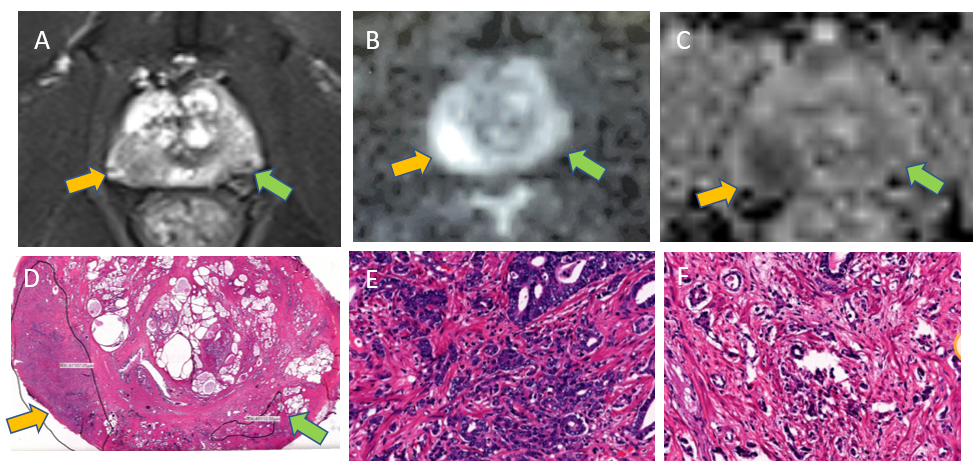
Fig 1. A 61-year-old patient with prostate
cancer of Gleason score 4+4 showing the dense tumor on the right PZ (orange
arrow) and the sparse tumor on the left PZ (green arrow)(A, T2WI; B, DWI; C, ADC;
D, the corresponding pathological slice; E, the dense tumor on the right PZ; F,
the sparse tumor on the left PZ)
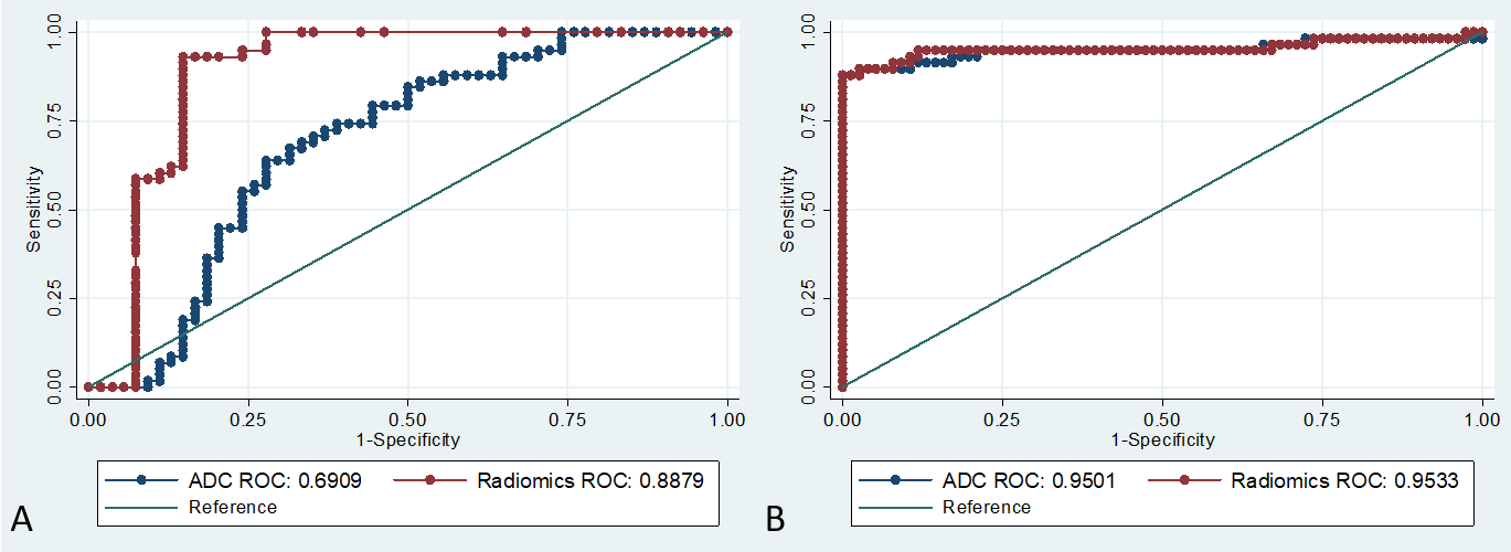
Fig 2. ROC curves of ADC and radiomics
models (A, sparse tumors vs. normal tissues; B, dense tumors vs. normal tissues)
DOI: https://doi.org/10.58530/2023/2057