2053
Liver regeneration of residual right lobe after left lateral sectionectomy in living liver donors: a pilot study based on IVIM and T2* mapping MRI
shuangshuang xie1, caixin qiu1, and wen shen1
1Tianjin First Central Hospital, Tianjin, China
1Tianjin First Central Hospital, Tianjin, China
Synopsis
Keywords: Liver, Liver, liver regeneration
This study investigated the dynamic changes of residual right liver lobe after left lateral sectionectomy (LLS) and the potential value of IVIM and T2* mapping in evaluation of liver regeneration in living liver donors. All IVIM indexes of right liver lobe showed no obvious changes after surgery, but liver T2* values increased significantly at 2 weeks after surgery, and kept increasing gradually from week 2 to 8. Liver T2* values correlated moderately with liver volume, it may be a good substitute to liver volume in evaluating liver regenerationBackground and purpose
The initiation mechanism of residual liver regeneration after partial hepatectomy (PH) is not clear. While, the main change that occurs immediately after PH is the liver hemodynamics. It speculated that hemodynamic change might have close relationship with liver regeneration. Intravoxel incoherent motion imaging (IVIM) can reflect blood flow and blood volume, and T2* mapping can reflect blood oxygen content of the liver parenchyma. The aim of this study was to investigate the dynamic changes of right liver lobe after left lateral sectionectomy (LLS) in living liver donors, and further explore the relationship between liver MRI quantitative parameters and liver volume within two months.Methods
Ninety-nine consecutive subjects with LLS of the liver (parent living liver donors) were collected prospectively. Twenty-one, 18, 20, 20, and 20 subjects received liver IVIM and T2* mapping before operation, 2 weeks, 3 weeks, 4 weeks, and 8 weeks after operation, separately. The other 11 cases completed MRI examination at all time points. D*, PF, D, T2* values and volume of right liver lobe were measured at each time point. Changing rate of MRI indexes and liver regeneration rate (LRR) were calculated. The changes of MRI indexes and volume of right liver lobe were observed dynamically and compared at each time point, and the relationship between MRI indexes and liver volume were analyzed.Results
After LLS, D*, D and PF values of right liver lobe derived from IVIM had no obvious changes from 2 to 8 weeks (p > 0.05), T2* values increased significantly at two weeks, and kept steady from week 2 to 8. ∆T2* showed no significant differences among all time points after surgery (p = 0.091). Both the volume and LRR of right liver lobe increased gradually after LLS, and significantly differences were found between the adjacent two time points. Liver D*, D and PF values showed no correlation with liver volume (all p > 0.05). Liver T2* values correlated moderately with changes of liver volume (r = -0.415, p = 0.005).Conclusion
In living liver donors after LLS, T2* values of right liver lobe increased significantly and correlated significantly with liver volume within 8 weeks after surgery. T2* values may be a good substitute to liver volume in evaluating liver regeneration.Acknowledgements
This study was supported by grants from National Nature Science foundation of China (81901710).References
No reference found.Figures
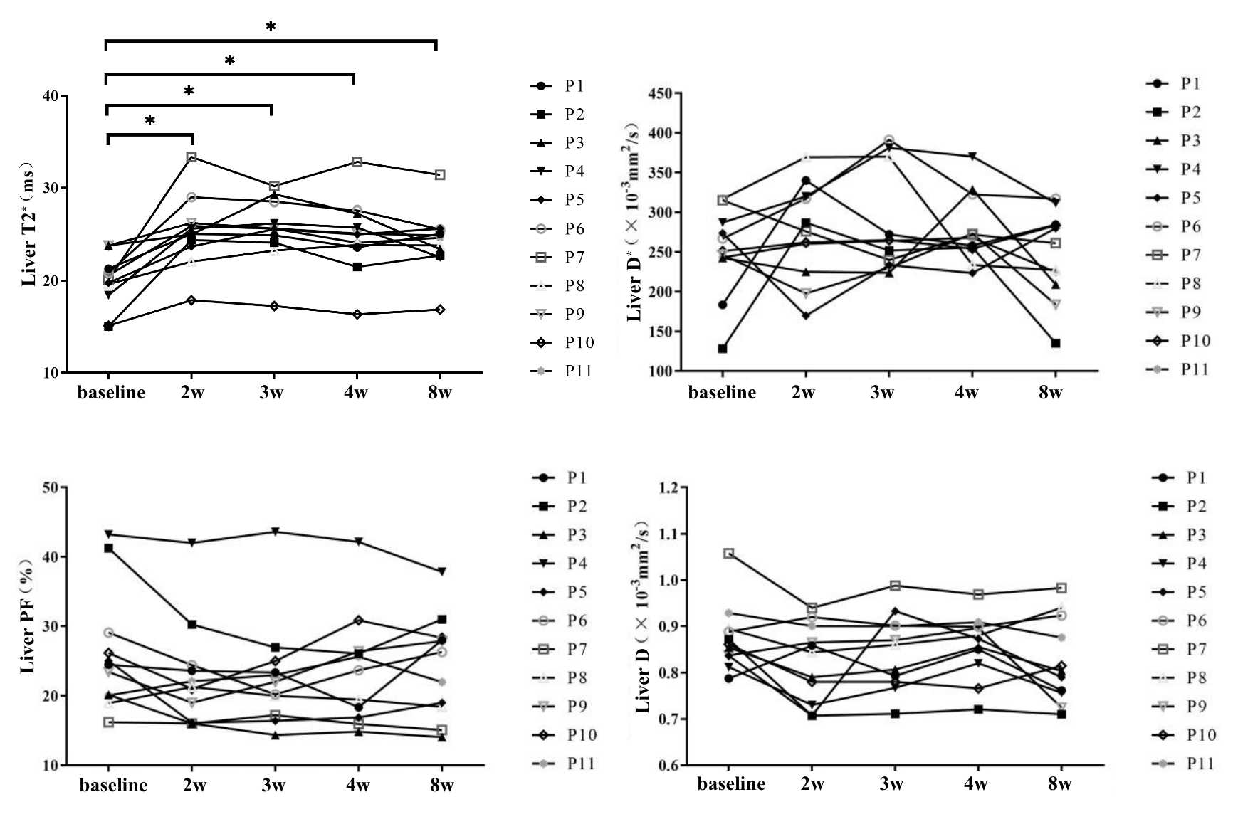
Figure 1 dynamic changes of IVIM and T2* mapping indexes of right liver lobe
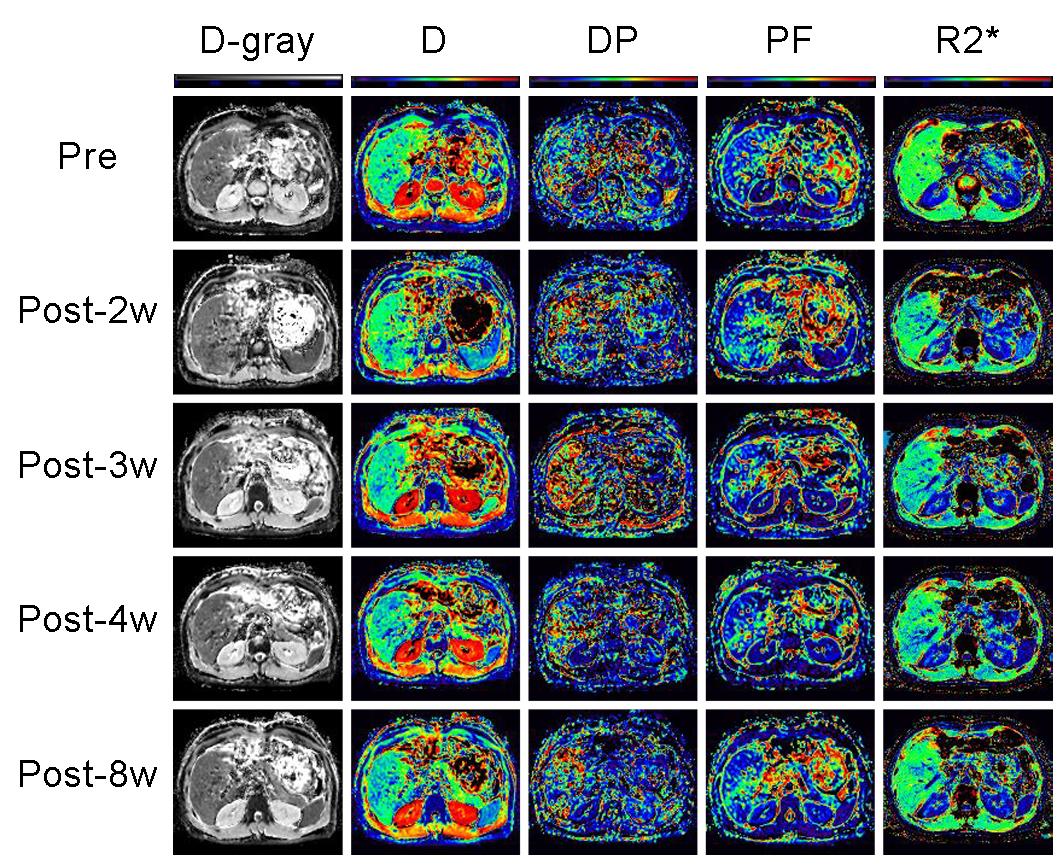
Figure 2 Liver changes showed in D, D*, PF and T2* maps.
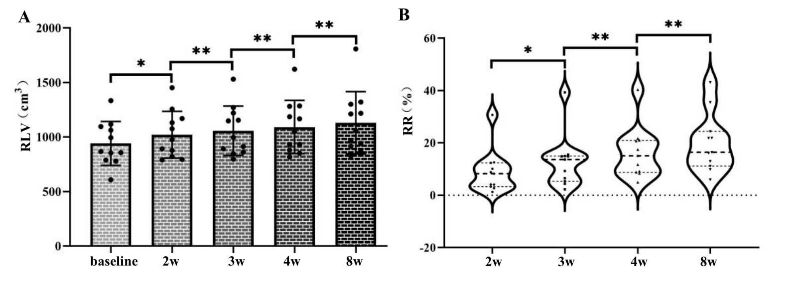
Figure 3 changes of liver volume and regeneration rate (RR) of right lobe. *p < 0.05; **p < 0.01.
DOI: https://doi.org/10.58530/2023/2053