2040
Whole Upper Abdominal T1 Mapping on Free-Breathing 3D Look-Looker Sequence using Radial Sampling Acquisition: A Phantom and Volunteer Study
Tomohiro Noda1, Keitaro Sofue2, Ryuji Shimada1, Yu Ueda3, Yoshiko Ueno2, Yuichiro Somiya1, Shintaro Horii1, Akiko Kusaka1, and Takamichi Murakami2
1Center of Radiology and Radiation Oncology, Kobe University Hospital, Kobe, Japan, 2Department of Radiology, Kobe University Graduate School of Medicine, Kobe, Japan, 3Philips Japan MR Clinical Science, Tokyo, Japan
1Center of Radiology and Radiation Oncology, Kobe University Hospital, Kobe, Japan, 2Department of Radiology, Kobe University Graduate School of Medicine, Kobe, Japan, 3Philips Japan MR Clinical Science, Tokyo, Japan
Synopsis
Keywords: Liver, Quantitative Imaging, T1mapping
We developed a free-breathing 3D Look-Locker sequence with radial sampling acquisition (3D-Radial LL) to acquire T1 map of the whole upper abdomen. To evaluate the quantitative accuracy, T1 values obtained from 3D-Radial LL and modified Look-Locker inversion recovery (MOLLI) in a phantom and volunteers were compared. The phantom study showed excellent correlation regarding T1 values between on the MOLLI and 3D-Radial LL. In the volunteer study, the difference in T1 values were within 6% for the liver, pancreas, spleen, and paraspinal muscle. The 3D-Radial LL enables accurate measurement of T1 values in the upper abdomen.Introduction
Accurate assessment of liver function is important to predict prognosis and clinical management of patients with chronic liver disease or who undergo liver resection. T1 mapping of the liver has been used to assess liver function and liver fibrosis by quantifying the uptake of gadoxetic acid1-3. Modified Look-Locker inversion recovery (MOLLI) or the dual flip angle method are widely used for T1 mapping of the liver. However, the MOLLI has a limitation on the acquisition number of slices, such as one slice per breath-hold, and the dual flip angle method measures T1 values from data at two different data, which may contain registration errors and is susceptible to B1 magnetic field inhomogeneity4. These methods cannot be available for patients who have breath-hold difficulty. Therefore, establishment of accurate and robust methods for the measurements of the T1 values in the whole upper abdomen are desirable. We developed a free-breathing 3D Look-Locker sequence with radial sampling acquisition (3D-Radial LL), which allows T1 map acquisition of the whole liver without breath-hold. The purpose of this study was to assess the quantitative accuracy on T1 measurements of the whole upper abdomen using developed free-breathing 3D-Radial LL sequence.Methods
All images were acquired with a clinical whole body MR scanner (Ingenia 3.0T, Philips Healthcare). Details of the sequence we developed in this study is presented in Figure 1. After applying a non-selective 180° pulse, T1W-TFE radial acquisition (stack-of-stars) was achieved. Continuous echoes were obtained during shot interval, and images were constructed for each inversion time (TI) by setting the phase interval and separating the k-space trajectory of the acquired data. This was repeated as several times as the number of shots to create an image. A recovery time was set to recover to its equilibrium state before the next 180° pulse applied. The T1 value for each pixel was calculated using the following formula:Mz = Mss - (M0 + Mss) * exp (- TI / T1*)$$
$$T1 = {((M0 + Mss) / Mss) - 1} * T1*
Mz: longitudinal magnetization, Mss: steady state condition (< M0),
M0: equilibrium magnetization, T1*: an apparent recovery time (< T1)
A phantom (106 type essential system phantom: Caliber MRI corp.) was imaged to determine sequence parameters followed by comparing correlation regarding eight different T1 values between on the 3D-Radial LL and MOLLI. Then, 15 healthy volunteers were imaged with both the MOLLI and 3D-Radial LL (Figure 2). MOLLI was imaged three slices including liver, pancreas, and spleen using electrocardiography synchronization and pulse wave synchronization with breath-hold. 3D-Radial LL was imaged in the whole upper abdomen in 5 minutes without synchronization and breath-hold. T1 values on the MOLLI and 3D-Radial LL were measured from the obtained T1 maps by placing region of interests (ROIs) set on the liver, pancreas, spleen and paraspinal muscle. Differences and correlations of the T1 values between on the MOLLI and 3D-Radial LL were examined.
Results
In the phantom study, T1 values showed excellent correlation between on the MOLLI and 3D-Radial LL (R2= 0.999, P < 0.0001) (Figure 3). In the clinical study, differences in T1 values obtained with MOLLI and 3D-Radial LL were within 6% for each organ in a total of 15 volunteers (liver; 2.53 ± 2.06%, pancreas; 3.19 ± 3.15%, spleen; 3.28 ± 1.98%, paraspinal muscle; 5.11 ± 2.08%). Furthermore, a moderated to excellent correlations were observed for each organ (healthy volunteers, liver; R2= 0.93, P < 0.001, pancreas; R2 = 0.70, P < 0.001, spleen; R2 = 0.91, P < 0.001, paraspinal muscle; R2 = 0.70, P < 0.001) (Figure 4).Discussion
Our phantom study showed excellent correlation regarding T1 values between on MOLLI and 3D-Radial LL. In our clinical study, differences in T1 values on MOLLI and 3D-Radial LL were within 6% for each organ. Additionally, measured T1 values for each organ on 3D-Radial LL were well correlated to those on MOLLI. The developed free-breathing 3D-Radial LL in this study has several advantages over MOLLI. 3D-Radial LL can cover whole upper abdomen because of radial sampling acquisition without breath-hold. This can eventually evaluate regional liver function within the whole liver, because it can quantify the amount of uptake per pixel based on the shortening rate of T1 values before and after injection of gadoxetic acid contrast agent. Furthermore, it is also suitable even when patients are unable to hold their breath. This newly developed free-breathing 3D-Radial LL sequence can contribute to accurate and robust measurements of the T1 values of the whole upper abdomen.Conclusion
Newly developed free-breathing 3D-Radial LL demonstrated similar ability for the quantification of T1 values to MOLLI in the phantom and clinical experiences. The free-breathing 3D-Radial LL sequence can measure T1 values in the whole upper abdomen, which may contribute to detailed imaging evaluation of liver function.Acknowledgements
I would like to thank MRI staffs in Center of Radiology and Radiation Oncology for help with data collection and useful discussions.References
- Katsube T, Okada M, Kumano S, et al. Estimation of liver function using T1 mapping on Gd-EOB-DTPA-enhanced magnetic resonance imaging. Invest Radiol. 2011 Apr; 46(4): 277-83.
- Jin K, Wang H, Zeng M, et al. A comparative study of MR extracellular volume fraction measurement and two-dimensional shear-wave elastography in assessment of liver fibrosis with chronic hepatitis B. Abdom Radiol (NY). 2019 Apr; 44(4): 1407-14.
- Bi XJ, Zhang XQ, Zhang T, et al. Quantitative assessment of liver function with hepatocyte fraction: Comparison with T1 relaxation-based indices. Eur J Radiol. 2021 Aug; 141.
- Lee Y, Callaghan MF, Nagy Z, Analysis of the Precision of Variable Flip Angle T1 Mapping with Emphasis on the Noise Propagated from RF Transmit Field Maps. Front Neurosci. 2017 Mar; 11.
Figures
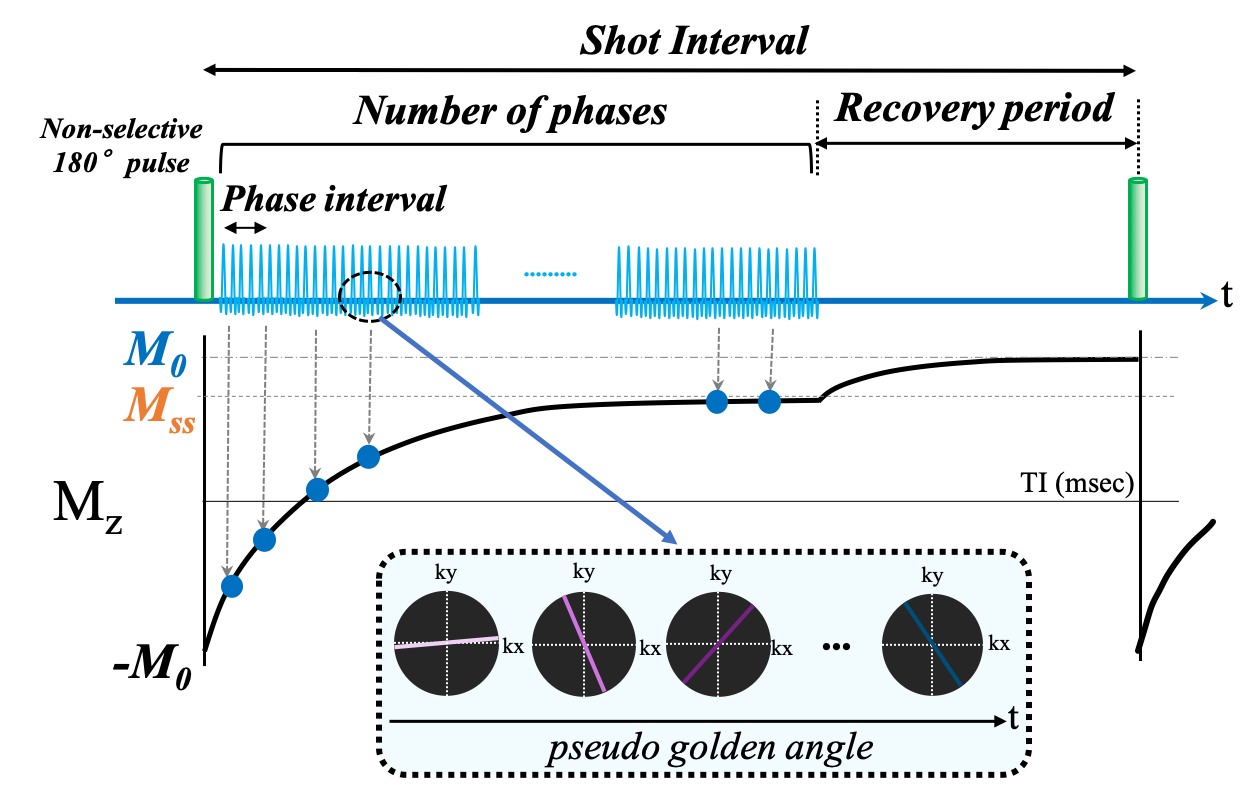
Figure 1: Detail of 3D-Radial LL. After applying a non-selective 180° pulse, T1W-TFE radial acquisition (stack-of-stars) was performed. Echoes were acquired continuously, but images were created for each TI by setting the phase interval and separating the k-space trajectory of the acquired data. A recovery time was set to recover to its equilibrium state before the next 180° pulse applied.
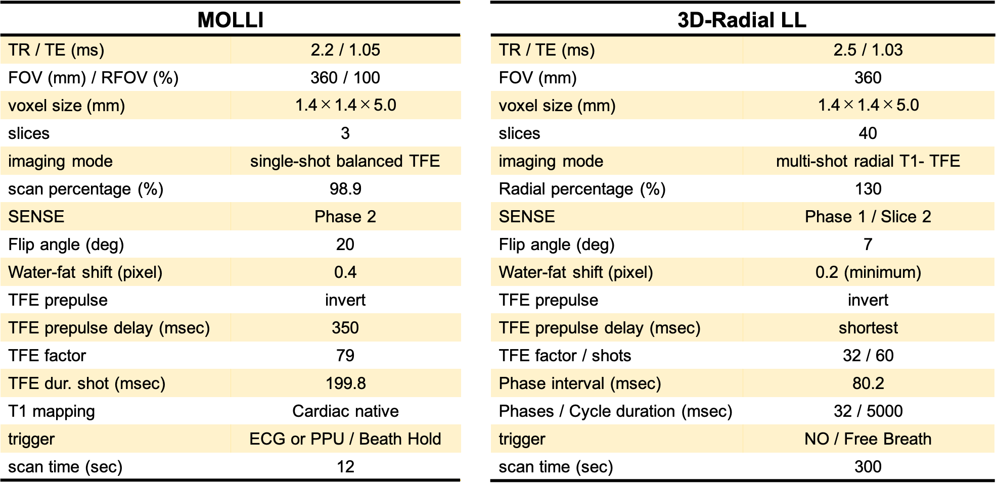
Figure 2: Imaging parameters for MOLLI and 3D-Radial LL. 3D-Radial LL allowed imaging without synchronization and breath-holding, and acquisition time was set to 5 minutes.
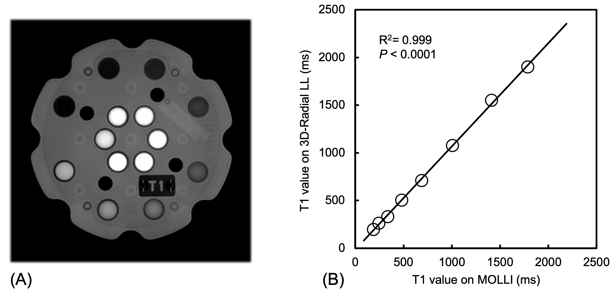
Figure 3: A phantom image (A) and comparison of T1 values obtained with MOLLI and 3D-radial LL in the phantom study (B). T1 values showed excellent correlation between on the MOLLI and 3D-Radial LL.

Figure 4: Comparison of T1 values obtained with MOLLI and 3D-radial LL in healthy volunteers. Correlations were observed in liver (A), pancreas (B), spleen (C) and paraspinal muscle (D).
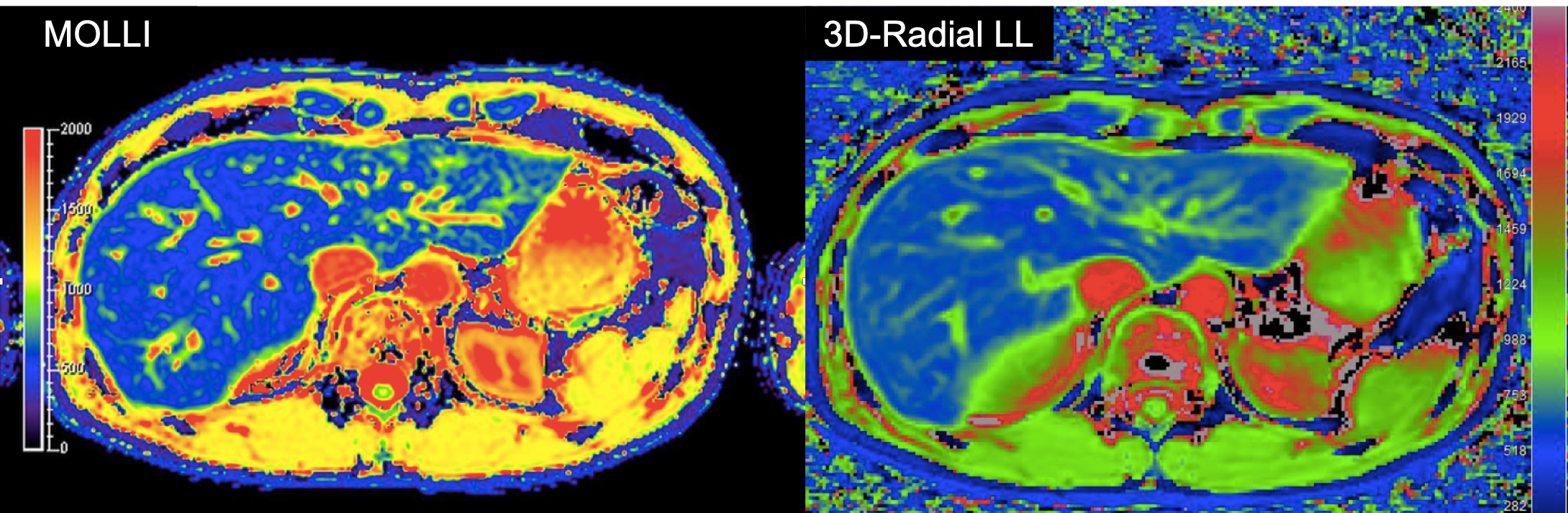
Figure 5: T1 map of a healthy volunteer obtained with MOLLI and 3D-Radial LL.
DOI: https://doi.org/10.58530/2023/2040