2034
An investigation of the performance of T2-weighted MR imaging with AI-assisted compressed sensing in routine clinical settings
Adiraju Karthik1, Apoorwa Devappa2, Aakaar Kapoor3, Dharmesh Singh4, and Dileep Kumar4
1Department of Radiology, Sprint Diagnostics, Jubilee Hills, Hyderabad, India, 2Department of Radiology, Mahadevappa Rampure Medical College, Kalaburagi, India, 3Department of Radiology, City X-Rays Scan & Clinical Private Limited, New Delhi, India, 4Central Research Institute, Global Scientific Collaborations, United Imaging Healthcare, New Delhi, India
1Department of Radiology, Sprint Diagnostics, Jubilee Hills, Hyderabad, India, 2Department of Radiology, Mahadevappa Rampure Medical College, Kalaburagi, India, 3Department of Radiology, City X-Rays Scan & Clinical Private Limited, New Delhi, India, 4Central Research Institute, Global Scientific Collaborations, United Imaging Healthcare, New Delhi, India
Synopsis
Keywords: Data Acquisition, Body, AI-assisted compressed sensing, T2-weighted Imaging
T2-weighted imaging (T2WI) is an essential diagnostic tool for several diseases. However, one of the challenges faced by patients and radiology departments is the longer scanning time of MR examinations. Recent advancements in artificial intelligence (AI) and deep learning techniques have made it able to acquire images quickly while preserving high-quality image resolution. In this study, the efficacy of a deep learning-based reconstruction technique termed AI-Assisted Compressed Sensing (ACS) was evaluated qualitatively and quantitatively using T2WI in routine clinical settings for brain, spine, knee, kidney and liver.Introduction
Magnetic resonance imaging (MRI) has been shown to be useful in the diagnosis of a variety of illnesses, including cancer, heart and vascular disease, and bone abnormalities. T2WI is an important MRI imaging technique that can enhance MRI's ability to diagnose various disorders. In recent years, significant work has been done to improve the field of view (FOV), resolution, and acquisition time of MRI sequences1. One of the issues faced by radiology departments and patients being tested is the longer examination duration, which makes it hard for certain patients to hold still during the examination, resulting to motion artifacts2. Longer scanning time not only introduces artefacts in acquired images but also considerably raises the cost and availability of health care, particularly in nations with a limited number of MR scanners. The signal-to-noise ratio (SNR) is primarily utilized in MRI for image interpretation and quality assurance; however, the SNR in MRI is inherently limited. As clinical examinations become more frequent, novel accelerated imaging is urgently needed to enable ultra-fast scanning while producing high-quality images3. AI-Assisted Compressed Sensing (ACS) is a recently-introduced deep learning-based reconstruction technique integrated with standard acceleration techniques to provide improved quality and faster scanning time4. ACS has previously been used successfully in a few body organs; however, its clinical performance in a standard clinical setting has not yet been evaluated to demonstrate its utility for other body organs. This study investigates the performance of T2WI with ACS in clinical settings for brain, spine, knee, kidney, and liver in terms of scanning duration, qualitative and quantitative parameters.Methods
MRI data acquisition In this study, 25 subjects were randomly recruited to undergo prospective MR examinations of the brain, spine, knee, liver, and kidney. Five people were selected for each body region, resulting in five MR datasets that were acquired for each body region, both with and without the use of the ACS approach utilizing various MR contrasts. All MR exams were performed on the 3T uMR780 system (United Imaging Healthcare Co. Ltd, Shanghai) at Sprint Diagnostics, Hyderabad, India. Before the examination, a consent form was signed by all subjects.Data processing
Qualitative Evaluation: A standard scoring system was developed to assess the quality of images in terms of artefacts in the images, the sharpness of tissue edges, the overall quality of the images, and the diagnostic effectiveness of the images. Table 1 provides more specific information about the scoring. Two radiologists (Radiologist 1 with over 5 years of experience and Radiologist 2 with over 12 years of experience) assessed all images and assigned ratings based on the parameters.
Quantitative Evaluation: The quantitative evaluation is done by measuring the Signal-to-Noise Ratio (SNR) and the Contrast-to-Noise Ratio (CNR) for each sequence and body region. To calculate these image quality measures, multiple areas of interest (ROIs) in different tissue locations were formed in the images to obtain average signal intensities and standard deviation.
All the statistical analysis was done in MedCalc,version-19.3 (MedCalc Software-Ltd). The Student's t-test was used to compare the qualitative and quantitative findings from the image assessments of both ACS and Non-ACS enabled images. Cohen's kappa coefficient (k) is also measured the inter-rater reliability for qualitative scores.
Results
According to a qualitative evaluation, the images obtained with ACS were either comparable or superior to images obtained without ACS in terms of overall quality, as shown in Figure-1. Figure-2 and 3 depict the mean score of all subjective assessments performed by the two radiologists for each body region. There was a fair agreement (k = 0.37) in the subjective scores between the two radiologists.Mean SNR (brain-38.6±1.32,spine-47.62±1.28,liver-45.51±2.20,kidney-45.36±2.56, knee-45.81±2.83) & mean CNR (brain-26.99±3.56,spine-31.90±4.08,liver-22.54±4.20, kidney-28.70±3.67, knee-31.56±2.45) with ACS are significantly higher (p<0.05) as compared to SNR (brain-36.35±0.81, spine-46.19±0.85,liver-43.25±1.37,kidney-43.41±1.62, knee-42.53±1.53) and CNR (brain-23.87±4.13,spine-29.30±4.37,liver-20.83±3.62,kidney-24.72±2.95,knee-29.48±2.33) obtained in images without ACS, as shown in Figure-4(a)&4(b). Figure-4(c) shows the total scanning time values for each body region.
Discussion
A clinical investigation of ACS technology developed by United Imaging Healthcare was conducted in this study to assess its utility in routine clinical settings for different body regions (brain, spine, liver, kidney,and knee) using T2WI. The current investigation found that the diagnostic quality of images obtained with ACS was comparable to or better than that of images obtained without ACS. In addition, the SNR and CNR of the ACS subgroup were greater than those of the non-ACS or conventional group, which is consistent with literature5. ACS has substantially shorter scan times for all body region sequences than non-ACS sequences, allowing for ultra-fast scans. It may be difficult for some patients with severe diseases to maintain a static condition for an extended amount of time during imaging studies due to discomfort, fluctuations in consciousness, and other variables, which lead to artefacts and reduces image quality. This problem can be overcome with ACS technology, which can also improve image quality and enable an ultra-fast scan.Conclusion
ACS technology not only significantly reduces scan time but also provides images with diagnostic quality and without artefacts, allowing this method to be clinically effective, particularly in routine clinical settings. A large cohort and multicenter study can provide more robust evidence for more extensive clinical applications.Acknowledgements
Authors would like to acknowledge the technical support of staff members at Sprint Diagnostics Private Limited and MR Application team members of United Imaging Healthcare for protocols optimization.References
- Wu W, Miller KL. Image formation in diffusion MRI: A review of recent technical developments. J Magn Reson Imaging. 2017; 46(3):646-662.
- Kozak BM, Jaimes C, Kirsch J, et al. MRI techniques to decrease imaging times in children. Radiographics. 2020; 40(2):485–502.
- Wang S, Su Z, Ying L, et al. Accelerating magnetic resonance imaging via deep learning. In: IEEE 13th International Symposium on Biomedical Imaging (ISBI). 2016: 514–517.
- Wang S, Cao G, Wang Y, et al. Review and Prospect: Artificial Intelligence in Advanced Medical Imaging. Frontiers in Radiology. 2021; 781868: 1:18.
- Zhao Y, Peng C, Wang S, et al. The feasibility investigation of AI-assisted compressed sensing in kidney MR imaging: an ultra-fast T2WI imaging technology. BMC Med Imaging. 2022; 4; 22(1):119.
Figures
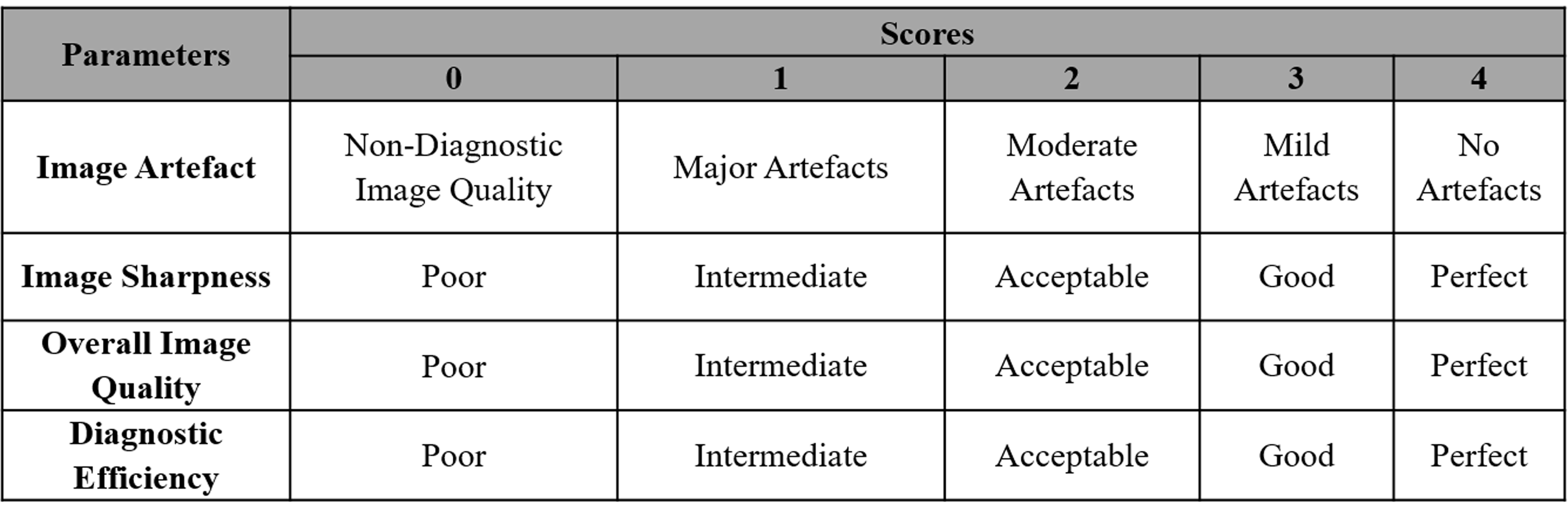
Table 1: Scoring criteria for
qualitative analysis
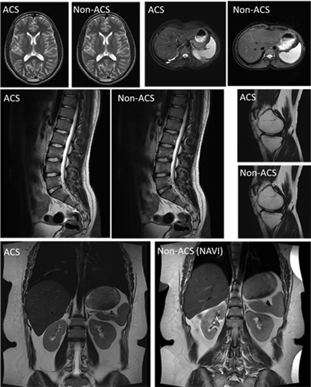
Figure
1: Example of T2-weighted FSE images acquired in various body regions with and
without ACS
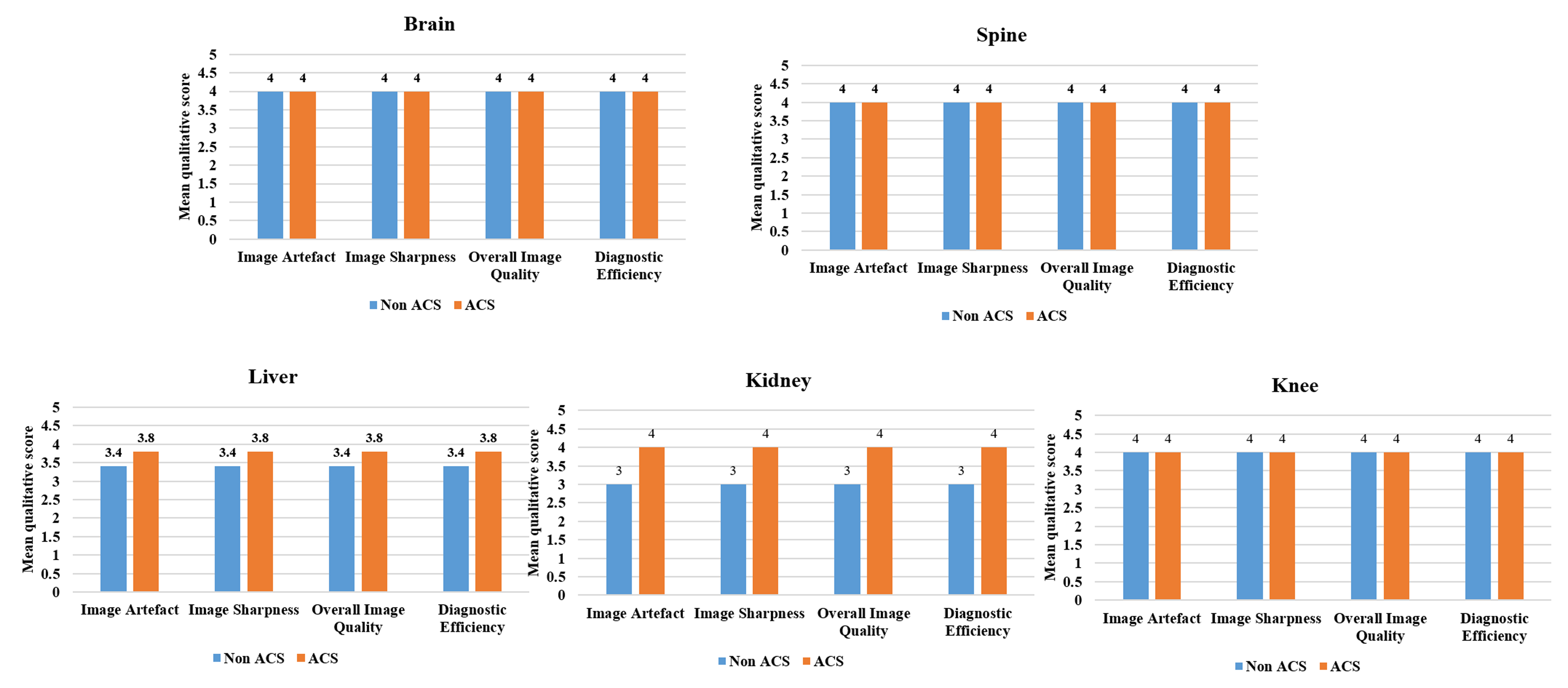
Figure 2: Mean qualitative score of ACS vs. Non ACS for
all body regions read by radiologist 1
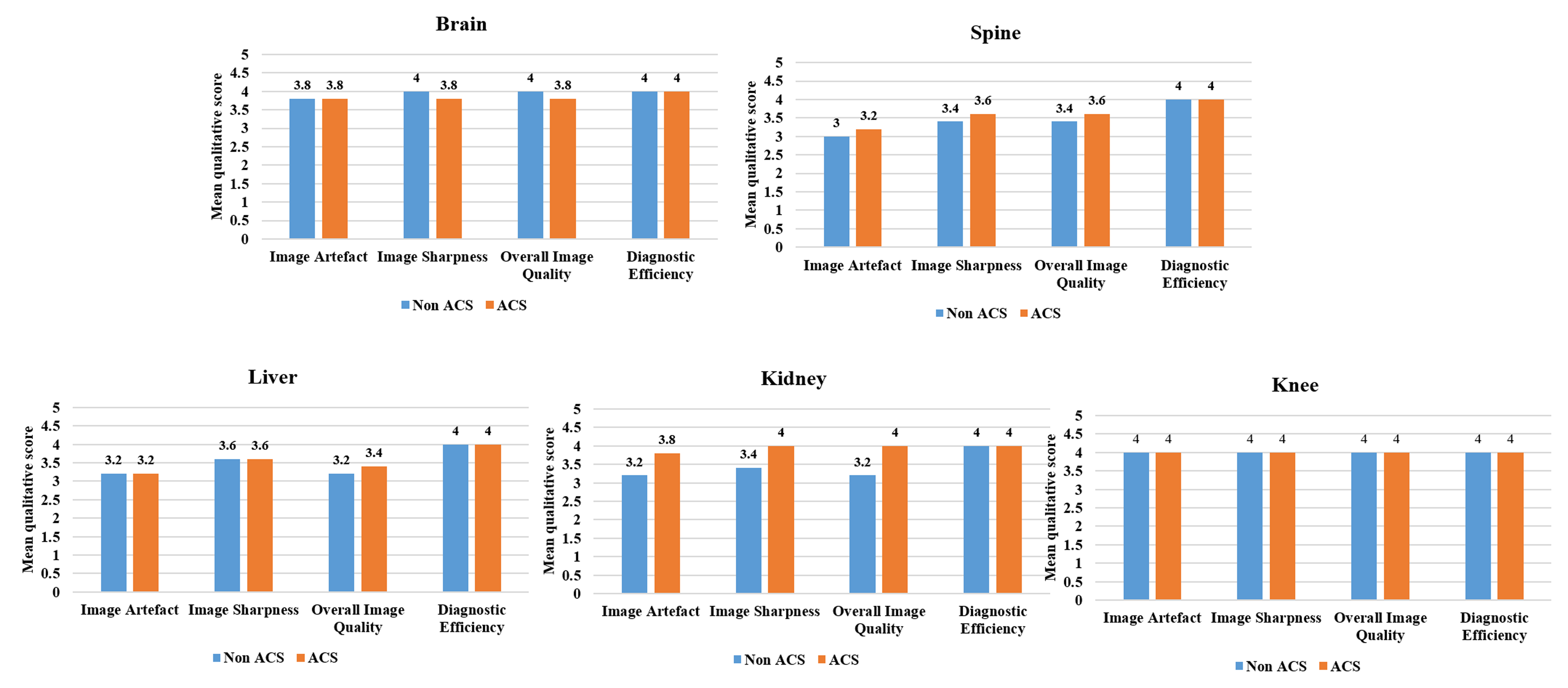
Figure 3: Mean qualitative score of ACS vs. Non ACS for
all body regions read by radiologist 2
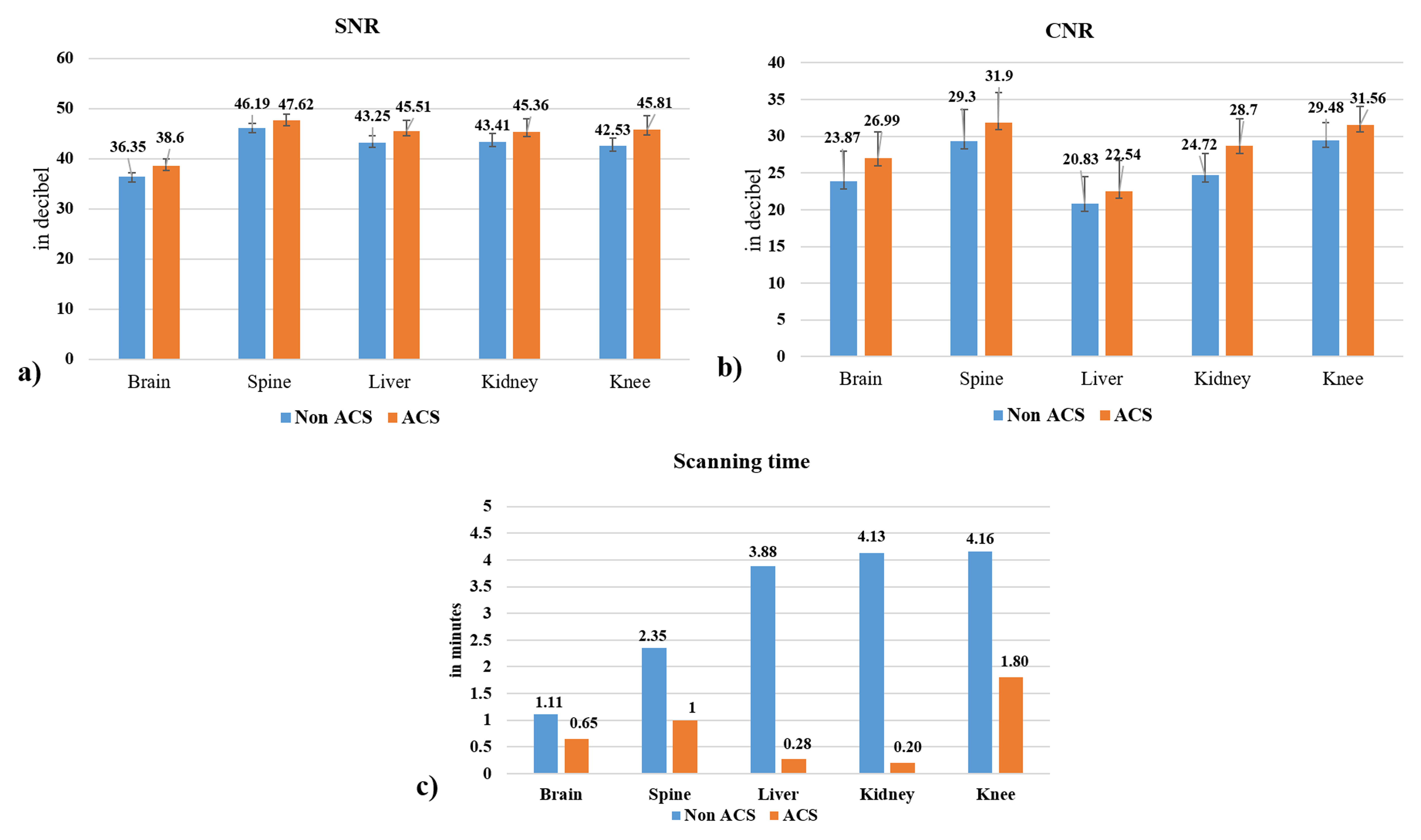
Figure
4
(a):
Mean
SNR values across all subjects of different body regions for Non-ACS vs. ACS T2
FSE sequence, (b): Mean
CNR values across all subjects of different body regions for Non-ACS vs. ACS T2
FSE sequence, and (c):
Total
scanning time across all sequences for different body regions
DOI: https://doi.org/10.58530/2023/2034