2032
Model-based image reconstruction for highly accelerated point spread function encoded echo planar imaging1Radiology, Mayo Clinic, Rochester, MN, United States, 2Mayo Clinic Graduate School of Biomedical Sciences, Rochester, MN, United States
Synopsis
Keywords: Image Reconstruction, Signal Representations
A model-based image reconstruction (MBIR) framework is developed for highly accelerated point spread function (PSF) encoded echo planar imaging (EPI). The reconstruction accounts for known physics, and utilizes a subspace representation with local low-rank (LLR) regularization along with a variable splitting solution. Comparisons with images obtained through standard methods demonstrate considerable artifact reduction and sharpness preservation through MBIR. MBIR accommodates arbitrary sampling trajectories along the PSF-encoding dimension, enabling reconstruction of quality images from less than one minute of scan time; and we demonstrate performance boosts with nonstandard trajectories precluded by conventional reconstruction methods.Introduction
Echo planar imaging (EPI)1 comprises fast acquisition facilitating advanced MR applications. Low effective phase encoding bandwidth and system non-idealities limit single-shot EPI practically to relatively low-resolution exams. Point spread function (PSF)-encoded EPI2-4 utilizes multi-shot auxiliary phase encoding, enabling projection into undistorted images following reconstruction (through standard methods) of an interim PSF volume. Despite in-plane and reduced field of view (rFOV) acceleration, the number of shots required for PSF-EPI remains relatively high with classical methods; scan time remains a limitation. While shots are considerably reducible with tilted CAIPI5, this requires PSF-encoded calibration. Extending to time-resolved acquisition with subspace reconstruction6 enables multi-contrast imaging, but in current form, requires algorithmic $$$B_0$$$ updates.This work extends prior preliminary efforts7 to investigate model-based image reconstruction (MBIR)8 for highly accelerated PSF-EPI, combining known physics, partial temporal separability, and regularization to directly estimate undistorted images with arbitrary sampling of the PSF space.
Theory
A scalar forward-mapped signal model for PSF-EPI is$$\mathbf{G}[u,p,v,c]\approx\sum_i\sum_l\mathbf{S}[i,l,c]\mathbf{f}[i,l]e^{-\mathbf{z}[i,l]\tau[v]}e^{-\jmath \mathbf{k}_x[u]\mathbf{x}[i]}e^{-\jmath \mathbf{k}_y[v]\mathbf{y}[l]}e^{-\jmath \mathbf{k}_a[p]\mathbf{y}[l]}+\mathbf{\epsilon}[ u,p,v,c ]$$
where $$$u,p,v,$$$ and $$$c$$$ respectively index measurements at readout, pass (for auxiliary PSF encoding), phase-encoding, and coils; $$$\mathbf{S}$$$ are coil sensitivity profiles; $$$\mathbf{f}$$$ and $$$\mathbf{z}$$$ are unknown magnetization and rate map8 arrays of interest, the latter encompassing relaxation and off-resonance effects; $$$\tau[v]$$$ denotes post-RF acquisition time for $$$v$$$th phase encodings; $$$\mathbf{k}_x, \mathbf{k}_y,$$$ and $$$\mathbf{k}_a$$$ respectively are $$$k$$$-space encoding along readout, phase, and auxiliary (PSF); $$$\mathbf{x}[i], \mathbf{y}[l]$$$ are spatial coordinates and associated indices; and $$$\mathbf{\epsilon}$$$ is a zero-mean complex Gaussian noise instance. An abstracted signal model written as the 3rd tensor unfolding9 of $$$\mathbf{G}$$$ is
$$\mathbf{G}_{[3]}=\sum_{v,c}\big(\delta_c\otimes\mathbf{\Lambda}_v\text{diag}(\mathbf{S}_{[3]}\delta_c)\big)\mathbf{M}\mathbf{X}\mathbf{\psi}_K^*\delta_v\delta_v^T+\mathbf{\epsilon}_{[3]}$$
where $$$\delta$$$ is Kronecker's delta, and the transform operator is
$$\mathbf{\Lambda}_v\overset{\Delta}{=}\big(\mathbf{\Xi}_\text{psf}\mathbf{F}_\text{psf}\otimes\mathbf{I}_{N_x}\big)\big(\text{diag}((\mathbf{\Xi}_\text{pe}\mathbf{F}_\text{pe})^T\delta_v)\otimes\mathbf{I}_{N_x}\big)\big(\mathbf{I}_{N_y}\otimes\mathbf{F}_\text{ro}\big),$$
where $$$\mathbf{F}_\text{ro},\mathbf{F}_\text{pe}$$$ and $$$\mathbf{F}_\text{psf}$$$ are respectively Fourier transforms along readout, phase-encoding, and PSF encoding, with associated subsampling operators $$$\mathbf{\Xi}$$$. $$$\mathbf{I}_{N}$$$ is an identity matrix of size $$$N$$$. $$$N_x$$$ and $$$N_y$$$ are image dimensions; $$$N_{v}, N_{c}$$$, and $$$N_{p}$$$, the number of phase encodings, channels, and PSF encodings.
This signal model has embedded representations to reduce the search space and/or exploit low-rank characteristics in reconstruction. First, $$$\mathbf{M}$$$ is an undersampled identity matrix confining unknown voxels to known spatial support10. Next, partial temporal separability11 is directly embedded: we target a composite unknown $$$\mathbf{\theta}\in\mathbb{C}^{N_i \times N_v}$$$ comprising both magnetization and rate map effects evolving temporally through phase-encodings, confined to known support where $$$N_i$$$ is the number of voxels of interest. We approximate this unknown with $$$\mathbf{\theta}=\mathbf{\theta}\mathbf{\psi}\mathbf{\psi}^*\approx\mathbf{\theta}\mathbf{\psi}_K\mathbf{\psi_K^*}\equiv\mathbf{X}\mathbf{\psi}_K^*$$$, where $$$\mathbf{\psi}$$$ is an orthonormal temporal basis and $$$\mathbf{\psi}_K=\text{span}\begin{bmatrix}\mathbf{\phi}_1&\dots&\mathbf{\phi}_K\end{bmatrix}$$$12. The reconstruction target is the set $$$\mathbf{X}\in\mathbb{C}^{N_i \times K}$$$ of basis expansion coefficients, and $$$\mathbf{\psi}_K\in\mathbb{C}^{N_v \times K},K<N_v$$$, is a matrix of precomputed temporal basis vectors6,12.
To facilitate subsampling operator decoupling in a variable splitting solution13,14, we rewrite $$$\mathbf{\Lambda}_v$$$ in an alternate form and approach the objective
$$\mathcal{J}(\mathbf{X})\overset{\Delta}{=}\big\Vert\sum_{c,v}(\mathbf{I}_{N_c}\otimes\mathbf{H})(\delta_c\otimes\mathbf{K}_v\text{diag}(\mathbf{S}_{[3]} \delta_c))\mathbf{M}\mathbf{X}\mathbf{\psi}_K^*\delta_v\delta_v^T-\mathbf{G}_{[3]} \big\Vert_F^2+\alpha\sum_{r\in\zeta}\big\Vert\mathbf{E}_r\mathbf{M}[\mathbf{X}\ |\ \overline{\mathbf{X}}]\big\Vert_*$$
where $$$\Vert\cdot\Vert_F$$$ and $$$\Vert\cdot\Vert_*$$$ are Frobenius and nuclear norms. We promote local low-rank (LLR)15,16 on weighting coefficients describing temporal evolution of images12, appended across columns with conjugates to encourage smooth phase17. $$$\mathbf{E}_r$$$ is an extraction operator for block $$$r$$$ from the set $$$\zeta$$$ for LLR promotion; $$$\alpha$$$ is a regularization parameter. We solve this with alternating direction method of multipliers (ADMM)18, applying variable splitting described in Figure 2.
Methods
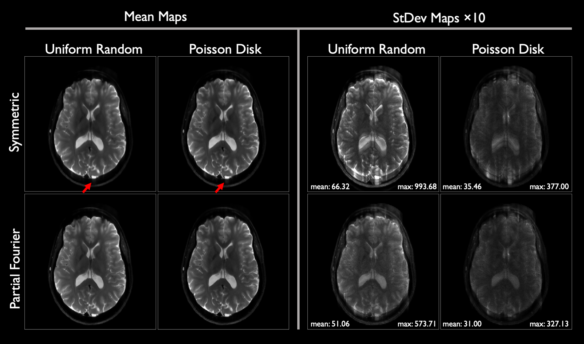
Following an IRB-approved protocol, volunteers were scanned on a compact 3T scanner19 with accelerated T2-weighted PSF-EPI (T2-DIADEM)20 with parameters in Figure 1. Partial Fourier acceleration was applied only along PSF. Coil sensitivities were generated with ESPIRiT21. In a conventional reconstruction, PSF volumes were reconstructed by unfolding in-plane images with SENSE22. Partial Fourier PSF reconstruction comprised zero-filling with rFOV unfolding4. Conversely, MBIR was run. Temporal basis vectors were precomputed following simulations6,12 using field map data from a prior study23; we set $$$K=31$$$. PSF-EPI data were retrospectively truncated from 28 (7/8 PSF-PF, 2:09 m:s actual scan time) to 18 shots (9/16 PSF-PF, 1:23 m:s simulated) and reconstructed for comparison. Additionally, in vivo brain data were acquired with full PSF-encoding, then retrospectively undersampled uniformly $$$12\times$$$ along PSF, then reconstructed. Further, data were undersampled with 50 distinct 12-shot random sampling instances (55.2 s simulated scan time) in uniform-random and 1D Poisson-disk patterns, with and without partial Fourier along PSF, and reconstructed with MBIR. Figure 1 depicts sampling variants.Reconstruction was completed using Matlab. $$$k$$$-space preprocessing comprised ramp sampling and Nyquist ghost correction using vendor-provided tools. Image post-processing steps included zero-padding, Fermi filtering, scaling, and vendor-provided gradient nonlinearity correction.
Results
Figure 3 illustrates reconstruction comparisons between full-duration and truncated data with prospective acceleration. Figure 4 demonstrates performance with subsampling to <1 minute of scan time. In both cases, for each sampling instance, MBIR exhibits fewer residual artifacts than standard. Sharpness is preserved despite aggressive partial PSF encoding. Random encoding suppresses artifacts furthermore. Figure 5 shows MBIR performance with distinct random sampling patterns, demonstrating best results with 1D Poisson disk and partial encoding.Discussion and Conclusion
Our MBIR framework for PSF-EPI directly reconstructs high-quality undistorted images. Reconstructions are superior relative to standard methods with uniform acceleration, exhibiting fewer artifacts and improved detectability. MBIR accommodates arbitrary PSF sampling, unachievable by classical reconstructions, producing quality images from <1 minute of scan data$$$-$$$with nonstandard sampling achieving best performance. MBIR thus extends PSF-EPI beyond previously possible capabilities. Ongoing work includes a radiologists' evaluation.Acknowledgements
This work is supported by NIH U01 EB024450, the National Science Foundation Graduate Research Fellowship Program, and Mayo Clinic Graduate School of Biomedical Sciences.References
1. Mansfield P. Multi-planar image formation using NMR spin echoes. J Phys C: Solid State Phys. 1977;10:L55.
2. Robson MD, Gore JC, Constable RT. Measurement of the point spread function in MRI using constant time imaging. Magn Reson Med. 1997;38(5):733-740.
3. Zaitsev M, Hennig J, Speck O. Point spread function mapping with parallel imaging techniques and high acceleration factors: Fast, robust, and flexible method for echo-planar imaging distortion correction. Magn Reson Med. 2004;52(5):1156-1166.
4. In MH, Posnansky O, Speck O. High-resolution distortion-free diffusion imaging using hybrid spin-warp and echo-planar PSF-encoding approach. Neuroimage 2017;148:20-30.
5. Dong Z, Wang F, Reese TG, et al. Tilted-CAIPI for highly accelerated distortion-free EPI with point spread function (PSF) encoding. Magn Reson Med. 2019;81(1):377-392.
6. Z Dong, F Wang, TG Reese, et al. Echo planar time-resolved imaging with subspace reconstruction and optimized spatiotemporal encoding. Magn Reson Med. 2020;84(5):2442-2455.
7. Meyer NK, In MH, Kang D, et al. Model-based PSF-encoded multi-shot EPI reconstruction with low-rank constraints. Proc Intl Soc Magn Reson Med 2022;3484.
8. Fessler J. Model-based image reconstruction for MRI. IEEE Signal Process Mag. 2010;27(4):81-89.
9. Kolda TG, Bader BW. Tensor Decompositions and Applications. SIAM Review 2009;51(3):455-500.
10. Le M, Ramani S, Fessler J. An Efficient Variable Splitting Based Algorithm for Regularized SENSE Reconstruction with Support Constraint. Proc Intl Soc Mag Reson Med 2013;2654.
11. Liang ZP. Spatiotemporal Imaging with Partially Separable Functions. Proc IEEE ISBI 2007;988-991.
12. Tamir J, Uecker M, Chen W, et al. T2 shuffling: Sharp, multicontrast, volumetric fast spin-echo imaging. Magn Reson Med. 2017;77(1):180-195.
13. Ramani S, Fessler J. Parallel MR image reconstruction using augmented Lagrangian methods. IEEE Trans Med Imaging. 2011;30(3):694-706.
14. Trzasko J, Borisch E, Weavers P, et al. An Efficient ADMM-based Sparse Reconstruction Strategy for Multi-Level Sampled MRI. Proc ACSSC 2014;411-414.
15. Trzasko J, Manduca A. Local versus Global Low-Rank Promotion in Dynamic MRI Series Reconstruction. Proc Intl Soc Mag Reson Med 2011;4371.
16. Candès E, Sing-Long C, Trzasko J. Unbiased risk estimates for singular value thresholding and spectral estimators. IEEE Trans Signal Process. 2013;61(19):4643-4657.
17. Levine E, Hargreaves B. An Image Domain Low Rank Model for Calibrationless Reconstruction of Images with Slowly Varying Phase. Proc Intl Soc Mag Reson Med 2015;3624.
18. Boyd S, Parikh N, Chu E, et al. Distributed Optimization and Statistical Learning via the Alternating Direction Method of Multipliers. Found Trends Mach Learn. 2010;3(1):1-122.
19. Foo TK, Laskaris E, Vermilyea M, et al. Lightweight, compact, and high-performance 3T MR system for imaging the brain and extremities. Magn Reson Med. 2018;80(5):2232-2245.
20. In MH, Campeau N, Huston III J, et al. Rapid T2-DIADEM Echo-Planar Imaging as an Alternative to T2-FSE: A Clinical Feasibility Study. Proc Intl Soc Mag Reson Med 2021;0838.
21. Uecker M, Lai P, Murphy M, et al. ESPIRiT - An eigenvalue approach to autocalibrating parallel MRI: Where SENSE meets GRAPPA. Magn Reson Med. 2014;71(3):990-1001.
22. Pruessmann KP, Weiger M, Scheidegger MB, et al. SENSE: sensitivity encoding for fast MRI. Magn Reson Med. 1999;42(5):952-962.
23. In MH, Tan E, Trzasko J, et al. Distortion-free imaging: A double encoding method (DIADEM) combined with multiband imaging for rapid distortion-free high-resolution diffusion imaging on a compact 3T with high-performance gradients. J Magn Reson Imaging. 2020;51(1):296-310.
Figures