2013
Motion robust multi-shot EPI of the abdomen using VFA-FLEET1Radiology, Harvard Medical School, Boston, MA, United States, 2Computational Radiology Laboratory, Boston Children's Hospital, Boston, MA, United States
Synopsis
Keywords: Motion Correction, Body
Multi-shot EPI can be a promising technique to improve the resolution limits of the single shot EPI. However, its application has been so far limited in the abdominal MRI, as phase errors between shots due to unavoidable breathing motion creates artifacts in the images. Here, we tested a Variable-Flip-Angle Fast Low-angle Excitation Echo planar Technique (VFA-FLEET) pulse sequence and showed that it can be used to create high-resolution motion-robust images with high geometric fidelity without breath holding.Introduction
Rapid MRI is crucial for abdominal imaging due to the critical limitation with regard to breath-holding [1,2]. Single-shot echo planar imaging (SS-EPI) can be used to acquire fast snapshots of the region of interest. However, the spatial resolution is inherently limited in SS-EPI since acquiring high resolution EPI images requires long readout times, resulting in geometric distortions and spatial blurring. Multi-shot EPI (MS-EPI) acquisition strategies can potentially achieve higher spatial resolution and fidelity, and they recently gained attraction in fast protocols for brain MRI [3]. However, MS-EPI is generally susceptible to motion-induced phase errors, which are unavoidable when imaging the abdomen. Recently, the Variable-Flip-Angle Fast Low-angle Excitation Echo planar Technique (VFA-FLEET) pulse sequence [4] was suggested to improve the motion robustness of MS-EPI, as segments of a given slice are acquired sequentially. In this work, we implemented and evaluated the VFA-FLEET sequence for motion-robust MS-EPI imaging of the abdomen.Methods
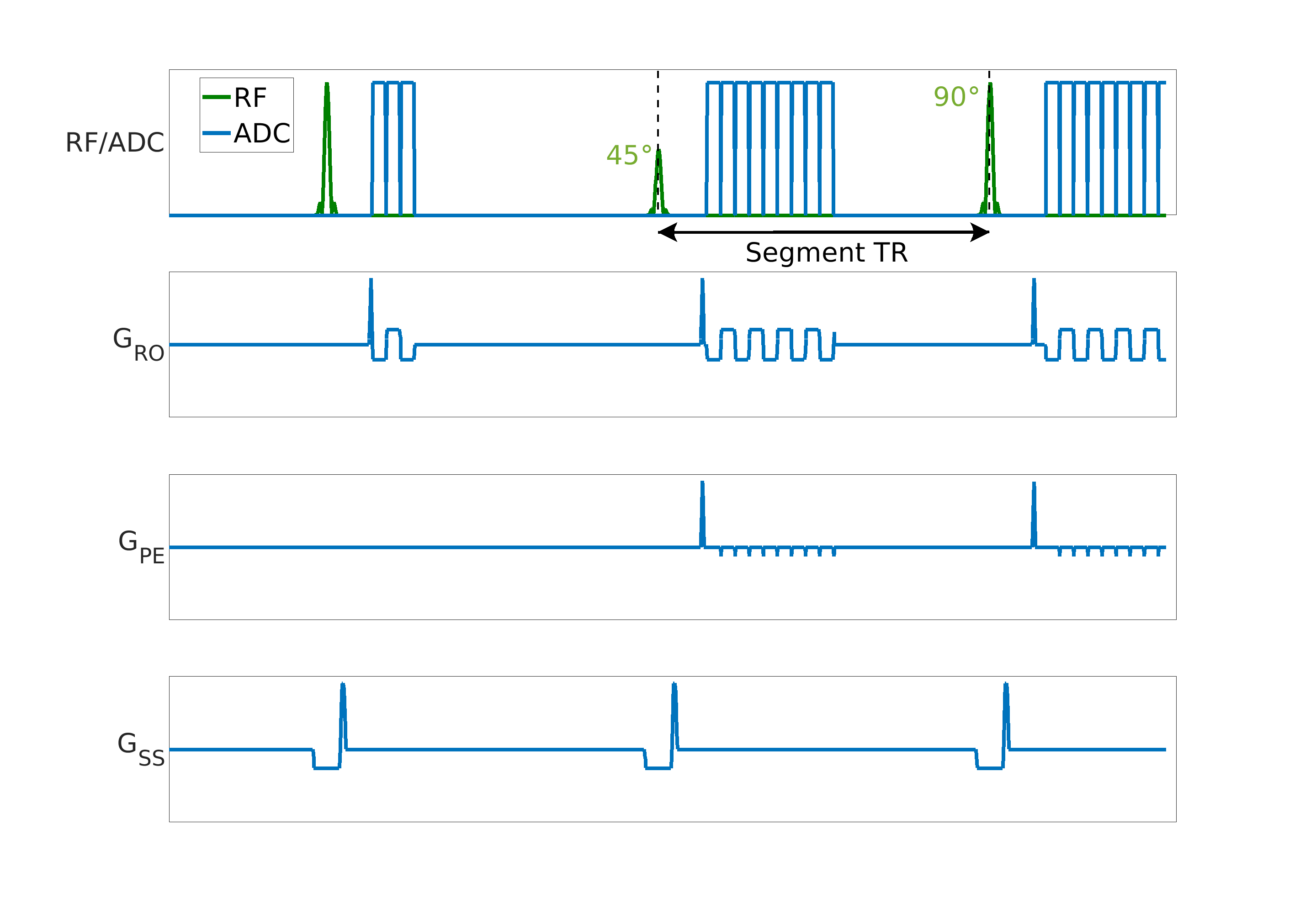
TheoryIn FLEET, the time between segments of data acquired in multiple shots is minimized by acquiring all segments of a given slice (i.e. multiple shots) sequentially, reducing susceptibility to motion and field changes between shots [5,6]. To maximize the SNR and avoid signal dropout between shots, a variable flip angle approach was suggested [4,5]. The ideal flip angles can be calculated by assuming that the longitudinal relaxation is negligible and the longitudinal magnetization available immediately before each RF excitation pulse is a function of both the previous excitation pulse’s rotation parameters and the longitudinal magnetization available immediately before the previous excitation pulse. Given that the flip angle of the last shot is 90° (i.e., 𝛼last=90°), previous flip angles can be recursively calculated as 𝛼i-1=tan-1(sin(𝛼i)) [4,5]. Figure 1 shows the pulse sequence diagram for a 2-shot VFA-FLEET sequence.
Phantom Study
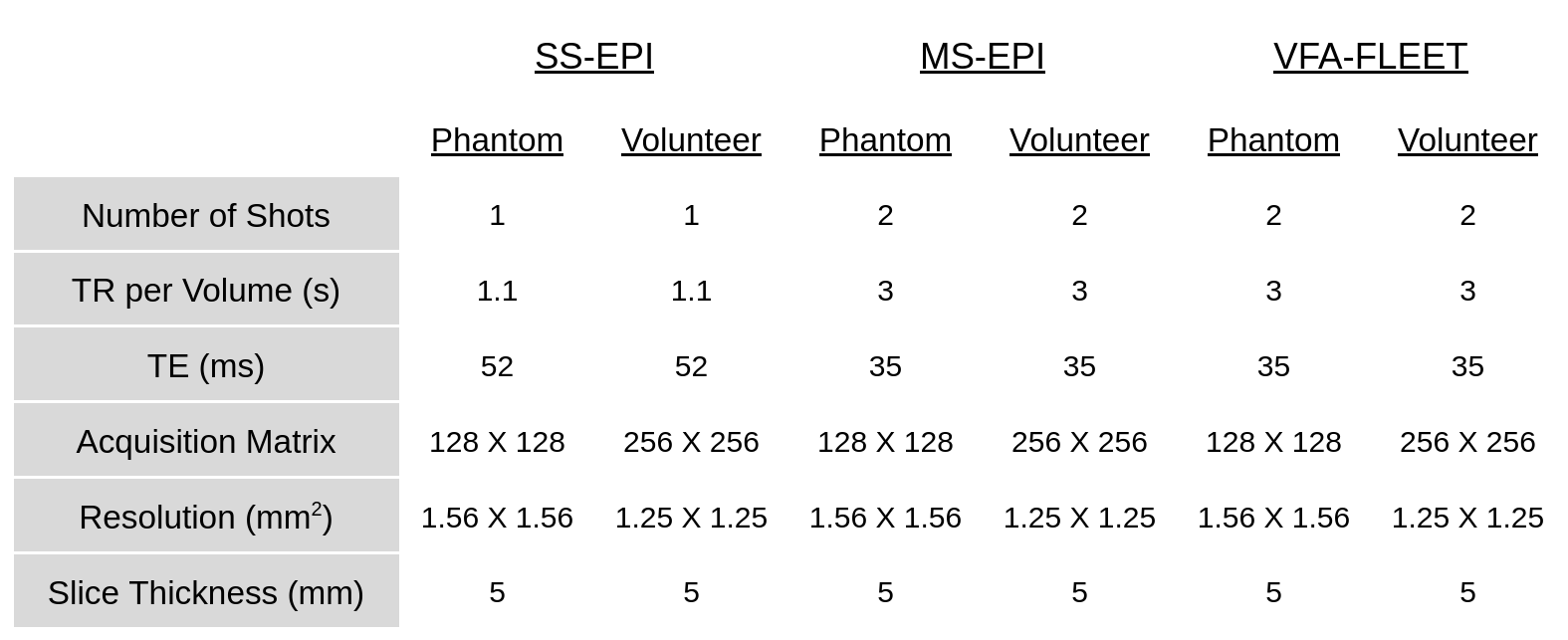
An ADNI phantom was scanned at 3T (MAGNETOM Prisma, Siemens Healthcare, Erlangen) using a 20-channel head coil. The imaging protocol consisted of SS-EPI, MS-EPI and two-shot EPI with VFA-FLEET. The imaging parameters are summarized in Table 1. The scans were acquired with (i) no disturbance and (ii) a water bottle rolling in and out at ~10 seconds intervals to induce phase errors between shots/images. We acquired 20 repetitions of each sequence.
Volunteer Study
Following written informed consent, three subjects (aged 22-41 years) were scanned at 3T (same as above) using a 30-channel body coil following the same protocol as in the phantom scans. Two sets of images were acquired: In the first set subjects were instructed to breathe normally, in the second set they were instructed to hold their breath. We acquired 20 repetitions of each sequence for the scans without breath holding (~1 min for MS-EPI sequences) and 5 repetitions for breath hold scans (15 seconds for MS-EPI sequences).
Results
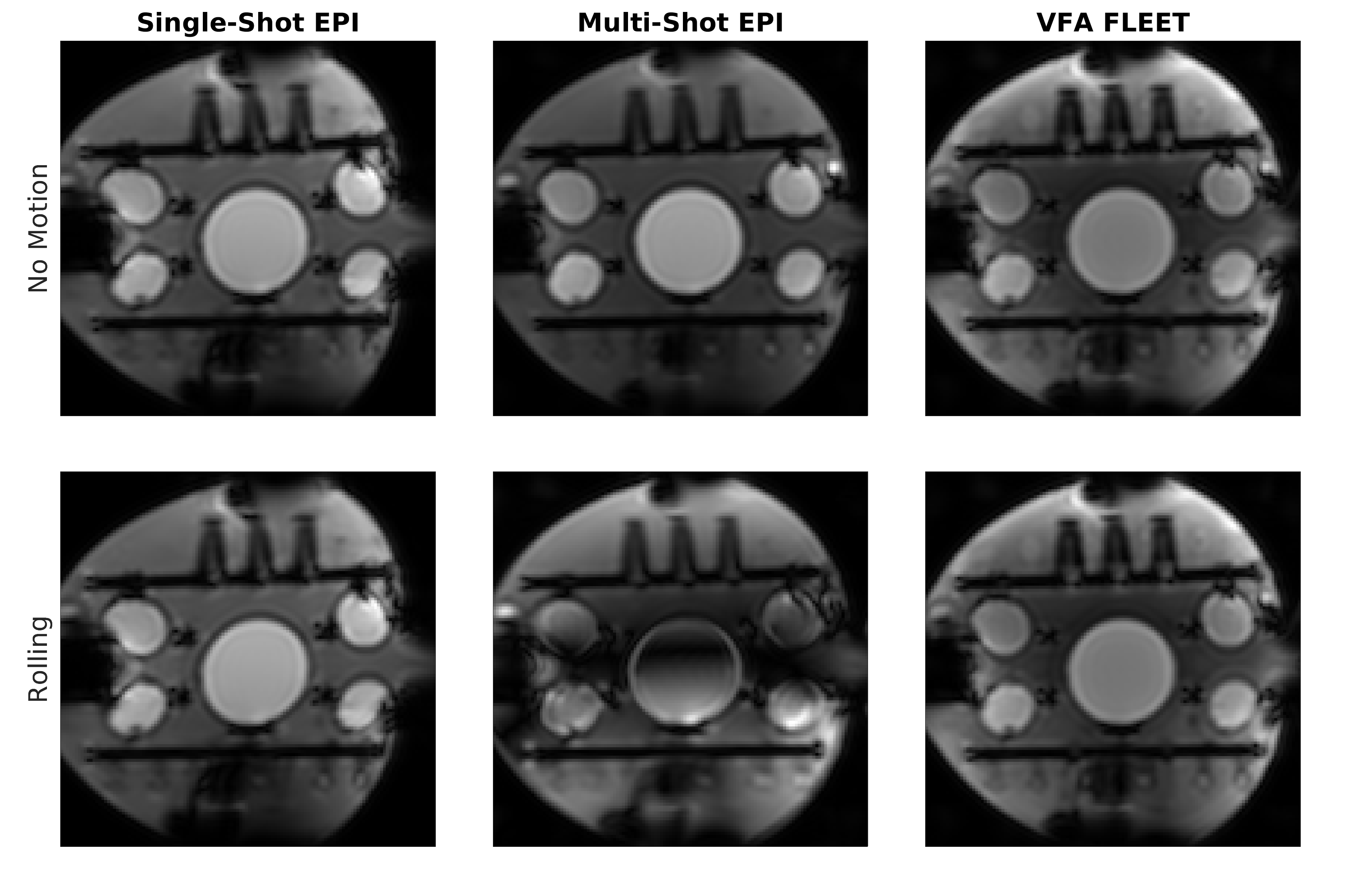
Figure 2 shows a sample slice from the phantom experiment with and without induced phase errors. Both VFA-FLEET and MS-EPI had reduced distortion artifacts as the readout time is halved with these techniques compared to SS-EPI. As can be seen, MS-EPI images have visible artifacts when there is a phase mismatch between shots, induced by the rolled water bottle. This artifact is minimized using the VFA-FLEET technique. Figure 3 shows results from a volunteer scan. When the subject performs a breath hold, all the sequences generate robust images. However, as expected during breathing, MS-EPI had large artifacts due to phase mismatch between shots. SS-EPI and VFA-FLEET show improved temporal stability compared to MS-EPI. VFA-FLEET generated motion robust images with reduced distortions.Discussion and Conclusion
In this work, we have implemented a VFA-FLEET sequence and performed an initial evaluation of the technique in improving motion robustness of MS-EPI acquisitions for imaging the abdomen. Our results demonstrate that the sequence is capable of generating images with minimal artifacts even when the subject is taking deep breaths, at the cost of a slight reduction in SNR (i.e. square root of number of shots). Future work will concentrate on creating a spin-echo based VFA-FLEET sequence to apply this technique to diffusion MRI of the abdomen, and comparison of our results with other multi-shot techniques that reduce the motion sensitivity such as MUSE [7].Acknowledgements
This work was supported in part by NIH grants R01 EB019483, R01 NS121657, R01 DK125561, R21 DK123569, R21 EB02962, S10 OD025111, and a pilot grant (PP-1905-34002) from the National Multiple Sclerosis Society.References
1. Gallo-Bernal, S., Bedoya, M.A., Gee, M.S. and Jaimes, C., 2022. Pediatric magnetic resonance imaging: faster is better. Pediatric Radiology, pp.1-15
2. Jaimes, C., Kirsch, J.E. and Gee, M.S., 2018. Fast, free-breathing and motion-minimized techniques for pediatric body magnetic resonance imaging. Pediatric radiology, 48(9), pp.1197-1208
3. Clifford, B., Conklin, J., Huang, S.Y., Feiweier, T., Hosseini, Z., Goncalves Filho, A.L.M., Tabari, A., Demir, S., Lo, W.C., Longo, M.G.F. and Lev, M., 2022. An artificial intelligence‐accelerated 2‐minute multi‐shot echo planar imaging protocol for comprehensive high‐quality clinical brain imaging. Magnetic Resonance in Medicine, 87(5), pp.2453-2463
4. Berman, A.J., Grissom, W.A., Witzel, T., Nasr, S., Park, D.J., Setsompop, K. and Polimeni, J.R., 2021. Ultra‐high spatial resolution BOLD fMRI in humans using combined segmented‐accelerated VFA‐FLEET with a recursive RF pulse design. Magnetic resonance in medicine, 85(1), pp.120-139
5. Mansfield, P., 1984. Spatial mapping of the chemical shift in NMR. Magnetic Resonance in Medicine, 1(3), pp.370-386
6. Polimeni, J.R., Bhat, H., Witzel, T., Benner, T., Feiweier, T., Inati, S.J., Renvall, V., Heberlein, K. and Wald, L.L., 2016. Reducing sensitivity losses due to respiration and motion in accelerated echo planar imaging by reordering the autocalibration data acquisition. Magnetic resonance in medicine, 75(2), pp.665-679
7. Chen, N.K., Guidon, A., Chang, H.C. and Song, A.W., 2013. A robust multi-shot scan strategy for high-resolution diffusion weighted MRI enabled by multiplexed sensitivity-encoding (MUSE). Neuroimage, 72, pp.41-47
Figures