2009
SPAMM Tagged EPI Acquisition for Assessment of Geometric Distortion Caused by an Endorectal Coil
Ken-Pin Hwang1, R. Jason Stafford1, and Tharakeswara K. Bathala2
1Department of Imaging Physics, The University of Texas M.D. Anderson Cancer Center, Houston, TX, United States, 2Department of Abdominal Imaging, The University of Texas M.D. Anderson Cancer Center, Houston, TX, United States
1Department of Imaging Physics, The University of Texas M.D. Anderson Cancer Center, Houston, TX, United States, 2Department of Abdominal Imaging, The University of Texas M.D. Anderson Cancer Center, Houston, TX, United States
Synopsis
Keywords: Artifacts, System Imperfections: Measurement & Correction
Diffusion weighted imaging is an essential sequence for diagnosis of prostate cancer but is often impacted by distortion. Current existing phantoms designed for assessing geometric accuracy do not sufficiently simulate the environmental factors that affect distortion in diffusion weighted EPI. In this work we apply a rectilinear tagging technique to visualize and understand patterns of distortion caused by an endorectal coil immersed in water when imaged with a PI-RADS compliant protocol. Maximum spatial distortion including regions of spatial collapse was observed near the coil element. Observed distortion pattern supports proper centering of the coil to avoid distortion.Introduction
Diffusion weighted imaging (DWI) is an essential sequence for diagnosis of prostate cancer and is one of the two core sequences in the PI-RADS reporting system. However, DWI is often impacted by distortion artifacts that confound the alignment of structures and modulate pixel intensities. Distortion in diffusion weighted EPI (DW-EPI) is induced by various causes, including magnetic field inhomogeneity, subject susceptibility, and eddy currents in the MR system. However, the amount of observed distortion also depends on the subject, imaging parameters, and sequence options. Current phantoms designed for assessing geometric accuracy do not sufficiently simulate the environmental factors affecting distortion in DW-EPI. A simple, multi-slice technique for visualizing distortion was designed for use with a wide variety of phantoms and sequence variations by using periodic saturation bands to create virtual landmarks1. Without the need for physical landmarks, phantoms can be designed to better simulate a wider range of clinical scenarios, enabling the investigation of the independent causes of distortion, including object-induced distortions. In this work, we apply this imaging technique to visualize and understand patterns of distortion caused by an endorectal coil.Methods
The proposed methodology utilizes a saturation technique known as Spatial Modulation of Magnetization (SPAMM) that tags the imaging subject with saturated grid lines2,3. Originally designed for tracking cardiac motion, these grids are applied here to assess spatial differences between diffusion weighting directions and/or b-values. The saturation preparation sequence as implemented in this work consisted of binomially weighted (e.g. 1-3-3-1) pulses interleaved with gradient blips along the frequency encode direction, followed by the same sequence with gradient blips in the phase encode direction. The gradient area of the blips was set to produce line spacings of 2.0 cm. Total time for the preparation sequence was about 9 msec.An endorectal coil was inflated and immersed in a container of tap water such that the coil element and air bladder were surrounded by water. The anterior side of the coil was facing down into the container, simulating a patient in the prone position. The entire apparatus was imaged in a 3T scanner using a torso array coil positioned above and below the water container in addition to the endorectal coil.
The phantom was imaged with three sequences: a conventional DW-EPI sequence (3 orthogonal diffusion weighting directions) with parameters identical to our PI-RADS compliant clinical prostate protocol, a SPAMM tagged DW-EPI with identical sequence parameters, and a B0 mapping sequence. Sequence parameters for the DW-EPI sequences were: FOV = 18x10.8 cm, thickness = 3 mm, matrix = 90x54, b-values = 50 and 800, FOCUS excitation, TR=4500ms, TE=55ms. Sequence parameters for the B0 mapping sequence were: FOV = 18x18cm, thickness = 3, matrix = 320x256, frequency range = +/- 500Hz. All sequences were acquired axially along the endorectal coil, and the B0 mapping sequence was also acquired in the sagittal orientation. Regions of high displacement were identified in the b=50 image on the SPAMM tagged images while areas of off-resonance were identified in the B0 mapping sequences. Measurements on the grid lines were performed to calculate displacement and spatial expansion or compression.
Results
Grid lines were visualized on tagged images and enabled the quantification of distortions. Displacements of up to 12 mm and spatial expansions of up to 70 percent were observed in areas close to susceptibility. Maximum spatial compression resulted in completely collapsed squares near the inferior turn of the coil element. Negative frequency shifts were concentrated close to the superior turn of the coil element and posterior side of the air bladder. Positive frequency shifts were observed at the superior and inferior ends of the air bladder.Discussion
SPAMM tagging provides a promising mechanism for assessing the spatial distribution of distortion in EPI sequences. The preparation sequence is relatively short compared to the long-TR EPI sequences and places few, if any, limitations on the sequences. Since the tagged grid provides landmarks for spatial measurements, phantoms do not need to be designed with landmarks for spatial measurements, significantly expanding the types of distortion that can be assessed. These measurements can easily be extended to evaluate various sequence options and reconstruction or correction methods designed for DW-EPI sequences. The use of tagged grids may even be applied in-vivo to characterize distortion.While a B0 map can predict displacement of signal, distortion in the form of spatial expansion or compression would not be easily visualized. Both spatial expansion and spatial compression could be directly assessed using the grid lines. Visualization is impossible within spatially collapsed regions, and spatial expansion can cause an artificial decrease in DWI signal. Generally, little distortion was observed at approximately the central third of the anterior face of the coil, but the locations of the most extreme distortions suggest that distortion is primarily determined by the susceptibility of the coil elements at the anterior face of the coil. While proximity to the coil element provides high signal on fast spin echo sequences, the observed pattern of distortion stresses the importance of properly centering the coil to avoid distortion of the prostate in DW-EPI sequences.
Acknowledgements
References
1. Hwang KP, Maier J, Yung J, Stafford RJ. Assessment of Geometric Distortion in EPI with a SPAMM Tagged Acquisition. ISMRM 2017.
2. Zerhouni EA, Parish DM, Rogers WJ, Yang A, Shapiro EP: Human heart: tagging with MR imaging-a method for noninvasive assessment of myocardial motion. Radiology. 1988, 169: 59-63.
3. Axel L, Dougherty L: MR imaging of motion with spatial modulation of magnetization. Radiology. 1989, 171: 841-845.
Figures
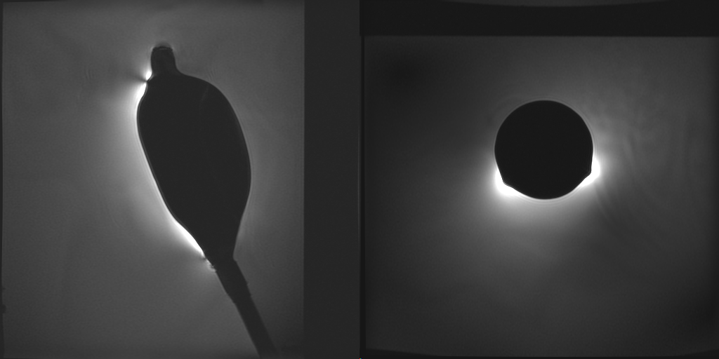
Sagittal and axial views of the endorectal coil in a water container. Orientation of the axial image is matched to that of an axial image of a clinical prostate MR exam.
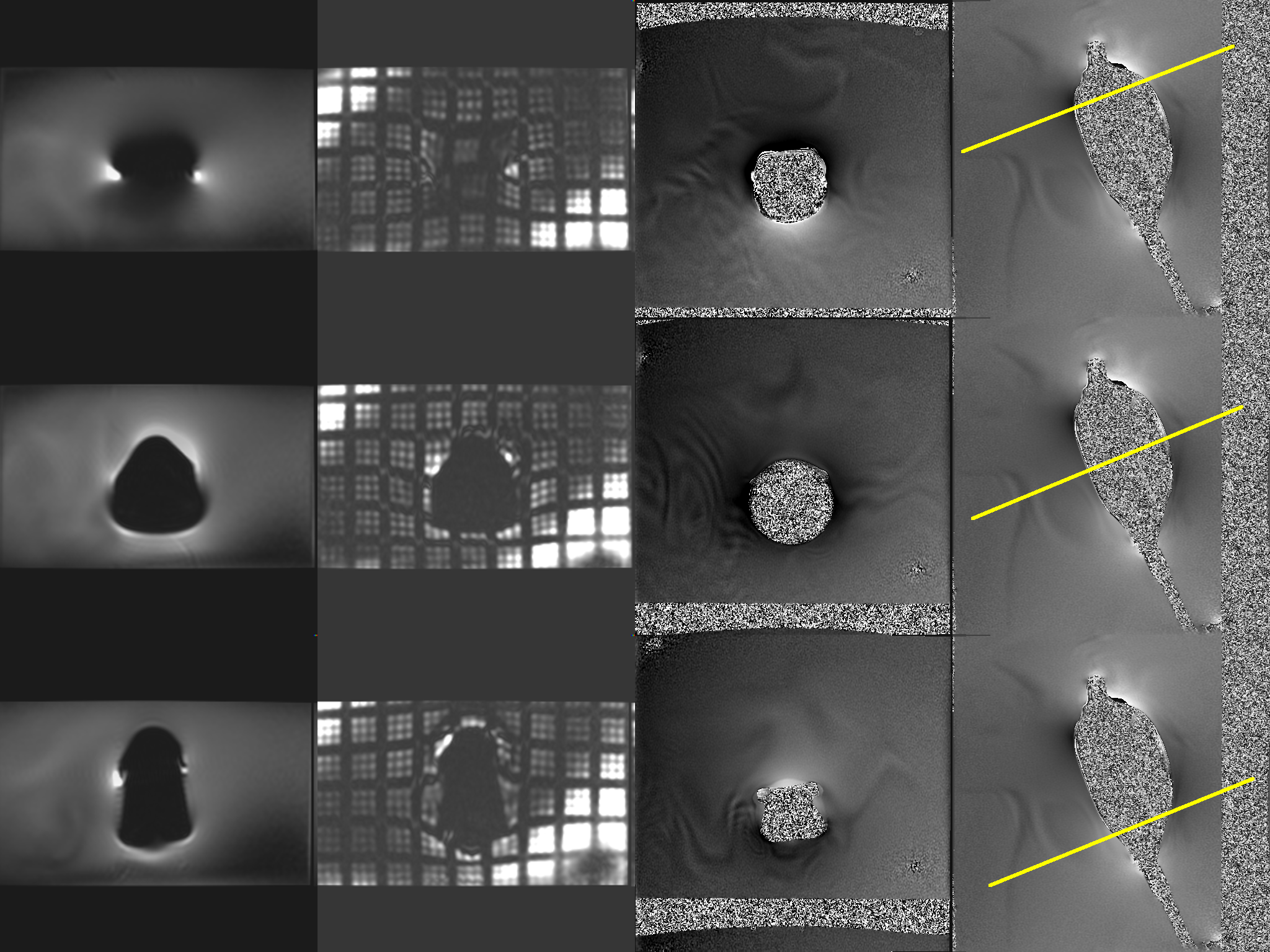
(left to right) Axial conventional DW-EPI, axial SPAMM tagged DW-EPI, axial B0 map, and sagittal B0 map showing a slice at the superior end (top row), center (center row), and inferior end of the coil.
DOI: https://doi.org/10.58530/2023/2009